New findings show the wide-ranging involvement of the brain when you feel sick
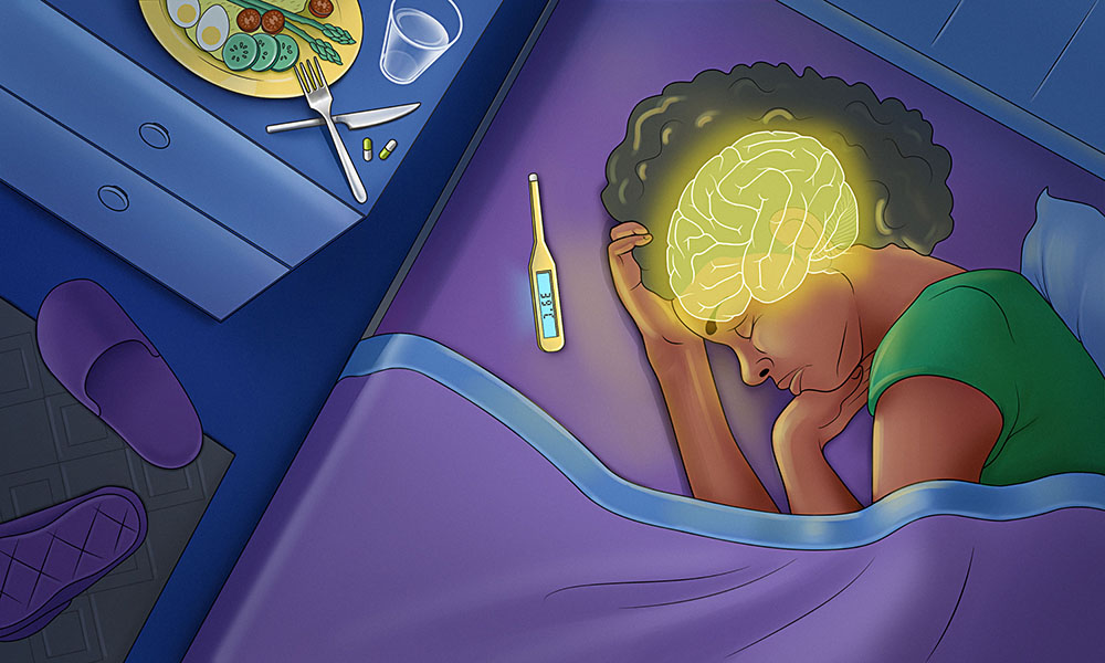
Typical symptoms such as loss of appetite, fatigue, and fever shape the experience of being sick. However, research shows that the brain plays a central role in creating and coordinating this overall sickness state. Credit: Daniela Velasco/EMBL
We are all familiar with that dreaded sensation: a whole-body achiness and fatigue, waves of both bone-chilling cold and acute sweating that cause one to pull cosy blankets closer and only moments later kick them away. A loss of appetite turns us off even our favourite foods. This state may stem from any number of infections, but one thing is certain: our brain is telling us we are sick.
Sickness is an evolutionarily old, protective response that helps the body recover better and faster from illness, and scientists have sought to determine where exactly in the brain these signals originate. Research from scientists at EMBL Heidelberg, applying a new methodology, has provided evidence to show the brain’s involvement is not localised to one or two regions, but widespread.
“Many things are happening. It’s not one specific isolated group of neurons in some hidden area of the brain. It probably requires engagement of large parts of the brain, or multiple brain areas, to achieve this state,” said Gretel Kamm, a former postdoctoral fellow in Robert Prevedel’s team at EMBL, who led the research and brought this hypothesis to the group. The findings have now been published in the journal Current Biology. “Our main hypothesis is that we can understand sickness as a distinct brain state, and that it changes our decisions and behaviour when we have an infection,” she said.
These findings expand the school of thought on brain involvement in sickness, while introducing an efficient, effective way to study this phenomenon further.
Old brain, new brain
Scientists have known for a while that the parts of the brain deep below the surface, such as the hypothalamus and brainstem, help control symptoms associated with infections, but they were unclear about the role the outer layer of the brain played.
The brain has evolved by inheriting foundational circuits from old, ancestor species. Natural evolution over millions of years has modified these circuits and added new structures, allowing the brain new functionalities such as higher order thinking. These older, foundational brain structures are involved in basic functions such as bodily regulation, movement, emotion, and threat response. Not surprisingly, scientists focused on these areas and pathways as they worked to better understand the brain’s involvement in detecting and reacting to infection.
In Kamm’s research, the scientists were specifically interested in the neocortex, the outermost layer of the brain associated with interpreting information, thinking, planning, and controlling voluntary behaviour. This section of the brain also constantly interacts with the older brain structures.
Finding a new way to study illness and the brain
The scientists already knew that when one develops an infection, the immune system naturally produces a small chemical messenger known as prostaglandin E2 (PGE2). This acts much like an alarm system, triggering the symptoms we associate with illness, such as fatigue, chills, fever, and loss of hunger. But it was not clear whether PGE2 produced these effects by activating some parts of the brain’s autonomic system or if the effects emerged from a distributed activation extending beyond it.
In this study, the researchers found that when they injected PGE2 into the mice’s brains, the onset of symptoms was much quicker than with classic methods that mimic infections in the lab. In fact, the onset was almost immediate, compared to hours or days with these other approaches. Additionally, the duration of symptoms decreased significantly as well – only 30-45 minutes.
The scientists then analysed the mice’s behaviour, mapped their brain activity, and made recordings of individual neurons to study how the whole brain’s activity changed during sickness.
“Gretel found an approach to study sickness with many technical advantages over previous techniques,” said Robert Prevedel, senior author on the paper. “We were able to essentially get a very comprehensive picture of sickness in a much shorter period of time.”
The mice quickly developed fever, became sluggish, and ate less. As the scientists looked at which areas of the brain were active, they found that PGE2 had activated many parts of a network known to monitor the body’s internal state. Specifically, they saw individual groups of nerve cells in the insular cortex engaged, suggesting the insular cortex’s central role in the brain as it responds to a state of illness.
The right place for this research
As Kamm described the work involved in this research, she also noted how essential the involvement of EMBL Rome was in this study.
“Our colleagues at EMBL Rome were crucial for our work. Cornelius Gross (Head of EMBL Rome) and Hiroki Asari (former EMBL Rome Group Leader) are well connected within the neuroscience research community, and thus provided important links to key people and resources,” she said. “Additionally, our close interactions with the Rome unit, for example, during seminars, led to important knowledge exchange.”
Gross notably introduced Kamm and her research team to Nicola Renier, who pioneered a method called iDISCO to visualise neuronal activation across the entire brain, using activity markers.
Prevedel also pointed to how EMBL’s EIPOD fellowship and the lab’s own expertise came together to support Gretel’s idea.
“Gretel is taking a different look at a common problem, and she’s a great example of what the EIPOD programme looks for: interdisciplinary postdocs who bring their own ambitious research ideas to EMBL,” Prevedel said. “In her case, the various methods we had established over time in our lab – imaging, electrophysiology, plus others – helped make her idea a reality.”
“The idea of looking at sickness as a brain state is relevant to the general public, and potentially medicine,” Kamm said. “Many people associate sickness with the bacteria or viruses attacking you, but most symptoms we associate with being sick are actually produced by the brain. So the main takeaway is that the whole brain is probably involved in changing our decisions and behaviour when we have an infection.”
After several years of living with HIV, some people are able to produce a special type of antibody known as broadly neutralising antibodies. (Photo: Pixabay)
By Catherine Tomlinson for Spotlight
HIV is known for its ability to outsmart our immune system’s normal defences. A small number of people living with the virus are however able to generate unusually effective immune responses. In this special briefing, Spotlight zooms in on broadly neutralising antibodies, the secret sauce in these immune responses, and their potential role in the future of HIV treatment and prevention.
Our immune systems are highly effective at identifying and fighting off foreign invaders, such as viruses. One way our immune systems does this is by producing antibodies. In short, antibodies recognise viruses and then latch on to them. This blocks the viruses from entering our cells and flags them for destruction by other parts of the immune system.
One of the most remarkable things about our immune system is that it is able to create an enormous variety of such antibodies tailored to each different virus and other disease-causing pathogen that we encounter over our lifetime.
The human immunodeficiency virus (HIV), however, outsmarts our bodies’ normal immune responses by constantly changing the parts of its surface that antibodies recognise. This makes HIV difficult for antibodies to attach to and neutralise.
After several years of living with HIV, some people are able to produce a special type of antibody, known as broadly neutralising antibodies, or bNAbs. These antibodies are more effective at neutralising HIV than regular antibodies because they recognise parts of the virus that change very little, known as ‘conserved regions’. By targeting parts of the virus that are less prone to change, bNAbs are more effective than regular antibodies in identifying and neutralising the constantly changing virus.
“About 20 percent of people living with HIV naturally develop bNAbs, after many years,” explains AVAC, a US-based NGO seeking to advance the development of HIV prevention tools. “By the time bNAbs have developed in these individuals, the constantly mutating HIV has outpaced these defenders, changing too fast and too significantly for bNAbs to be effective in that individual. But that same bNAb, or a combination of them, may work in someone else,” they say.
A vibrant area of research
Researchers first identified bNAbs in a person living with HIV in the 1990s. Since then, they have discovered many more bNAbs and papers and presentations on the topic have become a staple at HIV conferences. At the 2026 International AIDS Conference held in Rio de Janeiro, Brazil, in July, there were 21 abstracts related to the topic.
Since the 1990s, researchers have learned how to replicate and produce bNAbs in the lab. They have conducted early-stage trials showing that bNAbs can be safely administered to people and they have learned how to engineer bNAbs to increase their potency and make them last longer in our bodies.
Currently, researchers are studying whether bNAbs, given by infusion or injection, can prevent HIV infection in people who are HIV negative and control the virus in people who are already living with it. There is also an interesting cross-over with vaccine research, whereby researchers are trying to develop HIV vaccines that prompt the body into making bNAbs.
Before we dig into the details, it is worth stressing that all of this research is still at an early stage. Whereas bNAbs show promise, they have neither set the world alight, nor completely failed. For now, antiretroviral medicines remain the only effective form of HIV treatment, as well as being an extremely effective form of HIV prevention. It is not clear whether bNAbs will ever reach the high bar set by antiretrovirals.
bNAbs for HIV prevention
One of the big HIV stories of the last decade or so has been the use of antiretrovirals to prevent HIV infection. Antiretroviral tablets to prevent HIV infection are already widely available in the public sector, and since June this year, government has been rolling out the six-monthly lenacapavir HIV prevention injection to around 10% of clinics. Such pre-exposure prophylaxis, taking something to prevent infection, is commonly referred to as PrEP.
One of the big hopes for bNAbs is that an infusion of the cells could similarly work as a form of HIV PrEP. The thinking is that these ‘smarter’ immune responses will be more effective than our regular immune responses in recognising and neutralising the shape-shifting virus, and thus clearing it before it can get a foothold in the body.
Substantial research has already been done in this area with two landmark studies, the AMP trials, having garnered the most attention. In the two trials, researchers evaluated an infusion of a bNAb called VRC01 to prevent HIV acquisition in men and transgender people who have sex with men, as well as in cis-gender women. The trials were conducted by the HIV Vaccine Trials Network (HVTN) and the HIV Prevention Trials Network (HPTN).
The AMP trials found that VRC01 did not prevent HIV infection. While this was disappointing, the studies did make a breakthrough by showing that bNAbs could neutralise strains of the HIV virus under certain conditions. While HIV could shape-shift enough to get around VRC01 and cause HIV infection, VRC01 was able to neutralise the HIV strains that were vulnerable to this specific bNAb.
This pattern of bNAbs blocking some, but not all strains of HIV, has been seen in several other studies. It provides both reason for hope, since there is clearly some efficacy, but also frustration, since the efficacy is not nearly as good as what is achieved with antiretrovirals.
Learning from the AMP trials, scientists are now studying whether combining different bNAbs that target a broader range of HIV strains, as well as different regions of the virus’ surface, into a single infusion or injection can be used to prevent HIV.
HVTN and HPTN’s planned Combo-AMP trial will evaluate whether providing people with a combination of different bNAbs can prevent HIV, explained Fred Hutchinson Cancer Center’s Holly Janes at the recent AIDS Conference.
Beyond the AMP trials, the Durban-based research group CAPRISA has also led important studies on the use of bNAbs for HIV prevention. They recently announced the results of a trial called CAPRISA 012C that evaluated the use of a combination of two bNAbs to prevent HIV acquisition in young women in Southern Africa.
Disappointingly, the combination bNAb provided in this trial did not prevent HIV infection. However, CAPRISA reported that “a positive finding was that there was a trend towards protection when the viruses were sensitive to both or one of the two bNAbs compared to when the viruses were resistant to both bNAbs.” In other words, HIV infections occurred more frequently with strains of the virus that were resistant to the bNAbs studied than with strains that were sensitive to them.
“The CAPRISA 012C trial is a culmination of 22 years of research – while it has not led to a new HIV prevention product, it provides valuable information to guide further bNAb research,” said CAPRISA, adding that sensitivity to bNAbs in contemporary circulating viruses will need to be factored into planning future trials of bNAbs.
bNAbs for HIV treatment
bNAbs are also being evaluated as potential treatment for HIV. Researchers are trying to understand whether, under what circumstances, and for how long bNAbs can control the virus in people living with HIV, with the goal of developing products that can achieve long-lasting HIV control without antiretroviral treatment.
This is important because the emotional and psychological burden of having to adhere to a life-long daily pill regimen to treat HIV is a known cause of poor treatment adherence. For infants and young children there are also practical challenges to swallowing and keeping down daily treatment.
One of the main ways that researchers are evaluating the potential of bNAbs to treat HIV is through analytical treatment interruption (ATI) studies. In ATI studies, people living with HIV are given bNAb infusions or injections – sometimes in combinationwith long-acting injectable antiretroviral drugs – and then temporarily taken off their regular antiretroviral treatment under close medical observation.
Researchers then monitor how long HIV remains suppressed in order to learn whether and how well bNAbs can control HIV infection.
The results from ATI studies, including the RIO and FRESH trials, have been tantalizing. bNAb infusions have allowed some study participants to remain off antiretroviral treatment for more than a year without the virus rebounding in their bodies.
Yet, the studies have also raised questions about how and why bNAbs have such mixed efficacy. Researchers are still trying to understand why some people are able to maintain periods of viral control after receiving bNAbs, while others experience rapid viral rebound. The reasons for this appear to extend beyond a person’s sensitivity to the specific bNAbs being used to also include other factors related to the characteristics of one’s HIV infection and immune response.
At the 2026 AIDS Conference, Michel Nussenzweig, senior physician at the Rockefeller University, told delegates that research so far indicates that bNAb therapy is more likely to deliver periods of post-treatment control in individuals with a less diverse HIV reservoir, pre-existing autologous antibodies, and pre-existing stem cell like CD8+ T cells.
Scientists are now considering whether the factors associated with bNAb treatment success can be boosted through other interventions, said Nussenzweig.
Another important area of research is whether bNAbs can be used as a form of treatment for infants and young children living with HIV. An infusion or injectable treatment could be a gamechanger for this group, given the challenges faced by caregivers in getting infants and young children to swallow and keep down daily antiretroviral treatment.
The Tatelo and Tatelo Plus studies conducted in Botswana were set up to evaluate whether young children given bNAbs can maintain viral suppression after stopping antiretrovirals. Results from the Tatelo study reported in 2022 showed that some children (44%) who received a combination of two bNAbs were able to maintain a period of viral control (24 weeks) after stopping HIV treatment. The Tatelo Plus study, now underway, is evaluating whether and for how long a combination of three bNAbs can maintain HIV suppression in young children after antiretrovirals are stopped.
bNAbs for HIV vaccination
While bNAbs have not yet been shown to be a practical and effective form of HIV prevention or treatment, research has demonstrated that, under the right conditions, they can protect against and suppress HIV strains that are susceptible to them.
These findings have generated excitement about using bNAbs as a target for HIV vaccines. Unlike research into bNAbs for PrEP or HIV treatment, in which laboratory made bNAbs are infused or injected directly into our bodies, some HIV vaccine researchers are trying to figure out how to trigger our bodies to produce their own bNAbs.
In other words, vaccine researchers are trying to make our bodies, rather than laboratories, the factories that make bNAbs against HIV.
At this stage, scientists do not expect that a single vaccination will be able to trigger our bodies to produce mature bNAbs capable of combating HIV. Instead, they anticipate that a vaccine protocol that involves a series of vaccines will be needed to coax our immune systems to produce mature bNAbs.
While this branch of research remains at its early stages, many HIV researchers are hopeful that it may one day produce an effective vaccine protocol against HIV.
One study to watch is a Phase 1 safety and dosing trial launched by the International AIDS Vaccine Initiative (IAVI) and partners in South Africa at the end of 2025. “The hypothesis being tested is that highly specialized vaccine immunogens, delivered in a specific sequence, can target certain B cells within the immune system and coach them toward the production of broadly neutralizing antibodies against HIV,” says IAVI, adding “scientists widely believe that a vaccine inducing broadly neutralizing antibodies (bNAbs) could provide broad protection against many strains of HIV.”
Where to from here?
Since the first bNAbs against HIV were discovered in the 1990s, scientists have made important, but incremental, progress towards translating these immune responses into tools that can prevent and treat HIV.
As we’ve seen in this Spotlight special briefing, research into bNAbs for HIV treatment is arguably the furthest along, with bNAbs already demonstrating the ability to control HIV during extended periods of antiretroviral treatment interruption in some people. But why some people respond to this treatment and not others remains uncertain. This is an important area for future research.
In the HIV prevention space, bNAbs have delivered protection against HIV strains susceptible to the specific bNAbs studied, but this protection has not been broad enough to protect against HIV infection by the highly diverse, mutating virus. Hope however remains that combining different bNAbs that target different conserved regions of the HIV virus, as well as currently circulating viruses, could broaden protection enough to prevent HIV infection. Here too, as with attempts to develop vaccines that spark the production of bNAbs, it is imperative that the research continues.
Of course, even if scientists can crack the code and find a way to produce highly effective bNAbs, the road ahead might not be a smooth one. For these products to have an impact in the developing world, where they are most needed, they will have to be cost-effective compared to cheap antiretroviral therapy. They will also have to be easy to administer in often stretched and under-resourced healthcare systems.
While much remains to be done, the scientific leads are certainly there, waiting to be explored.
IL-1 pathway prevents a normally harmless fungus developing into a fatal infection
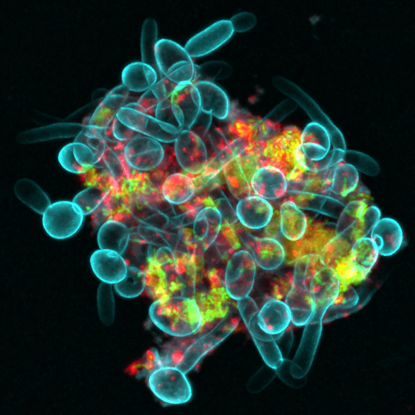
Bacterial-Fungal Clusters in Saliva. An interkingdom assemblage formed by fungi (Candida albicans in blue), bacteria (Streptococcus mutans in green), and bacteria-derived extracellular polymers (α-glucans in red) in human saliva. Credit: Zhi Ren, University of Pennsylvania. NIH support from: National Institute of Dental and Craniofacial Research (NIDCR)
A study from King’s College London provides the first potential clue as to why only certain patients with weakened immune systems, including those undergoing chemotherapy or living with HIV, are at risk of life-threatening Candida albicans infections. Candida albicans is a fungus that normally lives harmlessly in areas such as the mouth and gut but can sometimes spread through the body and cause fatal disease. If the results, published in Nature Microbiology, are confirmed in humans, the results could provide a test to understand who is at risk of developing fatal fungal infections and represent a potential therapeutic target to reduce the risk of developing the disease.
Fungal infections kill more than 2.5 million people each year, with Candida albicans alone killing almost a million. However, until now we knew little about why fungi escape their natural location in mouths and gut and cause life-threatening disease in around 10% of patients who have a weakened immune system.
The scientists focused on a signal produced by the immune system to trigger symptoms to fight off infection, IL-1. Mice which were genetically modified not to produce IL-1 experienced severe disease when exposed to Candida albicans. The findings suggest that the IL-1 immune pathway is critical in preventing Candida albicans from spreading around the body and causing life-threatening disease.
They investigated this further by injecting IL-1-deficient mice with a drug that removes neutrophils, a type of white blood cell that is among the first to respond to infections and help fight threats such as bacteria and fungi. This allowed the researchers to mimic the weakened immune system seen in some immunocompromised patients. By then introducing Candida albicans to the mouths of those mice, they for the first time observed the fungus spread throughout the body and cause fatal disease, confirming that IL-1 is critical in preventing disease spreading.
While the study focused specifically on Candida albicans, the researchers say the IL-1 immune pathway may be a broader defence mechanism that helps keep fungi normally found in healthy microbiomes from spreading and causing fatal disease, and further research is needed to confirm whether this applies to other fungal species.
Understanding what causes fungi that are naturally present in our microbiomes, such as Candida albicans, to cause life-threatening disease could help spot at-risk patients earlier. The researchers suggest that, if confirmed in humans, the findings could lead to a test that identifies which immunocompromised patients have low levels of IL-1 and so are at risk of Candida albicans escaping their microbiomes and causing disease before it happens.
While drugs such as antibiotics are currently used to treat life-threatening fungal diseases, more targeted therapies are needed that tackle the root cause of infection. The researchers suggest future clinical studies in humans could test whether drugs targeting IL-1 could work as a personalised therapy for preventing life-threatening Candida albicans infection.
A growing body of research has shown that autoimmunity influences certain psychiatric disorders. A new study by Nemani et al., currently in preprint, has shown that schizophrenia is strongly associated with an elevated level of autoantibodies that target the central nervous system. Using Rapid Extracellular Antigen Profiling (REAP) to screen 352 patients against 971 controls, the researchers found that schizophrenia is marked by an increased autoantibody burden that tracks with disease severity and duration, nearly doubling in the most chronic cases.
These immune responses are present near the start of the illness and tend to increase as the disease progresses, particularly targeting neuronal ion channels and synaptic proteins. Notably, certain autoantibodies appear to compromise the blood–brain barrier, which may further expose the brain to peripheral immune attacks.
The study also discovered that patients with a higher autoantibody burden respond less effectively to standard antipsychotic treatments like risperidone. However, clinical trials showed that these antibody levels significantly declined during successful treatment courses. These findings suggest that humoral autoimmunity is a core component of the disorder, potentially offering new pathways for immune-based therapies.
Strange “chimeric” RNA once thought to be the product of cancer is actually an important controller of women’s health, including influencing their susceptibility to infectious disease and autoimmune disorders, new University of Virginia School of Medicine research suggests.
UVA’s Hui Li, PhD, and colleagues have identified a chimeric RNA called UBA1-CDK16 that is found only in women. This RNA plays important roles in their blood cell development and in determining the severity of diseases such as COVID-19, the scientists found. The findings, published in Science Advances, could open the door to blood tests to help diagnose diseases or identify women at greatest risk for bad outcomes.
“Chimeric RNAs are RNA molecules composed of parts from different genes,” said Li, of UVA’s Department of Pathology and the UVA Comprehensive Cancer Center. “They were once believed to be cancer-specific. However, our research shows that they can also be part of normal physiology and play important roles in human health.”
Powerful Chimeras
RNA provides instructions for our cells, telling them what to do based on the genetic material, called DNA, that we inherit from our parents. Chimeric RNAs were long thought to be mistakes, as they are made up of instructions mashed together from different genes. This is why they were believed to be a byproduct of cancer; cancer itself is the result of cellular copying mistakes.
Li’s discovery, however, suggests that UBA1-CDK16 plays important roles in maintaining women’s health and in controlling their immune systems. This chimeric RNA is found only in women because women have two X chromosomes, while men have an X and Y. Normally, one of the two X chromosomes found in women’s cells are inactive. But Li found that the inactive X chromosome produces this peculiar chimeric RNA that he could identify in women’s blood.
Based on his findings, Li believes UBA1-CDK16 plays an important role in regulating blood cell formation. But his work also suggests the chimera may play an important role in the immune system’s response to infection. He found that the chimeric RNA was lost in 50% women who developed severe COVID-19 infections, while it was present in women who were asymptomatic. Further, the decrease in chimeric RNA correlated with the increasing severity of the infection.
Li suspects that the chimeric RNA may play an important role in governing the development of immune cells called neutrophils that act as the body’s first responders to infection. (Neutrophil count has already been identified as a way to predict how patients will fare against COVID-19.)
“As humans share similar number of genes with fruit flies and worms, gene number does not explain why we are much more sophisticated than these lower organisms” Li said. “We believe chimeric RNAs are another means to expand the functional genome, without an actual increase in gene number.”
Li’s findings suggest that the chimeric RNA also may serve as a natural brake to protect women from excessive autoimmune activity. Women are far more likely to suffer autoimmune disorders than men, and Li is urging additional research to better understand the role chimeric RNA could be playing – and how it could be targeted to improve patient outcomes.
“This finding highlights there is another layer of control for gene expression,” Li said. “These chimeric RNAs may represent a hidden repertoire for biomarkers and therapy targets as well.”
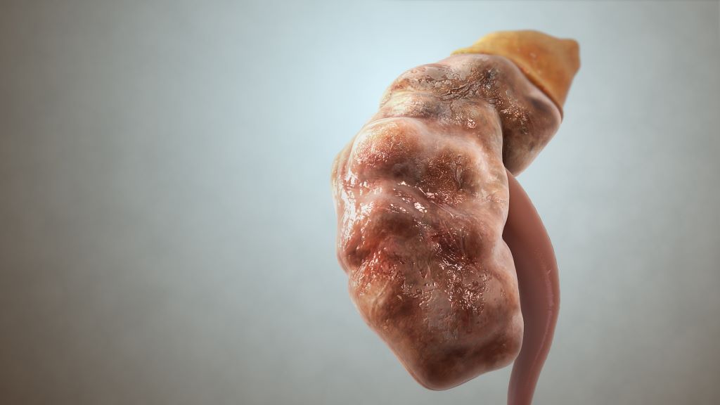
Kidney damage is a serious complication affecting individuals with lupus, an autoimmune disease where immune B cells malfunction and produce antibodies that attack the body’s own cells, tissues, and organs.
B cells, when they make autoantibodies, have been blamed for the illness, prompting the development of several FDA-approved medications that target them. However, lupus nephritis is inevitable in more than half of patients with lupus and B cell depletion is often ineffective therapeutically.
Now, in a study published April 20 in Immunity, Yale scientists have found that the kidney damage culprit is actually a specific T cell – the CD8 T cell.
“People have been, to some extent, ignoring CD8 T cells because of their focus on B cells and the production of autoantibodies,” says lead author Jafar Al Souz, an MD-PhD student in the lab of Joseph Craft, MD, Paul B. Beeson Professor of Medicine (Rheumatology) and professor of immunobiology at Yale School of Medicine. “But we need to think more deeply about why current therapies fail in some patients.”
T cells drive kidney damage in lupus
Previous work in the Craft Lab has shown that blocking T cells’ activity in mouse models of lupus could spare the animals from kidney injuries. That result prompted Al Souz to look closer at this subset of the immune cells.
“What I saw was that these T cells in the sick kidney had a killer phenotype; they were very activated with the potential to cause kidney damage,” he says. Nearly all of these cells were CD8 T cells – also known as cytotoxic T lymphocytes – which are generally tasked with identifying and killing pathogens. That they are aggravated in the kidneys of mouse models of lupus shows that the cells see the kidney as a foreign invader that needs to be destroyed, Al Souz says.
He also found that when the CD8 T cells were depleted, the kidney function was maintained.
Using single-cell multiomics, a high-resolution technique to simultaneously measure the various molecular characteristics of a cell, analysing simultaneous RNA production, gene regulation, and T cell identity, the researchers pinned down the origin of the CD8 T cells to the renal lymph nodes.
“What gave us the biggest clue was that the CD8 T cells in the kidney had very strong overlap with the cells in renal lymph nodes,” Al Souz says. T cells normally undergo rapid proliferation, where one T cell multiplies and generates identical offspring that target the same antigen proteins. “We know that once a T cell is activated in renal lymph nodes, it will leave, go to the circulation and then enter kidneys. So, the fact that we saw identical T cells in renal lymph nodes, circulation, kidneys tells us that the lymph nodes were actually the site of origin.”
What is unique about these CD8 T cells is their capacity to self-renew like stem cells, a discovery that sheds light on a long-held mystery of why it’s hard to stop treatment in patients with lupus. “Constant therapy is needed because these CD8 T cells in the lymph nodes continually supply the kidney with T cells that can damage the organ,” says Craft, who is also the director of the Colton Center for Autoimmunity at Yale.
Targeting T cells for treatment
To draw parallels in humans, the researchers used single-cell RNA sequencing to analyse biopsies from 156 patients with lupus nephritis and 30 healthy individuals. Just as they found in mice, the researchers identified kidney-infiltrating CD8 T cells with the stem-like property in the patients. Unlike in healthy individuals, there was a significantly higher proportion of CD8 T cells that were always active and ready to cause damage in the patients’ kidneys.
“Generally, CD8 T cells are good at pumping the brakes and limiting damage potential upon chronic activation. But we found that in lupus, even when molecules that should stop these T cells from damaging healthy cells are present, it didn’t make the T cells lose function and the capacity for kidney injury,” Al Souz says.
The tendency for T cells to attack a person’s own cells is a typical autoimmune response. For example, in type 1 diabetes – an organ-specific autoimmune disease – T cells mistakenly recognise pancreatic beta cells as foreign and destroy them, thereby stopping insulin production. In cancer and chronic diseases, the ability for T cells to replenish could be beneficial, leading to the elimination of pathogens and tumours. However, the role of T cells in systemic autoimmune diseases like lupus has not been fully described.
“Our results show that there’s more to it than what we understand,” Al Souz says.
By identifying the real culprit, the researchers hope that future treatment of lupus nephritis will be more targeted. “The first step is knowing that it’s abnormal in the first place,” Craft says. “Now, we can think about strategies to lower the number of active CD8 T cells back to the normal range.”
Memory cells in the nose slow the influenza virus as soon as it enters the body. They reduce viral levels and may help protect against more severe illness. A new study from the University of Gothenburg may help guide the development of better influenza vaccines.
Today’s influenza vaccines are given as injections in the arm and mainly stimulate immune responses in the blood. At the same time, researchers are working to develop influenza vaccines that can be administered through the nose – an effort this study helps inform. The goal is to strengthen the body’s defences where the virus first encounters the immune system.
Memory cells remain in the nose
The researchers identified a group of memory cells, known as CD4 memory T cells, that remain in nasal tissue after an influenza infection. When the body encounters the virus again, these cells can rapidly reactivate and help other parts of the immune system fight the infection. The study shows that these cells can reduce viral replication in the nose and thereby contribute to better protection against illness.
“We show that CD4 memory T cells can remain in nasal tissue after an influenza infection and rapidly reactivate when the virus returns. This means the immune system can respond directly at the site where the virus first enters the body,” says Nimitha R. Mathew, a researcher at the Sahlgrenska Academy, University of Gothenburg, and one of the study’s lead authors.
In studies in mice, the researchers showed that these immune cells help limit viral levels and reduce tissue damage in the nose during a subsequent infection.
Similar cells found in humans
The researchers also analysed cells from the nasal mucosa of healthy adults. There, they found the same type of influenza-specific memory cells, suggesting that a similar local immune defence may also exist in humans. The study is published in the Journal of Experimental Medicine.
“Many people likely already have these kinds of memory cells in their noses after previous infections, but they are not always enough to stop the virus completely. The important thing about our findings is that we now know which immune cells can limit the virus where infection begins. That knowledge can be used when developing future nasal vaccines,” says Davide Angeletti, professor at the Sahlgrenska Academy, University of Gothenburg, and also one of the study’s lead authors.
Article: Nasal CD4⁺ tissue resident memory T cells provide cross protective immunity to influenza; 10.1084/jem.20251793
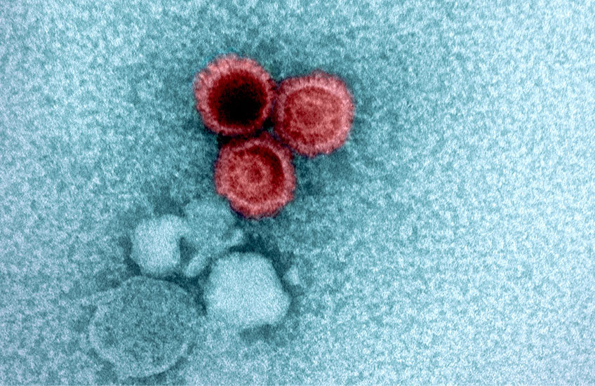
An electron micrograph showing three Epstein-Barr virions in red. Image: NIAID
Some viruses are cleared by the immune system within days, while others lurk in our bodies for a lifetime and reemerge later to cause new problems. How and why viral levels in the body change over time – and the health impacts of these changes – are only just starting to become clear.
A team led by scientists at Harvard Medical School, Massachusetts General Hospital, Brigham and Women’s Hospital, and the Broad Institute of MIT and Harvard recently reported the largest analysis to date of the human DNA virome – the collection of viruses in the body that have DNA as their genetic material.
The researchers tracked the viral load – the amount of viral DNA – for several common viruses in blood and saliva from over 900 000 individuals. They saw large variations in viral load from person to person depending on age, sex, lifestyle, and other factors, and discovered dozens of genetic factors strongly associated with viral load.
The team concluded that genetics plays a role in determining whether the effects of these viruses extend well beyond an initial infection.
“We’re getting to the point now where we can use human genetics to try to answer fundamental questions about pathology resulting from viruses,” including whether a virus is likely to play a role in causing cancer or other diseases later in life, said first author Nolan Kamitaki, research fellow in genetics in the Blavatnik Institute at HMS.
Study co-senior author Po-Ru Loh, HMS associate professor of medicine at Brigham and Women’s and an associate member at the Broad Institute, is broadly interested in the scientific information that can be mined from population-level datasets of DNA sequences that are readily available to researchers.
It turns out that these datasets capture information about the genomic material that people inherit, he said, as well as the makeup of their oral microbiomes, viruses hiding in their bodies, and acquired mutations in their DNA. Sometimes, scientists can link this information to downstream health consequences.
Recently, for example, Loh and Kamitaki analyzed whole-genome sequences from saliva samples collected from more than 12,500 individuals to investigate how genetics shapes the oral microbiome.
In their new study, the researchers analyzed whole-genome sequencing data from individuals in three biobanks: the UK Biobank, the National Institutes of Health’s All of Us Research Program, and Simmons Foundation Powering Autism Research for Knowledge. They tested blood and saliva samples for the viral loads of Epstein-Barr virus, two other human herpesviruses (HHV-6 and HHV-7), Merkel cell polyomavirus, as well as three common anelloviruses that are present in about 90% of people throughout life without causing disease.
They found that each virus had a markedly different trajectory over a lifetime. The viruses appeared most rapidly during the first several years of life, likely following primary infection. However, Epstein-Barr virus became more prevalent with age, while HHV-6 became less prevalent after childhood, possibly indicating more control by the immune system over time. The prevalence of HHV-7 similarly decreased sharply in middle age.
The team also found that Epstein-Barr viral load went up in the winter and down in the summer, while HHV-7 viral load showed the opposite pattern. Smoking was strongly associated with a higher Epstein-Barr viral load, nearly double in heavy smokers compared with nonsmokers, whereas smoking was associated with a lower HHV-7 viral load.
Notably, men consistently had a higher viral load in their blood and saliva than women across all seven viruses.
The role of genetics revealed
The researchers determined that many of the genetic factors most strongly linked to viral load were related to how the immune system responds to viruses and how infected cells dodge immune attacks.
Thus, the findings highlight the immune system’s role in controlling viral load in the body, showing how much immune responses can vary over time and between people.
“It’s amazing how much DNA can teach us about dynamic biological processes and the ways in which our habits, our genes, and our biology shape those processes,” said co-senior author Steven McCarroll, the Dorothy and Milton Flier Professor of Biomedical Science and Genetics at HMS and director of genomic neurobiology for the Stanley Center for Psychiatric Research at the Broad Institute.
The researchers identified the largest number of genetic associations for Epstein-Barr virus – which is thought to be a leading cause of multiple sclerosis and a risk factor for certain cancers – so they dug a little deeper. Their analysis revealed that the lifetime viral load for Epstein-Barr didn’t influence the risk of developing multiple sclerosis. Instead, the body’s immune response to Epstein-Barr is likely what increases the risk of the disease.
The researchers did find evidence that high Epstein-Barr viral load is a casual risk factor for Hodgkin lymphoma, a finding that needs further study in cell and animal models in the lab, they said.
“This finding is an example of why virus research in large genetic biobanks is important,” Kamitaki said.
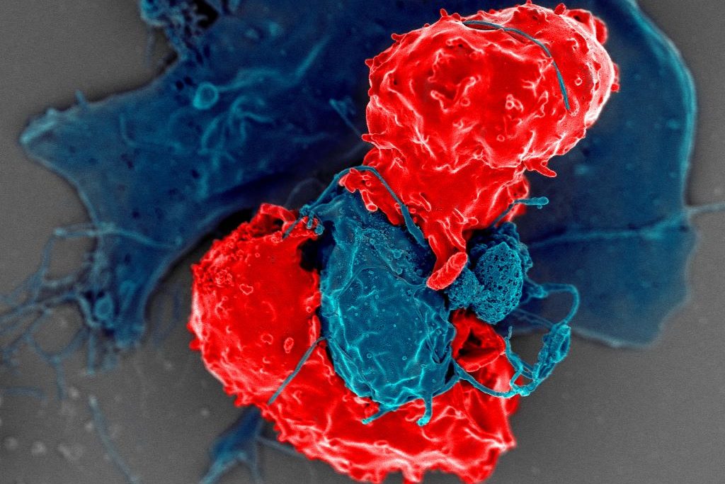
Scanning electron microscope image of T regulatory cells (red) interacting with antigen-presenting cells (blue). T regulatory cells can suppress responses by T cells to maintain homeostasis in the immune system. Credit: National Institute of Allergy and Infectious Diseases/NIH
Cedars-Sinai Health Sciences University investigators have identified for the first time a protein’s role as a “dimmer switch” that can calm an overactive immune system and restrain harmful inflammation. The protein, Butyrophilin 2A2 (BTN2A2), interacts with a key molecule that controls the strength of T-cell responses.
The findings, published inNature Communications, define a unique pathway that helps balance immune activity and could be harnessed to limit damage caused by a variety of autoimmune diseases.
In laboratory mice, loss of BTN2A2 led to exaggerated immune reactions and an increase in damaging kidney inflammation called glomerulonephritis. Treatment with BTN2A2 reduced disease severity by increasing immune-regulating T cells and lowering inflammation.
Supporting laboratory experiments in human T-cells demonstrated similar immune-calming effects.
“Glomerulonephritis remains a leading cause of chronic kidney disease and kidney failure worldwide, with limited treatment options,” said Ananth Karumanchi, MD, co-corresponding author of the study and director of the Renovascular Research Center at Cedars-Sinai. “Our findings provide a strong foundation for future studies aimed at modifying immune-driven kidney disease rather than simply managing its symptoms. The pathway could also be targeted in a range of autoimmune and inflammatory diseases including rheumatoid arthritis, multiple sclerosis, inflammatory bowel disease, and transplant rejections.”
Other Cedars-Sinai authors include Shafat Ali, Anders H. Berg, Michifumi Yamashita, Ambart E. Covarrubias, Jordan Mundell, Pranali N. Shah, Ruan Zhang, Vincent Dupont, Bong-Ha Shin, Shen Yang, Madhusudhanarao Katiki, Ramachandran Murali, Margareta D. Pisarska, Ravi Thadhani, Peter S. Heeger and Stanley C. Jordan
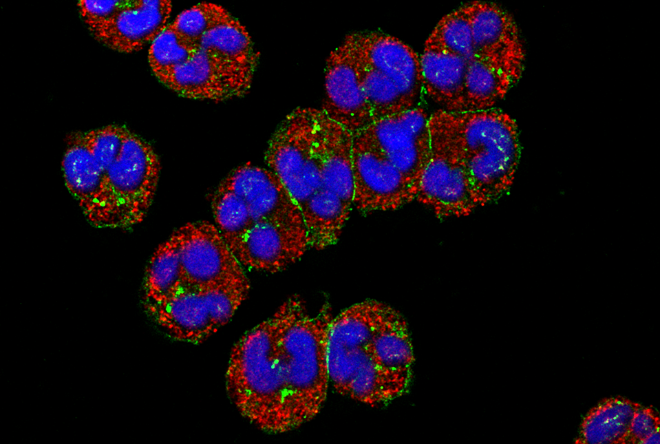
Human neutrophils visualised under a confocal microscope with cell membrane (red) and nucleus (blue). When faced with an infection during food scarcity, stress hormones trigger an immune response dependent on neutrophils, abundant cells that act as immediate, short-lived defenders. Credit: Thai Tran, National Institute of Arthritis and Musculoskeletal and Skin Diseases
When food is scarce, stress hormones direct the immune system to operate in “low power” mode to preserve immune function while conserving energy, according to researchers at Weill Cornell Medicine. This reconfiguration is crucial to combating infections amid food insecurity.
The answer could be important in helping those who are food insecure and face the risk of infectious diseases every day. “Mounting an immune response against infections requires a lot of energy. We have discovered a coordinated system that upholds immune function by shifting the composition and metabolism of immune cells,” Dr Collins said.
The results, published in Immunity, found that mice on a calorie-restricted diet fought off infection as well as mice that were fully fed, but did so while using very little glucose. This was possible thanks to glucocorticoids, stress hormones known for their role in regulating blood glucose. The researchers determined that glucocorticoids acted like master conductors, reorganizing immune cells and their energy usage to provide a survival advantage.
The research was co-led by Luisa Menezes-Silva, a visiting graduate student from the University of São Paulo, Brazil; Dr Mingeum Jeong, a postdoctoral associate; and Dr Seong-Ji Han, a research associate, all in the Collins lab at Weill Cornell.
Shifting Priorities
To understand the complex interactions involved in an immune response during scarcity, Dr Collins and his team put mice on a 50% restricted-calorie diet and then exposed the animals to bacteria that infect the gut. The mice that were fed a standard diet experienced a metabolic crash – their blood glucose levels and body weight plummeted.
The researchers had expected this would happen to all the animals because mounting an immune response can consume up to 30% of the entire body’s fuel reserves. But in the calorie-restricted mice, the immune system appeared to be functioning perfectly well without using much glucose.
To unravel this enigma, the researchers inventoried the immune cells of the infected animals and discovered that T cells, which normally target invading microbes, were depleted in the calorie-restricted mice. Instead, short-lived neutrophils, which serve as the body’s first responders to infection, were ramped up to twice the normal amount and had measurably enhanced pathogen-killing abilities. The cells seemed to be operating in energy-saving mode, consuming much less glucose than neutrophils from well-fed animals.
“So, this hormone rewires the immune system to eliminate the infection while keeping blood sugar from dropping, which rescues the calorie-restricted animals from malnutrition,” said Dr Collins.
Stress Hormones Lead the Charge
The researchers are breaking new ground by outlining how a sudden fall in food intake triggers glucocorticoid levels to rise, resulting in two major shifts. First, the body repositions certain immune cells – especially naïve T cells – into the bone marrow, which becomes a kind of “safe house” for when the cells are needed. Second, during an infection, glucocorticoids tilt the immune response away from energy‑intensive T cells toward neutrophils, abundant cells that act as immediate, short-lived defenders.
Beyond clearing a current infection, glucocorticoids prepare the immune system for repeat encounters with infectious agents. While the hormones direct killer T cells to stand down and neutrophils to step up, they also ensure memory T cells are preserved for future confrontations.
“Glucocorticoids reduce the immune cells that use up the most energy, while saving those that are critical for protection against future infections,” Dr Collins said. “So, these hormones are involved in every step of the infection-fighting process.”
“Since glucocorticoids are induced not only by nutrient restriction but also by any form of stress, our findings might have broader applicability,” said Dr Collins.
In the meantime, he and his team plan to explore what causes the system to fail when the degree and duration of calorie restriction are more severe. “We looked at reduced food intake over three weeks,” he said. “But when you cross the threshold into malnutrition, the whole system breaks down.” Understanding this collapse could inform better strategies to prevent infectious disease and infection-driven malnutrition in vulnerable populations.