Day: August 5, 2026

Fructose Identified as a Surprise Driver of Metastasis

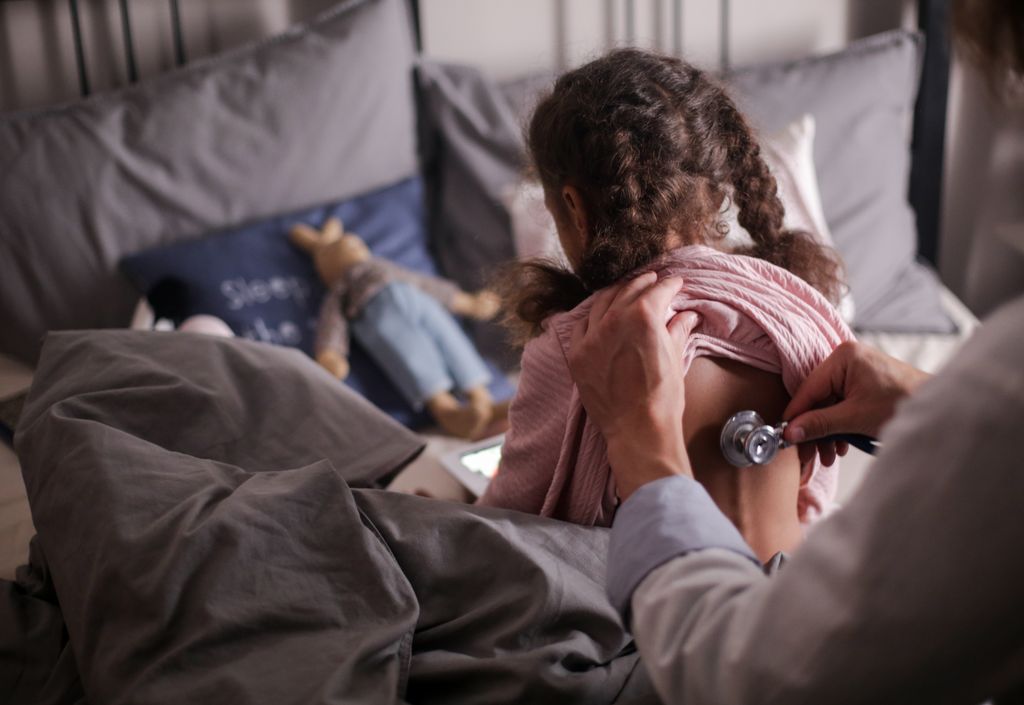
Photo by Sharon Mccutcheon on Unsplash

A new study from The Wistar Institute has uncovered an unexpected link between fructose and the spread of an aggressive form of ovarian cancer. Published in Nature Aging, the study found that cancer cells not killed by chemotherapy send signals to neighbouring tumour cells, driving metastasis. The researchers identified fructose as a key messenger in this process, revealing a previously unrecognised way that treatment-surviving cancer cells may promote metastasis.

“Some cancer cells that survive chemotherapy aren’t dividing anymore, but they’re still biologically active,” said Aidan Cole, PhD, a postdoctoral fellow in the lab of Katherine Aird, PhD, at The Wistar Institute and first author on the study. “Instead, they continue to release molecules that send signals to nearby cells. Our study is among the first to show that a nutrient – in this case, fructose – can act as one of those signals.”

Ovarian cancer is treated almost universally with platinum-based chemotherapy, and many patients respond well at first. However, the disease recurs in most patients and almost always spreads through the abdominal cavity. That spread, called metastasis, accounts for roughly 90% of deaths from the disease.

Prior research has suggested that cancer cells not killed by chemotherapy play a role in recurrence in part through the ability of these cells to release a complex mix of signalling molecules. Cole and his colleagues began their research by designing a unique experiment: they collected the molecules released by chemotherapy-surviving cells and found these factors alone could significantly increase the spread of cancer cells.

“As far as we know, this is the first time anyone has shown, in a preclinical model rather than just a dish, that it’s the molecules these cells release – not the cells themselves – that drive the cancer’s spread,” said Cole.

That finding sent the team looking for what was being released that caused the cancer cells to spread. Fructose, it turned out, was being made by the surviving cells and sent as the signal for increased spread. Even more interesting, they found that without chemotherapy treatment, consuming the high levels of fructose found in sugary drinks can also signal cancer to spread. The fructose finding is especially compelling because it raises the possibility that simple dietary changes could help shape how cancer progresses. This is particularly important given the prevalence of fructose consumption in the United States, with high fructose corn syrup accounting for ~8-20% of the daily caloric intake in some individuals. Unlike many cancer risk factors outside of patient control, fructose consumption can be modified by dietary choices. While the effectiveness of limiting fructose intake hasn’t yet been tested directly in patients, the study raises the possibility that nutrition could influence cancer progression in previously unrecognised ways.

The researchers next aimed to understand why fructose increases cancer cell spread. Through a variety of large-scale analytical techniques, including the use of a CRISPR screen, they discovered that fructose suppresses cholesterol within the neighbouring cells. Cholesterol is important for cells to stick to each other like a biological glue, so decreased cholesterol allows cells to more easily escape and spread.

The finding that fructose lowers cholesterol production has important clinical implications. Statins, which are taken by 39 million people in the United States, lower cholesterol production. The team found that statins alone decreased the glue between cells to promote escape. The research team is now examining whether these medications could be interfering with the effects of chemotherapy – though they stress this isn’t a reason for patients to stop taking them.

“We haven’t tested this effect in patients yet, but it raises questions about combining cholesterol-lowering drugs with chemotherapy, especially since ovarian cancer is most common in postmenopausal women who are often already on statins,” said Katherine Aird, PhD, professor and co-leader of the Molecular and Cellular Oncogenesis Program in the Ellen and Ronald Caplan Cancer Center at The Wistar Institute, and senior author of the study.

The researchers are also looking into how this mechanism might extend beyond ovarian cancer.

“We think other cancers that spread within the torso – pancreatic, colon, liver – could behave similarly. We can’t call it universal yet, but we think the effects are not just limited to ovarian cancer,” said Aird.

She and Cole have already started designing follow-up experiments to test the reproducibility of their findings in a variety of other cancer types.

Source: The Wistar Institute
Researchers Find Immune Pathway that Keeps Candida in Check

IL-1 pathway prevents a normally harmless fungus developing into a fatal infection

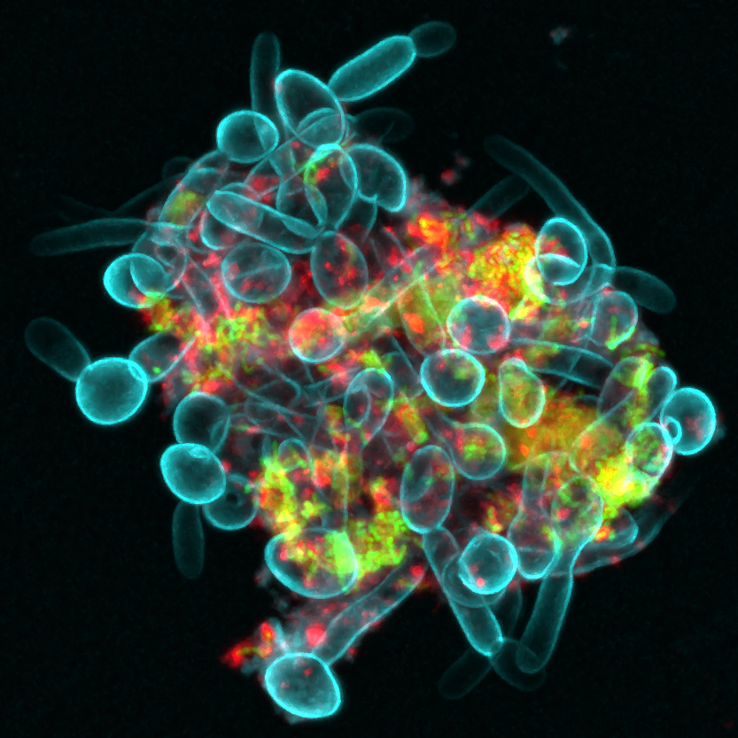
Bacterial-Fungal Clusters in Saliva. An interkingdom assemblage formed by fungi (Candida albicans in blue), bacteria (Streptococcus mutans in green), and bacteria-derived extracellular polymers (α-glucans in red) in human saliva. Credit: Zhi Ren, University of Pennsylvania. NIH support from: National Institute of Dental and Craniofacial Research (NIDCR)

A study from King’s College London provides the first potential clue as to why only certain patients with weakened immune systems, including those undergoing chemotherapy or living with HIV, are at risk of life-threatening Candida albicans infections. Candida albicans is a fungus that normally lives harmlessly in areas such as the mouth and gut but can sometimes spread through the body and cause fatal disease. If the results, published in Nature Microbiology, are confirmed in humans, the results could provide a test to understand who is at risk of developing fatal fungal infections and represent a potential therapeutic target to reduce the risk of developing the disease.

Fungal infections kill more than 2.5 million people each year, with Candida albicans alone killing almost a million. However, until now we knew little about why fungi escape their natural location in mouths and gut and cause life-threatening disease in around 10% of patients who have a weakened immune system.

The scientists focused on a signal produced by the immune system to trigger symptoms to fight off infection, IL-1. Mice which were genetically modified not to produce IL-1 experienced severe disease when exposed to Candida albicans. The findings suggest that the IL-1 immune pathway is critical in preventing Candida albicans from spreading around the body and causing life-threatening disease.

They investigated this further by injecting IL-1-deficient mice with a drug that removes neutrophils, a type of white blood cell that is among the first to respond to infections and help fight threats such as bacteria and fungi. This allowed the researchers to mimic the weakened immune system seen in some immunocompromised patients. By then introducing Candida albicans to the mouths of those mice, they for the first time observed the fungus spread throughout the body and cause fatal disease, confirming that IL-1 is critical in preventing disease spreading.

While the study focused specifically on Candida albicans, the researchers say the IL-1 immune pathway may be a broader defence mechanism that helps keep fungi normally found in healthy microbiomes from spreading and causing fatal disease, and further research is needed to confirm whether this applies to other fungal species.

Understanding what causes fungi that are naturally present in our microbiomes, such as Candida albicans, to cause life-threatening disease could help spot at-risk patients earlier. The researchers suggest that, if confirmed in humans, the findings could lead to a test that identifies which immunocompromised patients have low levels of IL-1 and so are at risk of Candida albicans escaping their microbiomes and causing disease before it happens.

While drugs such as antibiotics are currently used to treat life-threatening fungal diseases, more targeted therapies are needed that tackle the root cause of infection. The researchers suggest future clinical studies in humans could test whether drugs targeting IL-1 could work as a personalised therapy for preventing life-threatening Candida albicans infection.

Source: King’s College London

Earlier Discharge for Children with Severe Pneumonia After Switching to Oral Antibiotics

Children hospitalised with severe pneumonia can safely switch from injectable to oral antibiotics once they begin to recover, allowing many to return home sooner and complete treatment outside hospital, according to a major clinical trial involving 13 hospitals in Southern Africa.

The new results were published in The Lancet. The trial involved partners across Europe and Africa and was led with researchers at City St George’s, University of London.

Pneumonia remains one of the leading infectious killers of children worldwide, particularly in low- and middle-income countries. Current World Health Organization (WHO) guidelines recommend five days of injectable antibiotics for children hospitalised with severe community-acquired pneumonia, often requiring them to stay in hospital even after they have already substantially improved.

Longer hospital stays are more expensive, placing a higher burden on already pressurised healthcare systems and facilities, whilst increasing the risk of hospital-acquired antibiotic-resistant infections and impacting the wellbeing of the children and their families.

The PediCAP trial is one of the largest studies to assess antibiotic treatment for severe childhood pneumonia in Africa. The study enrolled 1101 children aged two months to six years with community-acquired pneumonia that developed outside hospital but was severe enough to require hospital treatment. Thirteen hospitals across South Africa, Uganda, Zambia, Zimbabwe and Mozambique contributed to the study.

All children in the trial began treatment with a WHO-recommended injectable antibiotic. Some were assigned to switch to either oral amoxicillin or oral amoxicillin-clavulanate when their condition had improved, as confirmed by a healthcare worker. Researchers compared these children to those who received the WHO-recommended injectable treatment for the full five days.

Children who switched to oral antibiotics recovered just as well as those who remained on injectable treatment for five days. Rates of hospital readmission or death within 28 days were similar across all groups – 6% for oral amoxicillin, 7% for oral amoxicillin-clavulanate and 6% for injectable antibiotics – showing that an early switch to oral treatment is a safe and effective strategy.

The standard amoxicillin performed just as well as the broader-spectrum antibiotic amoxicillin-clavulanate, supporting the use of a treatment that is cheaper and widely available.

Researchers also compared how well children recovered with different durations of antibiotic treatment, ranging from four to eight days in total. A total antibiotic course of four to five days was as effective as longer courses of seven or eight days, suggesting many children can be treated successfully with substantially less antibiotic exposure than is often used in practice.

Children who switched to oral antibiotics left hospital around one day earlier compared to those who remained on injectable treatment for the full five days.

Co-lead author Dr Michelle Clements, based at UCL Innovative Clinical Trials Unit, said: “PediCAP is the first large-scale study to use an innovative multi-arm trial design, which we developed here at UCL, to evaluate different antibiotics and treatment durations at the same time. Rather than simply comparing one short course with one longer course, this approach allowed us to establish that the shortest studied treatment strategy was effective and safe, while also helping us to understanding the relationship between treatment length and effect.

“By generating robust evidence more efficiently, this trial design has helped answer questions that we hope will support changes to global treatment guidelines and improve care for millions of children with pneumonia worldwide.”

Co-lead author Professor Julia Bielicki, from City St George’s, University of London, said: “Every year millions of children around the world are admitted to hospital with severe pneumonia. Our study shows that once a child is clinically improving, it is safe to switch from injectable to oral antibiotics, and complete treatment at home.

“This simple change could help children get back to their families sooner, reduce pressure on busy hospitals, lower healthcare costs and avoid sometimes catastrophic financial impacts on families from lost caregiver earnings. Because amoxicillin is affordable and widely available, these findings have the potential to change clinical practice and improve care for children around the world.”

The trial was funded by the European Union’s EDCTP2 programme and sponsored by the Penta Foundation.

Source: University of London

Exercise Helps Hip Arthritis Pain, but Perhaps Less than We Thought

Photo by RDNE Stock project


A new Cochrane review finds that exercise may improve pain and function, but the benefits may fall short of what patients would notice in daily life.

Hip osteoarthritis is a condition affecting millions of people worldwide and a leading cause of chronic pain and disability. Exercise is widely recommended as a first-line treatment for managing hip arthritis pain.

Led by researchers from University of Sydney and University of Melbourne, the findings show exercise produces small improvements in pain and physical function for people with hip osteoarthritis, but those improvements may not be large enough to make a meaningful difference to patients.

The review included 18 clinical trials involving 1368 people. Participants were mostly women (63%) and were aged between 53 and 74 years, meaning findings may not apply to younger people. Exercise programmes in the included studies varied widely, lasting between two and 52 weeks and covering a range of types including strengthening, aerobic, and mind-body approaches.

Compared with no treatment or usual care, exercise probably reduces pain by around 7 points on a 100-point scale. However, experts generally consider an improvement of at least 12 points necessary for patients to notice a meaningful difference in daily life. Physical function showed a similar pattern. The authors note, however, that these thresholds were derived largely from knee and mixed osteoarthritis populations, and may not entirely reflect the hip osteoarthritis experience.

Quality of life, arguably the outcome patients care about most, showed little to no improvement with exercise regardless of the comparison used.
 

“Exercise is recommended as a primary treatment for hip osteoarthritis, and this review doesn’t overturn that, but it does suggest we should be honest with patients that the average benefit may be modest, and that we need better-designed trials to understand who benefits most and from which type of exercise.”

— Michelle Hall, co-lead author from the University of Sydney. 


This update analysed results differently from its 2014 predecessor, which pooled all available data together and found high-quality evidence that exercise slightly reduced pain. The new review separated trials according to what exercise was being compared against.

When exercise was tested against a placebo or sham treatment, the evidence for pain relief weakened considerably. And adding exercise on top of another treatment made little difference either. While no single type of exercise came out on top, the authors caution that the evidence to answer that question properly simply isn’t there yet.

Not a recommendation away from exercise

The review stops short of saying exercise is ineffective or should be abandoned as a recommendation. Exercise carries broad health benefits beyond arthritis, is low-cost, and is unlikely to cause harm. 

Most studies included in this review were small and unblinded, which may have influenced outcomes. Because pain and function were largely self-reported, and participants knew whether they were exercising, exercise may appear more effective than it truly is.

The authors call for larger, better-designed trials to give patients and clinicians a clearer picture of what exercise can realistically achieve for hip osteoarthritis specifically.
 

“There just isn’t a huge body of evidence out there. For some people struggling with hip pain, exercise can really be their only hope, but I also don’t want to give patients false hope. It’s important future research is done with larger, better-quality trials, examining what types of exercise work specifically for different people.” 

— Belinda Lawford, co-lead author from the University of Melbourne. 

Read the review

By Mia Parkinson

Source: Cochrane

Company Successfully Bioprints Kidney and Liver Tissues in Space

Weightlessness of space allows tissues to be bioprinted without collapsing

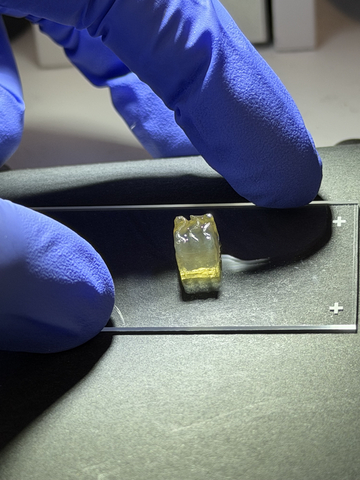
Bioprinted liver tissue
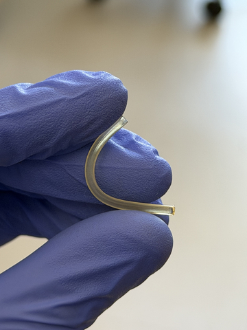
Bioprinted nerve implant

Auxilium Biotechnologies announced a major milestone in space biomanufacturing with the successful bioprinting of kidney and liver tissues aboard the International Space Station (ISS), marking the first time either tissue type has been manufactured in space. Auxilium’s bioprinted biological tissues and nerve repair implants returned to earth on Mission AXLM-3 that flew on a SpaceX capsule and returned to Earth on June 17th, 2026.

“Successfully bioprinting living liver & kidney tissue aboard the International Space Station marks an important step forward for regenerative medicine.” — Dr. Anthony Atala, MD, Professor & Director of the Wake Forest Institute for Regenerative MedicineShare

During the mission, Auxilium’s AMP-1 orbital bioprinter successfully manufactured kidney, liver, and cartilage tissues while also producing 28 nerve repair implants. The achievement represents the first demonstration of kidney tissue manufacturing in space, the first demonstration of liver tissue manufacturing in space, and the first mission to manufacture three distinct tissue types during a single spaceflight. The production of multiple tissue types and clinically relevant nerve repair implants represents the first demonstration of a scalable, multi-product biomanufacturing platform in space.

Equally important, the mission demonstrated the ability of a single autonomous manufacturing platform to produce both living tissues and implantable medical products during the same flight. The simultaneous production of multiple tissue types alongside 28 nerve repair implants highlights not only the versatility of the platform, but also its scalability and higher-throughput manufacturing in space.

The kidney and liver tissues were manufactured in support of research conducted by the Wake Forest Institute for Regenerative Medicine (WFIRM) using the institute’s cells and tissue designs. Auxilium provided the orbital manufacturing platform that enabled tissue fabrication in microgravity.

“This mission represents a significant milestone for both Auxilium and the future of space biomanufacturing,” said Jacob Koffler, PhD, MBA, CEO of Auxilium. “For the first time, we successfully bioprinted kidney and liver tissues in space, demonstrating that complex biological products can be manufactured in orbit. We also produced cartilage tissue and 28 nerve repair implants during the same mission using the same manufacturing platform. The ability to manufacture multiple tissue types alongside clinically relevant medical products highlights both the versatility and scalability of our technology. These results build on our previous demonstration of large-scale medical device manufacturing in space and represent another step toward establishing practical production capabilities for biomedical products beyond Earth.”

Dr. Anthony Atala, MD, Professor and Director of the Wake Forest Institute for Regenerative Medicine (WFIRM) commented, “Successfully bioprinting living liver and kidney tissue aboard the International Space Station marks an important step forward for regenerative medicine. The uniform cell distribution achieved aboard the space station points to real possibilities for manufacturing medical devices and tissues in space.”

Enabling the Next Generation of Biomedical Research

The successful bioprinting of kidney, liver, and cartilage tissues represents an important step toward enabling advanced biomedical research in space. One particularly promising application is the production of organoids, three-dimensional miniature tissue models that replicate key structural and functional characteristics of human organs. Organoids are increasingly used by researchers and pharmaceutical companies to study disease mechanisms, evaluate drug safety, screen new therapeutics, and predict responses to treatment.

Interest in organoid technologies has accelerated significantly as regulators and researchers seek more human-relevant alternatives to traditional animal testing. The U.S. Food and Drug Administration has identified organoids and other advanced tissue models as important components of its New Approach Methodologies initiative, while the National Institutes of Health has expanded efforts to advance and validate next-generation non-animal research platforms.

Today, organoids used for space-based research are manufactured on Earth and transported to orbit. The ability to manufacture these biological models directly in space could provide researchers with on-demand access to experimental systems while reducing dependence on launch schedules and Earth-based supply chains. As commercial space stations begin supporting larger research programmes, in-space production of organoids will create new opportunities for drug discovery, disease modelling, precision medicine, and human health research in microgravity.

By demonstrating the ability to manufacture multiple tissue types in orbit, Auxilium is helping establish the foundation for future space-based biomedical laboratories capable of producing advanced biological research tools whenever and wherever they are needed.

Source: Businesswire