A 1940s-era Drug Reveals a New Renal Water Regulation Pathway
Marla Broadfoot, PhD

Mayo Clinic researchers have identified a previously unrecognised way the kidneys regulate water balance – an advance that could lead to improved treatments for polycystic kidney disease (PKD) and other disorders. The study, led by Fouad Chebib, MD, a nephrologist at Mayo Clinic, is published in the Journal of Clinical Investigation.
The findings build on decades of scientific understanding by revealing an additional pathway the kidney uses to control water balance. Until now, the body’s ability to concentrate urine – and prevent dehydration – has been thought to depend primarily on the hormone vasopressin. Dr Chebib’s team discovered an alternative mechanism that operates independently of that system.
“The kidney’s ability to regulate water is one of the most fundamental processes in the body,” Dr Chebib says. “It’s not every day that you uncover a new way it carries out that function.”
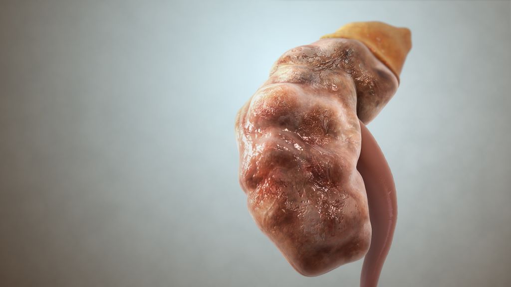
Polycystic kidney disease is a common inherited condition that causes fluid-filled cysts to grow in the kidneys over time, gradually reducing kidney function and often leading to kidney failure. It affects millions of people worldwide, including an estimated 140 000 people in the US with the most common form, autosomal dominant PKD (ADPKD). Many patients eventually require dialysis or a kidney transplant.
Watch: 1940s-era drug shows new promise for kidney disease
Dr Chebib’s team studies how kidney cysts grow in PKD using lab-grown cell models. As part of that work, they tested compounds expected to worsen the disease process by increasing cellular signals linked to cyst growth. One of those compounds was probenecid, a drug first used in the 1940s to conserve limited supplies of penicillin by reducing its urinary excretion.
“We thought this drug would make the disease process worse,” Dr Chebib says. “Instead, it did the opposite.”
Rather than promoting cyst growth, the drug slowed it. After repeating the experiments multiple times, the team realised they had uncovered something important.
Further investigation revealed that probenecid affects how kidney cells handle urate, a molecule commonly associated with gout. Inside the cell, urate acts as a signal – triggering a chain of events that helps move water channels to the cell surface. This allows the kidney to reabsorb water and concentrate urine without relying on vasopressin, the hormone traditionally thought to control this process.
“This represents a distinct pathway from what is described in traditional physiology models,” Dr Chebib says. “It demonstrates that the kidney has an additional mechanism to preserve water.”
For patients with PKD, the discovery could address one of the biggest challenges of current treatment. The only approved therapy, tolvaptan, works by blocking vasopressin, which slows cyst growth but causes patients to produce very large amounts of urine – often 6 to 7 litres a day. That side effect can be difficult to live with and leads some patients to stop treatment.
In preclinical studies and a small clinical trial, adding probenecid reduced urine volume and nighttime urination while preserving the treatment’s effectiveness.
After taking probenecid, patients’ urine volume dropped by about 30% on average, and they went from waking up several times a night to urinate to about once per night. Many also reported improved quality of life.
“The goal is to preserve the therapeutic benefit of tolvaptan while reducing its burden,” Dr. Chebib says.
Despite these promising results, researchers are not planning to rely on probenecid as a long-term solution. The drug is decades old, affects multiple systems in the body and is not widely available today. Instead, the team is using what they learned to design more targeted therapies.
“Probenecid helped us uncover the mechanism,” Dr Chebib says. “Our goal is to take this insight and develop therapies designed specifically for this pathway.”
For Dr Chebib, the work is rooted in early inspiration. He was drawn to kidney research after his father developed PKD.
“This has been a long and deeply purposeful journey,” he says. “It started with a personal motivation and led to something that could ultimately benefit patients.”
For a complete list of authors, disclosures and funding, see the study.
Source: Mayo Clinic