Category: Regenerative Medicine

Company Successfully Bioprints Kidney and Liver Tissues in Space

Weightlessness of space allows tissues to be bioprinted without collapsing
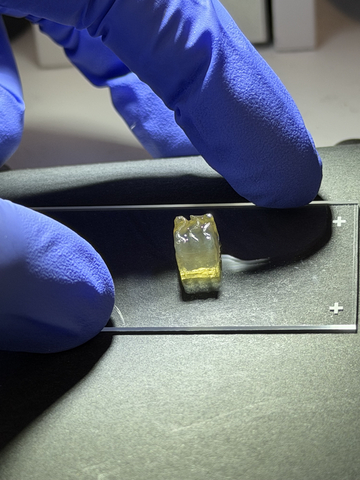
Bioprinted liver tissue
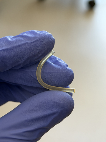
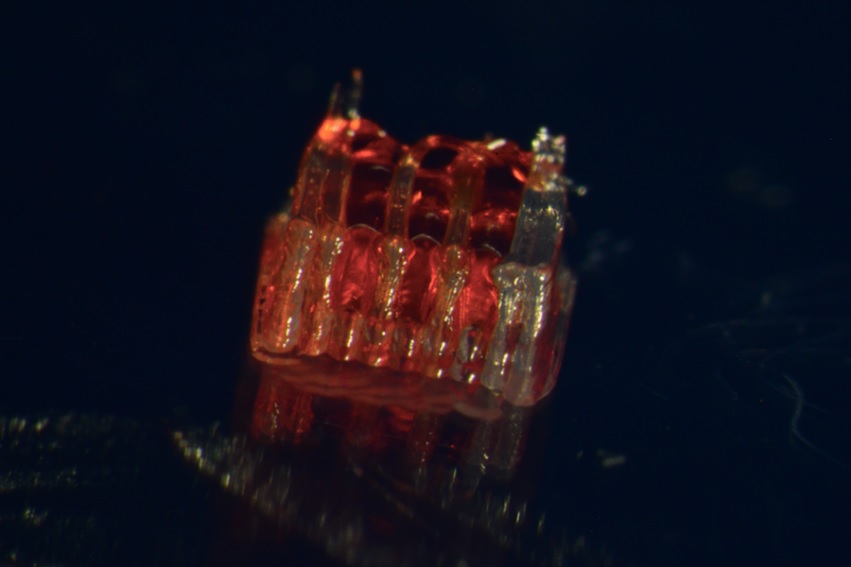
Bioprinted nerve implant

Auxilium Biotechnologies announced a major milestone in space biomanufacturing with the successful bioprinting of kidney and liver tissues aboard the International Space Station (ISS), marking the first time either tissue type has been manufactured in space. Auxilium’s bioprinted biological tissues and nerve repair implants returned to earth on Mission AXLM-3 that flew on a SpaceX capsule and returned to Earth on June 17th, 2026.

“Successfully bioprinting living liver & kidney tissue aboard the International Space Station marks an important step forward for regenerative medicine.” — Dr. Anthony Atala, MD, Professor & Director of the Wake Forest Institute for Regenerative MedicineShare

During the mission, Auxilium’s AMP-1 orbital bioprinter successfully manufactured kidney, liver, and cartilage tissues while also producing 28 nerve repair implants. The achievement represents the first demonstration of kidney tissue manufacturing in space, the first demonstration of liver tissue manufacturing in space, and the first mission to manufacture three distinct tissue types during a single spaceflight. The production of multiple tissue types and clinically relevant nerve repair implants represents the first demonstration of a scalable, multi-product biomanufacturing platform in space.

Equally important, the mission demonstrated the ability of a single autonomous manufacturing platform to produce both living tissues and implantable medical products during the same flight. The simultaneous production of multiple tissue types alongside 28 nerve repair implants highlights not only the versatility of the platform, but also its scalability and higher-throughput manufacturing in space.

The kidney and liver tissues were manufactured in support of research conducted by the Wake Forest Institute for Regenerative Medicine (WFIRM) using the institute’s cells and tissue designs. Auxilium provided the orbital manufacturing platform that enabled tissue fabrication in microgravity.

“This mission represents a significant milestone for both Auxilium and the future of space biomanufacturing,” said Jacob Koffler, PhD, MBA, CEO of Auxilium. “For the first time, we successfully bioprinted kidney and liver tissues in space, demonstrating that complex biological products can be manufactured in orbit. We also produced cartilage tissue and 28 nerve repair implants during the same mission using the same manufacturing platform. The ability to manufacture multiple tissue types alongside clinically relevant medical products highlights both the versatility and scalability of our technology. These results build on our previous demonstration of large-scale medical device manufacturing in space and represent another step toward establishing practical production capabilities for biomedical products beyond Earth.”

Dr. Anthony Atala, MD, Professor and Director of the Wake Forest Institute for Regenerative Medicine (WFIRM) commented, “Successfully bioprinting living liver and kidney tissue aboard the International Space Station marks an important step forward for regenerative medicine. The uniform cell distribution achieved aboard the space station points to real possibilities for manufacturing medical devices and tissues in space.”

Enabling the Next Generation of Biomedical Research

The successful bioprinting of kidney, liver, and cartilage tissues represents an important step toward enabling advanced biomedical research in space. One particularly promising application is the production of organoids, three-dimensional miniature tissue models that replicate key structural and functional characteristics of human organs. Organoids are increasingly used by researchers and pharmaceutical companies to study disease mechanisms, evaluate drug safety, screen new therapeutics, and predict responses to treatment.

Interest in organoid technologies has accelerated significantly as regulators and researchers seek more human-relevant alternatives to traditional animal testing. The U.S. Food and Drug Administration has identified organoids and other advanced tissue models as important components of its New Approach Methodologies initiative, while the National Institutes of Health has expanded efforts to advance and validate next-generation non-animal research platforms.

Today, organoids used for space-based research are manufactured on Earth and transported to orbit. The ability to manufacture these biological models directly in space could provide researchers with on-demand access to experimental systems while reducing dependence on launch schedules and Earth-based supply chains. As commercial space stations begin supporting larger research programmes, in-space production of organoids will create new opportunities for drug discovery, disease modelling, precision medicine, and human health research in microgravity.

By demonstrating the ability to manufacture multiple tissue types in orbit, Auxilium is helping establish the foundation for future space-based biomedical laboratories capable of producing advanced biological research tools whenever and wherever they are needed.

Source: Businesswire
Researchers Create Microrobots to Repair Spinal Cord

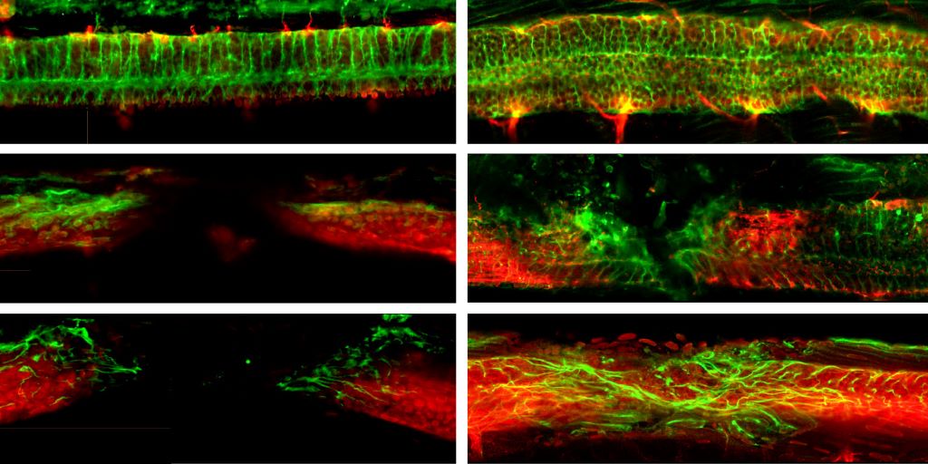
At the start and after three days: the top images show the uninjured spinal cord of a zebrafish; those in the middle show the injured spinal cord; and those at the bottom illustrate how the nerve cells grow thanks to the microrobots. (Image: ETH Zurich)

A research team from ETH Zurich and the University of Zurich (UZH) has developed a novel approach to treating spinal cord injuries: controllable microrobots deliver stem cells directly to the site of an injury, where they promote nerve cell regeneration. In animal experiments, this approach significantly improved mobility.

Spinal cord injuries can have devastating consequences for those affected. Nerve cells in the spinal cord rarely regenerate naturally, while scarring often prevents the regrowth of nerve fibres. Modern therapies attempt to influence implanted stem cells using electrical stimulation to promote the growth of new nerve cells. This approach has several drawbacks: it requires implanted electrodes, and the transplanted cells do not always survive or integrate properly into the existing tissue.

Cells and nanoparticles cleverly combined 

Researchers in Zurich are pursuing a new approach, which they have published in the journal Nature Materials. This involves combining therapeutic stem cells with magnetoelectric nanoparticles in such a way that the cells can be guided magnetically to the precise site of an injury and stimulate the stem cells to accelerate repair.

To achieve this, the researchers created a biohybrid microrobot, which combines living neural progenitor cells (NPCs) with a technical component in the form of specially engineered nanoparticles. The NPCs are derived from induced pluripotent stem cells (iPS cells), which are regular body cells reprogrammed in the laboratory to regain stem cell properties. These iPS cells have the potential to differentiate into various types of nervous system cells.

The nanoparticles consist of two layers: an inner layer that responds to magnetic fields and an outer layer that converts this response into electrical signals. By combining these special nanoparticles with the progenitor cells, the researchers fabricate what are known as NPCbots.

A lab the size of a chip

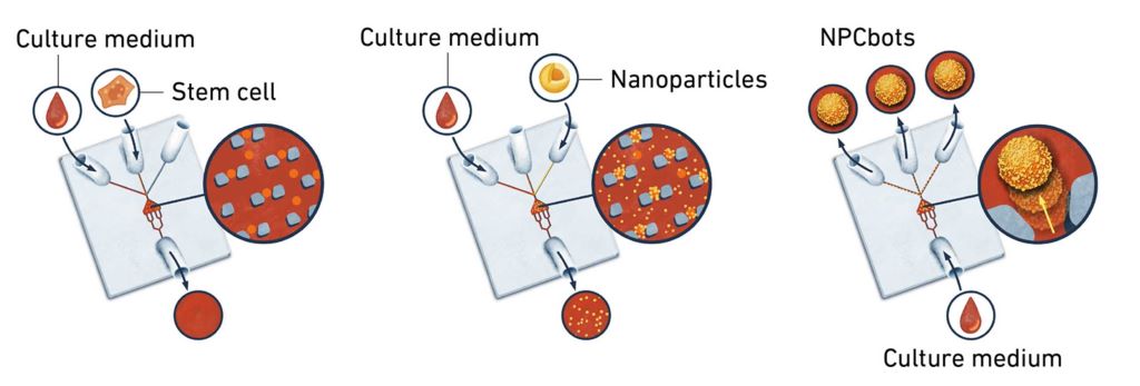
The researchers create the NPCbots in specialised labs on a surface measuring one square centimetre. This process can be illustrated graphically. “We place a reservoir in the centre where we trap the cells. Then we inject the nanoparticles and wait for the two components to bind,” explains Professor Salvador Pané i Vidal of the Multi-Scale Robotics Lab at ETH Zurich.

Illustration of how microrobots are fabricated on a lab-on-a-chip (LoC).  (Image: ETH Zurich)

After just thirty minutes, the NPCbots – each around six micrometres in size – are ready for use. “To scale up fabrication, we operate several lab-on-chip systems in parallel,” explains Hao Ye, senior scientist and the study’s first author. Depending on the test in question, the ETH researchers need hundreds of thousands of microrobots for cell-based studies and several million for animal experiments.

Injured zebrafish swim again

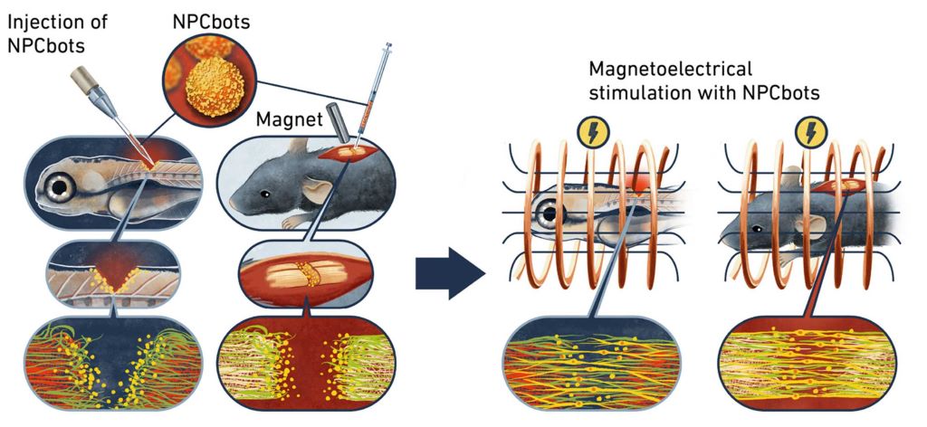
The team tested the NPCbots on zebrafish larvae with spinal cord injuries. The microrobots were injected precisely into the site of the fish’s injury, and electromagnetic fields were generated. For Pané Vidal, teamwork was vital to the experiment’s success: “Stephan Neuhauss and Jingjing Zang at the University of Zurich did extremely valuable work. They enabled us to demonstrate, in a well-characterised regenerative model system, how quickly cells differentiate using our method and how our bots repair the spinal cord.” In just three days, the zebrafish exhibited nearly normal swimming and exploratory behaviour.

Schematic illustration of nerve cell recovery in zebrafish and mice. (Image: ETH Zurich)

The researchers also tested the NPCbots on mice with completely severed spinal cords. Here, too, the results were very promising: after 28 days, the animals’ nerve cells had reconnected at the site of the injury. During this period, the treated mice exhibited increasingly normal movement patterns – their gait, stride length, coordination and exploratory behaviour improved significantly.

This result is particularly significant because, unlike in zebrafish, the mouse spinal cord does not normally regenerate. The treatment was well tolerated by the animals, with no evidence of any adverse effects or immune reactions. 

Success through minimally invasive stimulation 

These successes were made possible through electrical stimulation of stem cells, greatly enhancing their differentiation after transplantation. In this process, nanoparticles convert magnetic signals directly into electrical impulses that stimulate specific stem cells. When employing NPCbots, researchers need only apply external magnetic fields around the injury site, eliminating the need for implanted electrodes or cables in previous approaches. This is crucial because the spinal cord is extremely sensitive. “Microrobotic guidance makes the treatment more precise and minimally invasive,” Hao explains.

Magnetic fields are particularly well-suited for stimulating stem cells because they can penetrate tissue easily, and their frequency and field strength can be flexibly adjusted to the specific application. Once the progenitor cells have been stimulated and differentiated into nerve cells, the NPCbots essentially dissolve within the tissue. The researchers expect the nanoparticles to be stable and minimally reactive due to their barium titanate coating. Further studies will determine whether and how the particles are degraded or excreted over the long term.

The idea can be expanded as required

The results from animal experiments are extremely promising, but further research will be needed before NPCbots can be tested in humans. “In addition to many clinical aspects, we first need to test which magnetic fields work best in humans and determine the optimal stimulation duration,” Hao explains. Nevertheless, the researchers are already considering further applications: “The reproducible and scalable production of microrobots using our lab-on-a-chip system demonstrates that the platform’s application potential extends beyond basic research,” explains Professor Pané i Vidal. It could also be adapted for other biomedical applications – for example, in cardiology, oncology, wound healing and other targeted regenerative therapies. This could make these treatments safer, more controllable and more effective. 

By Franziska Schmid

Source: ETH Zurich

Researchers Find ‘Perfect Recipe’ to Regrow Bone and Blood Vessels

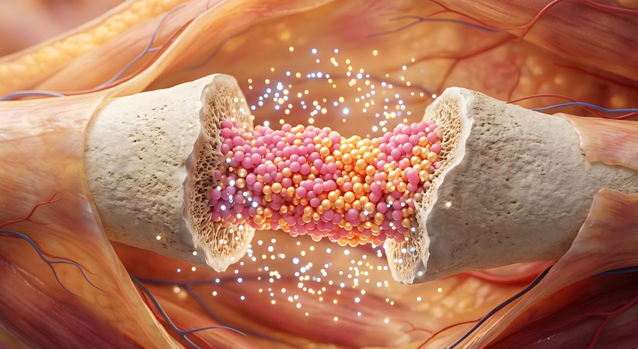
Medical illustration of bone regeneration.

For patients suffering from traumatic injuries that leave behind volumetric gaps – where significant bone and blood vessels are lost – the clock is always ticking. Without a nearby blood supply, cells in the centre of a large injury cannot survive, often leading to permanent tissue loss or failed grafts.

A team of eight scientists at The University of Texas at San Antonio has discovered a potential ‘perfect recipe’ to address this challenge. By blending two natural proteins found in the human body, the researchers created a specialised scaffold that allows bone and blood vessels to grow simultaneously at an accelerated rate.

The study, published in the journal Biomaterials Advances, identifies a 50:50 ratio of collagen and fibrin as the ideal environment for tissue repair.

The Lego blocks of healing

The technology relies on what scientists call interpenetrating polymer networks, or IPNs. In simpler terms, it is a microscopic support structure where different materials are entangled to create a stable foundation for new growth.

“An IPN network is two things that are entangled like a giant mess of Legos,” said Teja Guda, PhD, the Jacobson Distinguished Professor of Innovation and Entrepreneurship in the Department of Biomedical Engineering and Chemical Engineering at UT San Antonio and the study’s corresponding author. “We are leaving all the building blocks there and letting the cells build whatever Lego structure they like the most.”

In this biological ‘Lego’ set, one material is fibrin, the protein the body uses to form blood clots immediately after an injury. The other is collagen, the primary structural protein found in bones and other tissues.

Seeding the scaffold with MVFs and MSCs

To turn these protein gels into living tissue, the research team “seeded” the hydrogels with two critical types of biological starters: microvascular fragments (MVFs) and mesenchymal stem cells (MSCs). The MVFs have the capacity to grow into blood vessels, while the MSCs can, with the right environmental cues, grow into bone.

The researchers integrated these components by mixing the living MVFs and MSCs directly into the liquid protein solution before it underwent gelation. This 3D encapsulation ensured the cells were suspended throughout the entire depth of the scaffold rather than just sitting on the surface.

Balancing blood and bone

Standard medical treatments for severe bone loss typically involve autografts, where bone is harvested from another part of the patient’s body, or allografts, which use processed bone from a donor. These traditional grafts often fail to integrate because they lack an immediate blood supply to nourish the new tissue. Without rapid vascularisation, the transplanted bone can become necrotic, leading to a high rate of clinical failure in complex trauma cases.

The challenge for UT San Antonio researchers was finding the right balance between the two proteins to support both blood vessel and bone regeneration. Fibrin is excellent at recruiting the cells needed to form blood vessels, a process called angiogenesis. Collagen provides the mechanical strength needed to guide the development of bone, or osteogenesis.

“Whenever you have an injury where you are losing volume, you not only lose the tissue itself, but you’re also losing blood vasculature,” said Gennifer Chiou, a postdoctoral fellow at UT San Antonio and the study’s lead author. “We’re looking at how we can regenerate both the tissue and the vessel itself within specifically bone tissue.”

The team tested five different ratios of the two proteins. They found that while gels with more fibrin supported faster vessel sprouting, they lacked the stability needed for long-term bone growth. Conversely, high-collagen gels were too stiff for vessels to penetrate easily.

The 50:50 blend struck an ideal balance. The MVFs were able to sprout and branch out into a robust, interconnected network. Simultaneously, the MSCs developed in a stable environment, expressing the specific genetic markers needed to mature into bone-forming cells. This dual-growth approach ensures that as the new bone forms, it is continuously supplied with the blood and nutrients it needs to remain viable.

From the lab to the clinic

Because the materials used in the study – collagen, fibrin and the patient’s own blood vessels – are all naturally occurring in the body, the researchers believe the technology faces fewer regulatory hurdles than synthetic alternatives.

“There is almost nothing new in our material,” Guda said. “It’s your collagen, it’s your blood vessels, it’s your fibrin. The end goal is to provide evidence that will guide how clinicians think about healing wounds.”

The team hopes to proceed to preclinical trials in the near future, which will provide further support for the treatment to one day become standard practice.

By Audrey Gray

Source: UT San Antonio

How the Nervous System Activates Repair After a Spinal Cord Injury

View of the spinal cord. Credit: Scientific Animations CC4.0

After a spinal cord injury, cells in the brain and spinal cord change to cope with stress and repair tissue. A new study from Karolinska Institutet, published in Nature Neuroscience, shows that this response is controlled by specific DNA sequences. This knowledge could help develop more targeted treatments.

When the central nervous system is damaged – for example, in a spinal cord injury – many cells become reactive. This means they change their function and activate genes that protect and repair tissue. However, how this process is regulated has long been unclear.

Researchers at Karolinska Institutet have now mapped thousands of so-called enhancers; small DNA sequences that act like “switches” for genes, turning them on or boosting their activity. By analysing individual cell nuclei from mice with spinal cord injuries using AI models, the researchers discovered that these genetic switches are activated after injury and instruct specific cell types to respond. The main cells affected were glial cells such as astrocytes and ependymal cells – support cells that help protect and repair the nervous system.

New opportunities for precision treatments 

“We have shown how cells read these instructions through a code that tells them how to react to injury. This code combines signals from general stress factors with the cell’s own identity,” explains Enric Llorens-Bobadilla, researcher at the Department of Cell and Molecular Biology at Karolinska Institutet.

“This opens up the possibility of using the code to target treatments specifically to the cells affected by the injury,” says Margherita Zamboni, researcher at the same department and first author of the study.

The study is a collaboration between researchers at Karolinska Institutet and SciLifeLab, supported by the European Research Council (ERC), the Swedish Research Council, and the Swedish Foundation for Strategic Research. Some researchers have reported consultancy roles and patent applications related to the technology.

Source: Karolinska Institutet

Study Reveals the Dual Role for a Protein Critical for Healing Nerve Damage

Sarm1 appears to be essential for regeneration

Source: CC0

Nerve damage can be an unfortunate side effect from an accident, illness or even certain treatments, like chemotherapy. Fortunately, the peripheral nervous system can heal itself to a certain extent, albeit very slowly. Researchers are still trying to understand this natural healing process in order to improve it. A recent study published in Science Translational Medicine sheds new light on this.

This mouse-based study from the University of Michigan adds to the evidence regarding a specific protein inside of the nerves, called Sarm1, that seems key for regeneration. Previous studies have revealed that when Sarm1 is activated, it sets off the degenerative process in nerves. The thinking has been that for conditions like chemotherapy induced peripheral neuropathy, diabetes, or nerve trauma, blocking Sarm1 would beneficially block the breakdown of nerves.

But what else would blocking Sarm1 effect?

“We know that nerve breakdown after an injury is quite efficient, and the breakdown is what Sarm1 controls. So, there must be a biological reason for this breakdown to be so quick and efficient,” said Ligia B. Schmitd, PhD, of the Department of Cell and Developmental Biology, lead author of the study.

Schmidt is a research fellow in the lab of Roman Giger, PhD, co-senior author with Ashley Kalinski of the University of South Carolina.

Using mice bred to lack Sarm1 and subjecting them to peripheral nerve injury, the team could observe drastic changes to the distal nerve environment, including fewer blood-borne immune cells resulting in reduced nerve inflammation.

“These cells are important because they have to enter the injured nerve to clean up all of the debris,” said Schmitd.

More importantly, their study revealed a critical effect on Schwann cells, which line and support the peripheral nerves.

Normally following an injury, Schwann cells will convert to a repair state in which they express different genes and proteins to migrate and proliferate in order to regrow the axon, the long projecting portion of the neuron.

But without Sarm1, “the Schwann cells are just stuck there,” said Schmitd.

In essence, Sarm1 controls both nerve degeneration and regeneration through its effect on Schwann cells.

The team also noted that a lack of Sarm1 seemed to boost the nerve’s efforts to regrow, but without activating the repair Schwann cells, these efforts were much less efficient.

“For a long time, we’ve thought that simply preventing nerve breakdown would be a good thing. What our study now shows is that this early breakdown also sends powerful signals to Schwann cells and immune cells that are needed for efficient repair, so any future therapy that targets Sarm1 will have to preserve that delicate balance between protection and regeneration,” said Giger, professor in the Department of Cell and Developmental Biology.

Schmitd notes that the study needs to be done in other animal models and with other proteins involved in nerve repair, “but if this proves to be an important mechanism for triggering the repair Schwann cell state, then down the road, fixing this response could help humans regenerate peripheral nerves.”

Source: University of Michigan Medicine

Nanorobots Transform Stem Cells into Bone Cells with a Little Push

New method for the targeted production of specific cells

Figure 1
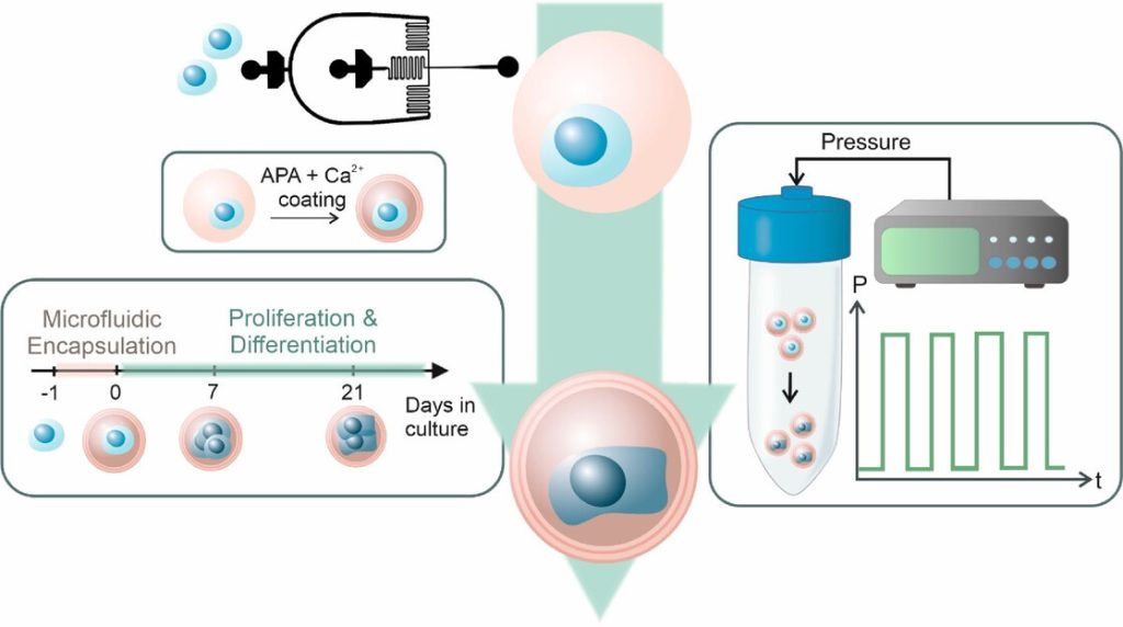
Schematic overview of the experimental workflow. MSCs (blue) were singly encapsulated using a microfluidic approach within calcium-crosslinked, RGD-functionalized alginate microgels (pink), followed by a secondary APA and calcium coating to enhance stability. Encapsulated cells were cultured for 21 days and subjected to cyclic hydrostatic pressure in regular cell culture media without any growth factors. Source: İyisan et al., Small Science, 2025.

For the first time, researchers at the Technical University of Munich (TUM) have succeeded in using nanorobots to stimulate stem cells with such precision that they are reliably transformed into bone cells. To achieve this, the robots exert external pressure on specific points in the cell wall. The new method offers opportunities for faster treatments in the future.

Prof Berna Özkale Edelmann’s nanorobots consist of tiny gold rods and plastic chains. Several million of them are contained in a gel cushion measuring just 60 micrometres, together with a few human stem cells. Powered and controlled by laser light, the robots, which look like tiny balls, mechanically stimulate the cells by exerting pressure. “We heat the gel locally and use our system to precisely determine the forces with which the nanorobots press on the cell – thereby stimulating it,” explains the professor of nano- and microrobotics at TUM. This mechanical stimulation triggers biochemical processes in the cell. Ion channels change their properties, and proteins are activated, including one that is particularly important for bone formation.

The research is described in Advanced Materials and Small Science.

Heart and cartilage cells: finding the correct stress pattern

If stimulation is carried out at the right rhythm and with the right (low) force, a stem cell can be reliably triggered to develop into a bone cell within three days. This process can be completed within three weeks. “The corresponding stress pattern can also be found for cartilage and heart cells,” asserts Berna Özkale Edelman. “It’s almost like at the gym: we train the cells for a particular area of application. Now we just have to find out which stress pattern suits each cell type,” says the head of the Microbiotic Bioengineering Lab at TUM.

Mechanical forces pave the way for transformation into bone cells

The research team produces bone cells using mesenchymal stem cells. These cells are considered to be the body’s ‘repair cells’. They are approximately 10 to 20 micrometres in size and are generally capable of developing into bone, cartilage or muscle cells, for example. The challenge: The transformation into differentiated cells is complex and has been difficult to control until now. “We have developed a technology that allows forces to be applied to the cell very precisely in a three-dimensional environment,” says TUM scientist Özkale Edelmann. “This represents an unprecedented advance in the field.” The researchers believe that this method can even be used to produce cartilage and heart cells from human stem cells.

Automation is the next step

For treatments, doctors will ultimately need far more differentiated cells – around one million. “That’s why the next step is to automate our production process so that we can produce more cells more quickly,” says Prof Özkale Edelmann.

Source: Technical University Munich

Repurposed Multiple Sclerosis Drug Could Help Bones Heal Faster

Photo by Tima Miroshnichenko on Pexels

Researchers at the University of Arizona College of Medicine – Tucson found evidence that a drug that improves the ability to walk in people with multiple sclerosis can also make bone fractures heal faster.

The findings help further the understanding of specific factors involved in the bone healing process, and potentially open avenues for new therapeutic approaches.

“Broken bones are typically slow to heal in many people, and they can impact lives for months and in different ways. People lose time at work and daily activities at home with family and friends are impacted,” said senior author John Elfar, MD, professor, surgeon and chair of the Department of Orthopaedic Surgery at the U of A College of Medicine – Tucson. “This drug has the potential to change that.” 

Elfar partnered with Prem Kumar Govindappa, PhD, DVM, an assistant professor in the department, on the preclinical study that showed treatment with the drug 4-aminopyridine, or 4-AP, resulted in leg fractures healing faster and stronger than without the drug. The paper was published in The Journal of Bone and Joint Surgery.

“Mice with bone fractures healed quicker and were stronger after they healed after treatment with 4-AP,” said Elfar said, who is a member of the university’s BIO5 Institute. “We saw more bone mass and less intermediate cartilage, meaning there was accelerated bone healing.” 

The drug is approved for use in chronic neurological conditions, where it helps with walking by improving how signals from the brain and spinal cord reach limbs.

The team also saw improvements in bone mass and the ability to bear weight after treatment with 4-AP. Collagen deposition and bone mineralization, both of which are necessary for bone healing, also received a boost. Collagen forms the structural foundation of bones. In bone mineralization, minerals like calcium and phosphate join the newly forming bone matrix, strengthening and hardening the bone.

“We found that every fine-tuned measure of the strength of bone was better after administering 4-AP to mice,” Elfar said. “We also found more BMP2 protein in bone-forming cells at the fracture site, which again told us we found something that could accelerate the process.”

Examining human bone cells exposed to 4-AP in a dish, the scientists saw increased production of bone morphogenetic protein, or BMP2, a bone-building substance used clinically to help with some kinds of bone repair. BMP2 prompted the production of stem cells that become cells called osteoblasts, which are essential to form new bone.

The research team also measured 4-AP’s effects on human bone narrow mesenchymal stem cells and human osteoblast cells in the lab. 4-AP increased the conversion of the stem cells into osteoblasts and the latter’s ability to migrate and grow, which are essential to the healing process.

Elfar said that 4-AP’s role in driving BMP2 gene and protein activity is key to its bone healing effects, and using 4-AP to prompt BMP2 production in the body could be especially important.

“BMP2 is a hormone the body makes to accelerate bone healing,” Elfar said. 

BMP2 is known to modulate bone healing and is approved for use in certain medical procedures, including spinal fusion and sinus reconstruction surgery. An artificial version that has orthopedic medicine uses can have side effects, though, including bone resorption and cervical spine swelling. Finding a way to channel naturally produced BMP2 could improve bone healing while avoiding such problems.

The scientists previously showed that 4-AP could prevent bone and muscle loss in a mouse model of nerve damage. Similarly, they saw indications of 4-AP’s healing effects for wound, nerve and limb injuries. 

The researchers plan to eventually test 4-AP’s potential use in healing bones in a clinical trial. They also want to better understand the drug’s effects on BMP2 production, and more broadly, on the biology of healing bone.

Source: University of Arizona

Groundbreaking Spinal Scaffold Allows Nerve Fibres to Regrow

New research combines 3D printing, stem cell biology, and lab-grown tissues for possible treatments of spinal cord injuries. Photo provided by: McAlpine Research Group, University of Minnesota

For the first time, a research team at the University of Minnesota Twin Cities demonstrated a groundbreaking process that combines 3D printing, stem cell biology, and lab-grown tissues for spinal cord injury recovery. 

The study was recently published in Advanced Healthcare Materials. Currently, there is no way to completely reverse the damage and paralysis from the injury. A major challenge is the death of nerve cells and the inability of nerve fibres to regrow across the injury site. This new research tackles this problem head-on.

The method involves creating a unique 3D-printed framework for lab-grown organs, called an organoid scaffold, with microscopic channels. These channels are then populated with regionally specific spinal neural progenitor cells (sNPCs), which are cells derived from human adult stem cells that have the capacity to divide and differentiate into specific types of mature cells.

“We use the 3D printed channels of the scaffold to direct the growth of the stem cells, which ensures the new nerve fibres grow in the desired way,” said Guebum Han, a former University of Minnesota mechanical engineering postdoctoral researcher and first author on the paper who currently works at Intel Corporation. “This method creates a relay system that when placed in the spinal cord bypasses the damaged area.”

n their study, the researchers transplanted these scaffolds into rats with spinal cords that were completely severed. The cells successfully differentiated into neurons and extended their nerve fibres in both directions – rostral (toward the head) and caudal (toward the tail) – to form new connections with the host’s existing nerve circuits. 

The new nerve cells integrated seamlessly into the host spinal cord tissue over time, leading to significant functional recovery in the rats.

“Regenerative medicine has brought about a new era in spinal cord injury research,” said Ann Parr, professor of neurosurgery at the University of Minnesota. “Our laboratory is excited to explore the future potential of our ‘mini spinal cords’ for clinical translation.”

While the research is in its beginning stages, it offers a new avenue of hope for those with spinal cord injuries. The team hopes to scale up production and continue developing this combination of technologies for future clinical applications.

Source: University of Minnesota

Scientists Discover the Mechanism for Peripheral Nerve Regeneration

Weizmann Institute scientists have discovered hundreds of molecules that promote nerve regeneration in mice – and may even encourage growth in brain neurons

Top: Overexpression of genes from the B2-SINE family in retinal ganglion neurons led to accelerated growth after injury. Bottom: Ganglion cells after injury without B2-SINE overexpression. Credit: Weizmann Institute of Science

Unlike the brain and spinal cord, peripheral nerve cells, whose long extensions reach the skin and internal organs, are capable of regenerating after injury. This is why injuries to the central nervous system are considered irreversible, while damage to peripheral nerves can, in some cases, heal, even if it takes months or years. Despite decades of research, the mechanisms behind peripheral nerve regeneration remain only partially understood.

In a new study published in Cell, researchers from Prof Michael (Mike) Fainzilber’s lab at the Weizmann Institute of Science discovered that a family of hundreds of RNA molecules with no known physiological function is essential to nerve regeneration. Remarkably, the study showed that these molecules can stimulate growth not only in the peripheral nervous system of mice but also in their central nervous system. These findings could pave the way for new treatments for a variety of nerve injuries and neurodegenerative diseases.

For a peripheral nerve to regenerate, it must maintain communication between the neuron’s cell body and its long extension – the axon – which in humans can reach more than a meter in length. In a series of studies over the past two decades, Fainzilber’s lab has revealed key components of this communication: proteins that act like postal couriers, delivering instructions for the production of growth-controlling factors and other proteins, from the cell body to the axon. These molecular couriers also help assess the distance between the cell body and the axon tip, allowing the neuron to modulate its growth accordingly. Yet one central issue remained: What triggers the regenerative growth after injury, and why does this not happen in central nervous system cells?

“While the growth acceleration observed in our study is not yet sufficient to address clinical paralysis, it is definitely significant”

In the new study, Dr Indrek Koppel of Fainzilber’s lab, in collaboration with Dr Riki Kawaguchi of the University of California, Los Angeles (UCLA), examined a specific kind of gene expression in the peripheral nerves of mice following injury. The researchers were surprised to find that one day after damage, the neurons increased the expression of an entire family of short genetic sequences called B2-SINEs, whose role was previously unknown. These sequences do not encode any proteins, and because they are known for “jumping” around the genome, meaning that they can appear at the wrong place or time, they have a bad reputation. But the researchers found that after injury, the neurons began expressing many B2-SINE RNA transcripts, in parallel with other processes preparing the cell for regeneration and repair.

However, B2-SINE is an enormous family, comprising some 150 000 sequences scattered throughout the mouse genome. The initial analysis could not determine which of these were responsible for promoting growth. Dr. Eitan Erez Zahavi, also of Fainzilber’s lab, who led the new study alongside Koppel, used bioinformatics tools to identify 453 B2-SINE sequences that are highly expressed after injury, promoting nerve growth. Collaborating with international research teams, the scientists showed that this overexpression after injury is unique to peripheral nerve cells and does not occur in the central nervous system.

The periphery leads, the center follows

The researchers then tested whether B2-SINEs from peripheral nerve cells could also stimulate neuronal growth in the central nervous system. They induced retinal neurons in mice to overexpress RNA molecules of the B2-SINE type and observed faster regeneration after injury. A similar experiment in the mouse motor cortex – the brain region that controls muscle movement via long axons projecting to the spinal cord – showed that neurons expressing high levels of B2-SINE also regenerated faster than control neurons.

“There are still no effective treatments to accelerate nerve cell growth and regeneration,” Fainzilber notes. “While the growth acceleration observed in our study is not yet sufficient to address clinical paralysis, it is definitely significant. Of course, the path from basic research to clinical application is long, and we must make sure that enhancing growth mechanisms does not, for example, increase the risk of cancer.”

One final mystery remained: How do B2-SINE RNA molecules actually promote regeneration? With help from Prof Alma L. Burlingame’s group at the University of California, San Francisco, the researchers discovered that these RNAs promote a physical link between the molecular “couriers” carrying instructions for producing growth-associated proteins and the ribosomes that read these instructions and carry them out. This means that production of the critical factors takes place closer to the cell body rather than to the tip of the axon. The researchers believe that this signals to the neuron that it is “too small,” triggering a growth response.

“There are over a million sequences called Alu elements in the human genome, the human equivalent of B2-SINEs in mice,” says Fainzilber. “These molecules had been previously shown to bind to ribosomes and mail couriers, but why this happens was unknown. We’re now trying to determine whether Alu or other noncoding RNA elements are involved in nerve regeneration in humans.”

“Recovery from peripheral nerve injuries, or from systemic diseases like diabetes that affect these nerves, can be very slow,” he adds. “That’s why we’re now testing a therapy that might speed up regeneration by mimicking B2-SINE activity. This therapy involves small molecules that connect the couriers to ribosomes while keeping them close to the nerve cell body, promoting faster growth. We are conducting this research in collaboration with Weizmann’s Bina unit for early-stage research with applicative potential.”

Beyond promoting peripheral nerve regeneration, the new study also hints at an even broader prospect: regeneration in the central nervous system. “We are currently working with UCLA on a study showing that the mechanism we discovered plays a role in recovery from stroke in mouse models,” Fainzilber says. “Additionally, we’re collaborating with Tel Aviv University, Hebrew University and Sheba Medical Center to study its possible role in ALS, a progressive neurodegenerative disease. Neurodegenerative conditions affect many millions of people worldwide. While the road ahead is long, I truly hope we’ll one day be able to harness our newly discovered regeneration mechanism to treat them.”

Science Numbers

After injury, the axon of a peripheral nerve cell regrows at a rate of around 1 millimetre a day.

Source: Weizmann Institute of Science

MIT Engineers Turn Skin Cells Directly into Neurons for Cell Therapy

A new, highly efficient process for performing this conversion could make it easier to develop therapies for spinal cord injuries or diseases like ALS.

Anne Trafton | MIT News
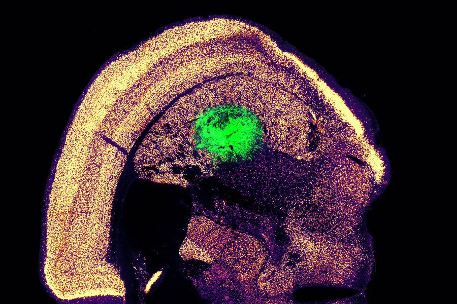
Researchers at MIT have devised a simplified process to convert a skin cell directly into a neuron. This image shows converted neurons (green) that have integrated with neurons in the brain’s striatum after implantation. Credits :Image: Courtesy of the researchers

Converting one type of cell to another – for example, a skin cell to a neuron – can be done through a process that requires the skin cell to be induced into a “pluripotent” stem cell, then differentiated into a neuron. Researchers at MIT have now devised a simplified process that bypasses the stem cell stage, converting a skin cell directly into a neuron.

Working with mouse cells, the researchers developed a conversion method that is highly efficient and can produce more than 10 neurons from a single skin cell. If replicated in human cells, this approach could enable the generation of large quantities of motor neurons, which could potentially be used to treat patients with spinal cord injuries or diseases that impair mobility.

“We were able to get to yields where we could ask questions about whether these cells can be viable candidates for the cell replacement therapies, which we hope they could be. That’s where these types of reprogramming technologies can take us,” says Katie Galloway, the W. M. Keck Career Development Professor in Biomedical Engineering and Chemical Engineering.

As a first step toward developing these cells as a therapy, the researchers showed that they could generate motor neurons and engraft them into the brains of mice, where they integrated with host tissue.

Galloway is the senior author of two papers describing the new method, which appear today in Cell Systems. MIT graduate student Nathan Wang is the lead author of both papers.

From skin to neurons

Nearly 20 years ago, scientists in Japan showed that by delivering four transcription factors to skin cells, they could coax them to become induced pluripotent stem cells (iPSCs). Similar to embryonic stem cells, iPSCs can be differentiated into many other cell types. This technique works well, but it takes several weeks, and many of the cells don’t end up fully transitioning to mature cell types.

“Oftentimes, one of the challenges in reprogramming is that cells can get stuck in intermediate states,” Galloway says. “So, we’re using direct conversion, where instead of going through an iPSC intermediate, we’re going directly from a somatic cell to a motor neuron.”

Galloway’s research group and others have demonstrated this type of direct conversion before, but with very low yields – fewer than 1 percent. In Galloway’s previous work, she used a combination of six transcription factors plus two other proteins that stimulate cell proliferation. Each of those eight genes was delivered using a separate viral vector, making it difficult to ensure that each was expressed at the correct level in each cell.

In the first of the new Cell Systems papers, Galloway and her students reported a way to streamline the process so that skin cells can be converted to motor neurons using just three transcription factors, plus the two genes that drive cells into a highly proliferative state.

Using mouse cells, the researchers started with the original six transcription factors and experimented with dropping them out, one at a time, until they reached a combination of three – NGN2, ISL1, and LHX3 — that could successfully complete the conversion to neurons.

Once the number of genes was down to three, the researchers could use a single modified virus to deliver all three of them, allowing them to ensure that each cell expresses each gene at the correct levels.

Using a separate virus, the researchers also delivered genes encoding p53DD and a mutated version of HRAS. These genes drive the skin cells to divide many times before they start converting to neurons, allowing for a much higher yield of neurons, about 1100 percent.

“If you were to express the transcription factors at really high levels in nonproliferative cells, the reprogramming rates would be really low, but hyperproliferative cells are more receptive. It’s like they’ve been potentiated for conversion, and then they become much more receptive to the levels of the transcription factors,” Galloway says.

The researchers also developed a slightly different combination of transcription factors that allowed them to perform the same direct conversion using human cells, but with a lower efficiency rate – between 10 and 30 percent, the researchers estimate. This process takes about five weeks, which is slightly faster than converting the cells to iPSCs first and then turning them into neurons.

Implanting cells

Once the researchers identified the optimal combination of genes to deliver, they began working on the best ways to deliver them, which was the focus of the second Cell Systems paper.

They tried out three different delivery viruses and found that a retrovirus achieved the most efficient rate of conversion. Reducing the density of cells grown in the dish also helped to improve the overall yield of motor neurons. This optimised process, which takes about two weeks in mouse cells, achieved a yield of more than 1000 percent.

Working with colleagues at Boston University, the researchers then tested whether these motor neurons could be successfully engrafted into mice. They delivered the cells to a part of the brain known as the striatum, which is involved in motor control and other functions.

After two weeks, the researchers found that many of the neurons had survived and seemed to be forming connections with other brain cells. When grown in a dish, these cells showed measurable electrical activity and calcium signaling, suggesting the ability to communicate with other neurons. The researchers now hope to explore the possibility of implanting these neurons into the spinal cord.

The MIT team also hopes to increase the efficiency of this process for human cell conversion, which could allow for the generation of large quantities of neurons that could be used to treat spinal cord injuries or diseases that affect motor control, such as ALS. Clinical trials using neurons derived from iPSCs to treat ALS are now underway, but expanding the number of cells available for such treatments could make it easier to test and develop them for more widespread use in humans, Galloway says.

The research was funded by the National Institute of General Medical Sciences and the National Science Foundation Graduate Research Fellowship Program.

Reprinted with permission of MIT News