Large study used wrist-worn accelerometer data to map associations between real-world sleep patterns and the incidence of more than 1000 health conditions
Photo by Cottonbro on Pexels
Greater amounts of REM and deep sleep were associated with lower risks of dozens of diseases, and less than 5 hours of total sleep was associated with high risks of disease, according to a new study published September 17th in the open access journal PLOS Medicine by Shengzhi Sun of Capital Medical University, China, and colleagues.
There remains limited understanding of how sleep stages and other real-world sleep patterns relate to health outcomes. In part, that is because self-reported sleep measures often show poor correlation with objective assessments, making it difficult to capture real-world sleep patterns.
In the new study, researchers analysed data from 95 559 UK Biobank participants who wore wrist accelerometers for seven consecutive days and nights. The researchers used a deep-learning algorithm to calculate sleep stages (REM, deep, and light sleep), total sleep duration, wakefulness after sleep onset, and night-to-night sleep irregularity. Participants were followed for a median of 8.9 years, and health records were available to test for associations with more than 1000 disease outcomes.
Greater REM sleep (per interquartile range, 47.6 minutes) was associated with a lower risk of 83 diseases, including heart failure (hazard ratio 0.74), dementia (hazard ratio 0.54), and Parkinson’s disease (hazard ratio 0.20), while greater deep sleep was linked to lower risk of 7 conditions, including type 2 diabetes and major depressive disorder. Greater sleep irregularity and wakefulness after sleep onset were each linked to higher risk of several conditions, including anxiety and substance use disorders. Total sleep duration showed a non-linear relationship with disease risk for many conditions, with the lowest risk concentrated in a 6-to-8-hour window; people sleeping less than 5 hours faced the most elevated clinical vulnerability, with an increased risk of 37 conditions. However, as an observational study, this research cannot establish that sleep patterns directly cause disease risk.
“The findings provide additional evidence supporting the role of a 6-8 hours’ sleep duration as a health safeguard for middle-aged and older adults, likely attributable to more favourable distributions of sleep stages,” the authors say. “Maintaining a sleep duration of 6-8 hours can effectively reduce the risk of multiple diseases, providing new insights for health promotion and preventive practice.”
The authors add, “Phenome-wide association analysis identified 156 significant associations between sleep patterns and incident diseases after Bonferroni correction.”
“Sleep duration exhibited significant non-linear associations with 86 disease phenotypes, with the minimum-risk duration for the majority of these conditions (69 phenotypes) precisely concentrated within a 6-8 hour window.”
This is a pseudo-colored image of high-resolution gradient-echo MRI scan of a fixed cerebral hemisphere from a person with multiple sclerosis.
Credit: Govind Bhagavatheeshwaran, Daniel Reich, National Institute of Neurological Disorders and Stroke, National Institutes of Health
A small-scale clinical trial has demonstrated that in vivo CAR-T-cell therapy can effectively alleviate symptoms of multiple sclerosis and other autoimmune disorders by reprogramming the immune system from within. Using a modified virus to deliver genetic instructions directly into the bloodstream, researchers successfully prompted the body to produce specialised cells that eliminate disease-causing B cells.
This in vivo approach promises to be cheaper and faster than current CAR-T-cell therapies. While participants showed significant functional improvements and manageable side effects, the researchers stress the need for long-term monitoring to assess potential risks like tumour development.
This trial, published in The New England Journal of Medicine, used a modified virus to transfer genetic instructions for making chimaeric antigen receptors (CARs) on T cells. The CAR T cells target autoantibodies expressed by B cells, which in autoimmune diseases, attack the body’s own healthy tissue.
“This is a very exciting proof-of-concept study” for in vivo CAR-T-cell therapy, which is made inside the body, says David Simon, a clinician-researcher at the Charité –University Medicine Berlin. In vivo therapy is cheaper and faster to produce than is conventional CAR-T-cell therapies that are made in a laboratory, he adds.
The trial involved people with multiple sclerosis and other autoimmune conditions. The participants received a single injection of the viral cells into their bloodstream and were monitored for about six months.
The team used a virus called a lentivirus, which last year was used for another CAR-T-cell therapy which was shown to effectively treat blood cancer.
The team reported that, following treatment, the participants generated more CAR T cells over time. These helped to deplete the number of B cells and levels of autoantibodies that attack healthy tissue.
The team says the participants’ replacement B cells did not produce such autoantibodies, suggesting that their immune systems had been reset. The people with multiple sclerosis showed improved motor and cognitive function, as well as reductions in fatigue.
Those with other conditions affecting their muscles showed improved scores for muscle strength and decreased inflammation. Simon says the efficacy seems promising and the risk of side effects was manageable.
A mild inflammatory response was experienced by participants, but lasted no longer than two weeks, and three participants had mild to moderately low levels of white blood cells, which later recovered to a typical level. However, it will be necessary to follow participants for ten years to properly assess the risk of long-term side effects.
If these promising results are validated in larger studies, this technique could represent a revolutionary shift in treating chronic autoimmune conditions.
Sickle cell disease. Credit: National Institutes of Health
A daily 20 milligram dose of zinc reduced infections by nearly 40% among Ugandan children younger than 5 with sickle cell anaemia, according to new research led by scientists at the Indiana University School of Medicine and their study partners in Uganda. The findings, recently published in the Journal of the American Medical Association (JAMA), point to an affordable, well-tolerated strategy to decrease hospitalisations and save more lives of children living with the genetic blood disorder.
Sickle cell anaemia causes misshapen red blood cells that block oxygen from smoothly travelling through the body, which can lead to serious health complications. Zinc deficiency is common in affected children, and because zinc supports the body’s immune response, this deficiency leaves children with the disease especially vulnerable to infections that can cause illness, hospitalisation and even death.
“At large volume prices, it can cost less than $3 a year to give a child daily zinc supplementation,” said Chandy John, MD, the Ryan White Professor of Pediatrics at the IU School of Medicine and co-lead investigator on the study. “If further studies confirm our latest research, this could be an important new intervention for children with sickle cell anaemia that is so low-cost and safe that it’s rapidly implementable in almost all health settings.”
In a randomised, double-blind, placebo-controlled pilot study, the team evaluated 100 children between 1 and 5 years old with sickle cell anaemia who received care at Jinja Regional Referral Hospital in Uganda. This clinical trial followed an earlier report that tested a 10 mg daily dose of zinc and found no reduction in infections, indicating the lower dose was insufficient.
Over six months of follow-up, children treated with 20 mg of daily zinc experienced a 38% reduction in all-cause infections – like upper respiratory tract infections, diarrhoea and bacterial infections – compared to the placebo group.
“Zinc given at 20 mg daily provides a readily available and safe medication with the potential to reduce the suffering and deaths of thousands of children in Africa,” said Ruth Namazzi, MMEd, lecturer in the Department of Pediatrics and Child Health at Makerere University in Uganda, research director at Global Health Uganda and co-lead investigator on the study. “However, while our study shows a 38% reduction in all-cause infections, we are not yet recommending this as standard of care for children with sickle cell anaemia as our results need to be validated in larger multi-site studies.”
Moving forward, the researchers expect to confirm their findings in a larger population across multiple sites. Future trials will also include older children with sickle cell anaemia and test the most effective zinc dosage with the fewest side effects.
If validated, researchers believe incorporating low-cost zinc into standard clinical care could be rapidly implemented in health settings globally.
IU researchers who contributed to the study include John, Kagan Mellencamp, Andrea Conroy, Michael Goings and Jie Ren. They collaborated with Namazzi, Irene Bagala, Charles Kato, Isaac Birungi, Priscilla Kasembo and Gloria Kyarisiima of Global Health Uganda; Emmanuel Tenywa of Jinja Regional Referral Hospital and Sarah Cusick of the University of Minnesota Medical School.
This research was supported by funding from the Riley Children’s Foundation, IU Dance Marathon, the Herman B Wells Center for Pediatric Research and Cures Within Reach.
Angiogenesis. Credit: Scientific Animations CC BY-4.0
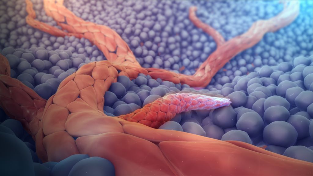
In the rare disease progeria, blood vessels deteriorate prematurely. A study from Karolinska Institutet shows how different cell types in the vascular wall undergo progressive changes and accumulate mutations over time. The findings are published in the journal Genome Medicine.
Hutchinson–Gilford progeria syndrome (HGPS) is a genetic disorder that causes remarkable premature ageing. Most patients die during their teenage years from cardiovascular disease, but the precise mechanisms underlying vascular damage remain unclear.
In the new study, researchers analysed cells from the aorta of mice carrying the same genetic mutation found in people with progeria. Using single-cell RNA sequencing, which enables gene activity to be studied in individual cells, they tracked how the vascular wall changes over time. In total, nearly 9000 cells from mice of different ages were analysed.
“This approach allows us to follow, step by step, how different cell types are affected throughout the course of the disease,” says Maria Eriksson, professor at the Department of Medicine, Huddinge, Karolinska Institutet.
Reduced numbers of smooth muscle cells
The researchers focused particularly on vascular smooth muscle cells, which provide blood vessels with strength and elasticity and are essential for normal vascular function. They observed that these cells gradually declined in number.
“Smooth muscle cells are progressively lost both in HGPS and during normal ageing. As these cells die, the vessel wall becomes weaker and more susceptible to disease,” says Lara Garcia Merino, doctoral student at the same department and first author of the study.
The study also showed that smooth muscle cells accumulated higher numbers of so-called somatic mutations, meaning genetic alterations that arise during an individual’s lifetime. The mutation burden was associated with increased cellular stress and activation of genes involved in DNA damage responses.
“This is the first evidence that the accumulation of somatic mutations is a hallmark of vascular disease in HGPS,” says Maria Eriksson.
Reveals a new mechanism
The findings link DNA damage to cellular stress, loss of cellular identity and cell death, thereby revealing a previously unrecognised mechanism driving irreversible vascular injury.
The researchers also found evidence that changes in cell behaviour may be influenced by signalling between different cell types within the vessel wall, suggesting that the process is not driven solely by alterations within individual cells.
“We see that cells undergo multiple changes over time, from stress to identity changes and ultimately cell death. Our results suggest that several different mechanisms interact in the development of vascular damage in progeria,” says Lara Garcia Merino.
The researchers believe that the findings may contribute to a better understanding of how vascular damage develops in progeria and underline the importance of initiating treatment early, before irreversible DNA damage has accumulated.
“New gene-editing approaches can correct the disease-causing mutation in HGPS, but correcting the mutation alone is unlikely to reverse damage in cells that have already accumulated a large number of somatic mutations. Early intervention is therefore essential,” says Maria Eriksson.
The study also provides new insights into the biological processes underlying normal vascular ageing. Several important similarities exist between HGPS and the cardiovascular disease that affects the general population. HGPS is therefore widely used as a model for understanding normal ageing and vascular disease.
The researchers emphasise that further studies are needed to confirm the findings in humans.
The study was conducted in collaboration with researchers from, among others, the Indian Institute of Technology in India and the University of Bergen in Norway. The research was funded by the Swedish Research Council, the European Research Council (ERC), the Swedish Cancer Society and the Center for Innovative Medicine, among others.
For decades, researchers studying myotonic dystrophy type 1 (DM1) have focused on the disease’s underlying genetic cause: a mutation that produces a toxic form of RNA, disrupting the normal processing of thousands of genetic messages inside cells. While scientists have known this widespread disruption contributes to disease, it has remained unclear which changes are most responsible for the progressive muscle weakness and wasting experienced by people living with DM1.
Now, a new study published in Nature Communications suggests that one hallmark symptom of the disease – muscle stiffness, known as myotonia—may play a much larger role in driving muscle damage than previously recognised.
“Our findings suggest that myotonia isn’t simply an uncomfortable symptom people experience,” said John Lueck, PhD, associate professor of Pharmacology and Physiology at University of Rochester Medicine and senior author of the study. “It appears to amplify the harmful effects of the disease in muscles. When we eliminated myotonia in our mouse model, we didn’t just improve muscle relaxation; we saw healthier muscles overall.”
The findings suggest that therapies aimed at reducing myotonia could help preserve muscle function while complementing emerging treatments designed to address the disease’s underlying genetic cause.
A disease caused by toxic RNA
DM1 is the most common form of adult muscular dystrophy. The inherited disorder causes progressive muscle weakness, muscle wasting, slow relaxation after muscle contraction, heart rhythm abnormalities, cataracts, excessive daytime sleepiness, and a range of other symptoms.
The disease begins with an abnormal expansion of repeated DNA segments in the DMPK gene. Rather than producing a faulty protein, this mutation creates a toxic RNA molecule that traps proteins needed to correctly process genetic instructions. As a result, hundreds to thousands of genes are improperly “spliced,” producing abnormal protein versions throughout the body. Decades of research led by URochester Medicine neurologist Charles Thornton, MD, a co-author of the study, helped establish how this toxic RNA disrupts normal RNA splicing and drives the disease.
One of the most important affected genes expresses a chloride channel that helps muscles relax after they contract. When that channel is disrupted, muscles become electrically overactive, producing the delayed relaxation known as myotonia.
Looking beyond the root cause
Most research has focused on eliminating the toxic RNA itself, with several RNA-targeted therapies now advancing toward clinical use. However, Lueck and his colleagues wanted to answer a different question: once myotonia develops, does it simply reflect the disease, or does it actively worsen muscle damage?
Previous work from the URochester Medicine team had hinted at the answer. They found that when myotonia occurred alongside another splicing defect affecting calcium channels, muscle disease became dramatically worse in mice. Treating those mice with calcium channel-blocking drugs reversed many of the effects. That finding suggested that muscle hyperexcitability might be directly contributing to muscle degeneration.
“We’ve spent years trying to understand which of the many splicing changes actually matter most,” Lueck said. “This study allowed us to isolate one of those changes and ask what happens when you permanently remove myotonia while leaving the underlying disease process in place.”
Turning down the disease’s “volume”
To answer that question, the researchers genetically corrected a single critical portion of the chloride channel gene in a mouse model of DM1. The researchers expected to reduce muscle stiffness. Instead, they saw improvements throughout the muscle.
The mice no longer developed muscle stiffness, but they also generated greater muscle force, showed healthier muscle tissue under the microscope, and experienced broad improvements in abnormal gene expression and RNA splicing. The findings suggest myotonia may act as what Lueck describes as a “volume knob” on the disease.
“The toxic RNA is still present,” he said. “But myotonia appears to turn up the damage happening in muscles. When we turned myotonia down, many aspects of muscle health improved, even though we hadn’t corrected the original genetic mutation.”
Implications for future treatments
The findings could influence how researchers think about treating DM1. Several experimental therapies currently in development are designed to eliminate the toxic RNA that causes the disease. Researchers have long used improvements in myotonia as an early sign that these therapies are working because the chloride channel is particularly sensitive to correction.
The new study suggests that reducing myotonia may itself contribute significantly to improved muscle health. In other words, treating myotonia may do more than relieve stiffness – it may actually help slow or reduce the muscle damage caused by the disease.
At the same time, existing medications that reduce myotonia – including drugs such as mexiletine and ranolazine – may deserve renewed attention. Although these medications can improve muscle stiffness, side effects often limit their long-term use, and many people with DM1 never receive them.
“If we can develop safer, better-tolerated myotonia drugs, they could become an important complement to RNA-based therapies—or provide meaningful benefit for patients who don’t have access to those advanced treatments,” said Lueck.
At 25, Kaitlin should be living independently. At 18, Lihle should be finishing school. Instead, both are fighting for their lives against aplastic anaemia (AA), a rare blood disease that leaves patients vulnerable to infections, uncontrolled bleeding, and severe anaemia. A stem cell transplant gives approximately 80% of patients a real chance at recovery, but for around 70% of those patients, that match will not come from within their family. It will come from a generous stranger.
“AA strikes hardest between 15 and 25 – the years nobody expects to spend fighting for their life,” says Palesa Mokomele, Head of Community Engagement and Communication at DKMS Africa. “We want South Africans to understand that registering as a stem cell donor is a simple act that could give someone like Kaitlin or Lihle their life back. Every person who registers increases their chances of finding a match.”
A long road to the right diagnosis: Kaitlin’s story
For years, nobody could tell Kaitlin from KwaZulu-Natal what was wrong. She experienced prolonged and excessive bleeding and severe fatigue, which was repeatedly misattributed to gynaecological issues. She kept going back to the hospital and kept being sent home. It was only in August 2025, when her condition deteriorated dramatically, and the bleeding would not stop despite ongoing treatment, that she was finally referred to a haematologist. A bone marrow biopsy told them what years of tests had missed: Kaitlin had AA.
Before this, she was working full-time and living independently. Today, she cannot work. She cannot manage basic daily tasks. She requires weekly blood transfusions simply to stay alive. Medication trials have yielded no response, and her doctors have been clear: a stem cell transplant is her only path to recovery.
Through it all, Kaitlin has held on. “I draw strength from my faith and from the people I love most – my nephews and siblings, who show up for me even on the hardest hospital days. I just want my life back, and a matching donor could make that possible.”
Sudden illness, endless resilience: Lihle’s story
Lihle was 14 years old when his life changed overnight. It started with severe nosebleeds in November 2021. Then one night, the bleeding became uncontrollable. He lost consciousness. After two months in hospital, the diagnosis came: Severe Aplastic Anaemia (SAA). That same year, his father passed away.
The eldest of four children, Lihle grew up fast. Hailing from Butterworth in the Eastern Cape and raised in Carletonville, Gauteng, he has always felt the weight of being the firstborn – the one his younger siblings look up to. Their mother cares for them all – while also carrying the emotional weight of losing her husband and watching her son fight for his life.
Lihle shares that he is determined to finish his education, set an example, and one day return to the football pitch. Like Kaitlin, all he needs is a matching donor to make that possible.”
How you can help
“No family should have to face what Kaitlin’s and Lihle’s are going through – knowing that a cure exists, but that the donor hasn’t been found yet. For patients from Black, Coloured and Indian/Asian backgrounds, that search is even harder, because the registry does not yet reflect the diversity of our population. We are calling on all South Africans to register. It costs nothing. It takes minutes. And it could mean everything,” concludes Mokomele.
Signing up could be the most important thing you ever do. If you are aged 17 – 55 and in good health, please register today at: https://www.dkms-africa.org/save-lives
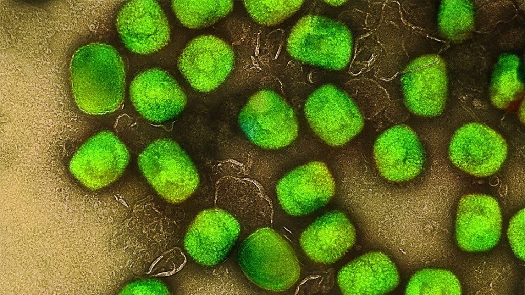
An international, randomised, double‑blind, placebo‑controlled phase 3 study, the largest of its kind for mpox, found that tecovirimat did not improve clinical outcomes for adults with clade II mpox compared with placebo, while demonstrating a similar safety profile. Results of the STOMP/A5418 trial, published in the New England Journal of Medicine underscore both the urgent need for alternative therapeutics and the critical importance of randomised trials during public health emergencies.
This Phase 3 study randomised 412 participants (344 with laboratory‑confirmed mpox), to receive either tecovirimat or a matching placebo for 14 days. Randomisation was stratified by early versus later symptom onset and by the presence of severe pain. Participants had active skin or mucosal lesions and self‑reported daily symptoms, pain scores, and lesion status through Day 29, with confirmatory clinical assessments at scheduled visits. Biospecimens, including lesion swabs, oral and rectal swabs, and blood samples, were collected at multiple time points to assess viral DNA clearance. The primary endpoint was time to clinical resolution of all lesions, and key secondary endpoints included pain reduction, complete lesion healing, and virologic response.
Conducted across seven countries at 49 sites, the phase 3 study showed that tecovirimat did not shorten the time to lesion resolution, reduce pain, or speed viral clearance compared with placebo. These results align with interim findings released in December 2024, which led the trial’s independent Data and Safety Monitoring Board to halt further enrolment due to statistical futility.
“In the midst of a global public health emergency, the ACTG team rapidly conducted this randomized controlled trial to deliver a clear answer for patients and clinicians,” said William A. Fischer II, MD, associate professor of pulmonary and critical care medicine at the UNC School of Medicine, and director of emerging pathogens research at the UNC Institute for Global Health and Infectious Diseases. “These findings advance our understanding of mpox and help the field refocus efforts on identifying safe, effective and accessible treatment strategies, particularly for people at highest risk of severe disease.”
Although the trial did not demonstrate efficacy, tecovirimat demonstrated a favourable safety profile, with no major safety concerns identified – an important confirmation as thousands of patients worldwide have already received the drug under expanded access protocols.
“The STOMP trial provides essential evidence at a critical time and demonstrates why randomized controlled trials are an indispensable part of outbreak response,” said Joe Eron, MD, chief of infectious diseases and chair of the ACTG network. “But now we must keep going to find safe and effective treatment for people as this virus continues to circulate globally.”
The study’s conclusions are expected to influence clinical practice and public health guidance worldwide. With mpox still causing outbreaks in multiple regions, researchers emphasise that developing and evaluating new antiviral candidates remains a top priority.
Ivermectin was originally celebrated as a revolutionary treatment for parasitic disease in humans and animals. It has since evolved into a focal point of misinformation and heated debate.
During the early part of the COVID pandemic, it was touted on social media as a miracle cure for the virus, despite a lack of robust evidence.
The drug is a small organic chemical that can be extracted from the bacterium Streptomyces avermitilis. This bacterium grows in the soil, and was first found near the grounds of a Japanese golf course.
It was first approved for use in animals in 1981 and in humans in 1987. It’s now available in various brands as tablets and creams you apply to the skin.
Assessing the evidence
Governments use human clinical trials to decide whether to approve a medicine for sale.
But clinical trials aren’t the highest level of evidence to inform best practice and guide decisions. For that, there are Cochrane reviews.
A Cochrane review brings together a panel of experts who collate and assess all the relevant evidence on a medication. It takes data from multiple clinical trials, and other studies, and evaluates it following clear and structured steps. It’s able to examine and critique study designs to identify bias and reject bad data.
Cochrane reviews are also regularly updated to take into account new information. The result is a summary that is considered the highest level of evidence to guide decision-making.
So what do Cochrane reviews say about ivermectin for different conditions?
Ivermectin is used to treat a variety of parasitic worm infections. These include the round worms Ascaris lumbricoides, Strongyloides stercoralis, Wuchereria bancrofti, and Brugia malayi.
The latter two worms cause the disease lymphatic filariasis (or elephantiasis) which causes severe swelling in the arms, legs, breasts and genitals.
When ivermectin is used to treat Strongyloides stercoralis, the Cochrane panel found it is better than albendazole and had fewer side effects than thiabendazole.
For Ascaris lumbricoides, the panel concluded ivermectin was as good as albendazole and mebendazole.
For treating lymphatic filariasis, a Cochrane review found ivermectin or diethylcarbamazine should be standard treatment in combination with albendazole.
Rosacea
The Cochrane review for rosacea evaluated 22 different treatments for this skin condition, including a variety of drugs, as well as light therapy, cosmetics and reducing the intake of spicy food.
It concluded that ivermectin applied to the skin was more effective than a placebo, and a bit better than the other standard medication, metronidazole.
Scabies
Cochrane has two reviews on the use of ivermectin for scabies. One specifically evaluated ivermectin and permethrin as treatments. The other evaluated all available treatments for scabies.
The first review concluded both permethrin and ivermectin were just as effective, regardless of whether the ivermectin was administered orally or directly onto the skin.
In contrast, the second review concluded ivermectin does work but topical permethrin appeared to be the most effective treatment.
Malaria
The Cochrane panel looked specifically at whether ivermectin could reduce transmission of the malaria parasite, rather than as a treatment.
Unfortunately there was just a single clinical trial to use as evidence. In that trial, residents of eight villages were given ivermectin and albendazole together, with follow up doses of just ivermectin. The researchers then looked at the rates of child infection over 18 weeks.
Even though the trial didn’t show ivermectin prevented infection, due to the high risk of bias in it, the Cochrane panel couldn’t conclude either way whether ivermectin worked or not.
River blindness
River blindness is caused by another parasitic worm called Onchocerca volvulus.
The Cochrane review concluded there was a lack of evidence either way to know whether it works to prevent infection-based visual impairment and blindness.
It evaluated the data from four clinical trials and two large community-based studies.
One of the reasons the panel was unable to make a firm conclusion was because it thought the drug may work differently against different strains of the parasite and in people of different ethnicity.
Cancer
There are no Cochrane reviews on ivermectin’s use for cancer because clinical interest in the drug for this condition is just starting.
There is a current clinical trial that is evaluating ivermectin in combination with antibody-based drugs for breast cancer.
Early results showed the combination of antibody drugs with ivermectin was safe to patients, but no efficacy data has been published.
COVID
The Cochrane panel rejected the data for seven clinical trials and included 11 other trials. Rejected trials included those which compared ivermectin against other drugs which were known to not be effective against COVID, such as hydroxychloroquine.
The review concluded there was no evidence to support the use of ivermectin for the treatment or prevention of COVID. In making that conclusion, it evaluated treatments that used invermectin or placebo in combination with standard care and whether treatment reduced death, illness, or the length of the infection.
Red Blood Cell Infected with Malaria Parasites Colourised scanning electron micrograph of red blood cell infected with malaria parasites (teal). The small bumps on the infected cell show how the parasite remodels its host cell by forming protrusions called ‘knobs’ on the surface, enabling it to avoid destruction and cause inflammation. Uninfected cells (red) have smoother surfaces. Credit: NIAID
A new clinical trial led by QIMR Berghofer, in collaboration with University of Sunshine Coast Clinical Trials Network has found a medication currently used for some blood disorders could help the body fight malaria more effectively.
The findings mean the drug, ruxolitinib, could potentially be used alongside standard treatment to boost recovery and strengthen people’s immune systems against future infections.
Malaria kills more than 600 000 people each year and three quarters of those deaths are in children under the age of five.
Current treatments for malaria work by killing the parasite that causes most malaria deaths, Plasmodium falciparum. However, even with these treatments, fatality rates from severe malaria remain high.
Furthermore, while patients develop some immunity after infection, this protection is often incomplete, leaving many vulnerable to reinfection.
“While antimalarial treatments are effective at killing the parasite, they don’t directly address the inflammation that contributes to severe illness and death. These findings suggest that we may be able to improve clinical outcomes by targeting the host inflammatory response as well as the parasite itself,” she said.
The research, published in Science Translational Medicine, looked at how the immune system responds to malaria via the body’s ‘early warning system’ known as type 1 interferon signalling.
To do this, researchers enrolled 20 healthy adult volunteers who had never been exposed to malaria. Participants were deliberately infected with Plasmodium falciparum under closely monitored conditions. Eight days later, all participants received standard malaria treatment (artemether-lumefantrine), while 11 were also given ruxolitinib. Three months later, participants were re-infected with malaria to test how their immune systems responded to a second infection.
The research revealed ruxolitinib was safe and well-tolerated, compared with the placebo group, and participants who received ruxolitinib showed a lower inflammatory response, and favourable changes in markers linked to disease severity.
QIMR Berghofer’s Program Director of Infection and Inflammation Professor Christian Engwerda says the results are encouraging.
“One of the biggest challenges in efforts to eliminate malaria is the limited efficacy and duration of protection provided by current vaccines. By boosting the immune system without causing detrimental inflammation with drugs like ruxolitinib, we may be able to overcome these challenges,” he said.
The researchers say it’s important to note that the study was conducted in healthy volunteers who did not live in malaria-endemic regions. Further studies in malaria-endemic regions will be needed to determine whether these findings translate into improved outcomes for patients most affected by the disease.
Lab tests confirm that antiviral class known as Pin1 inhibitors could reduce and stop outbreaks of herpes simplex virus-1
Photo by Cottonbro on Pexels
A class of antivirals called Pin1 inhibitors could reduce or stop outbreaks of herpes simplex virus 1 (HSV-1), the common infection behind oral herpes, according to new research published in Antiviral Research.
HSV-1 causes sores around the mouth, commonly called cold sores or fever blisters. Most people are infected with HSV-1 in childhood, and between 50% and 90% of people worldwide have HSV-1. After the initial infection, HSV-1 remains in the body and can reactivate throughout a person’s life. While HSV-1 infections are usually mild, they can be serious and even deadly for people with suppressed immune systems. Finding new, more effective antivirals for this common illness is essential.
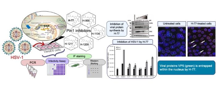
Pin1 inhibitors suppress HSV-1 replication by inhibiting viral protein synthesis and preventing nucleocapsid egress from the nucleus. (Takemasa Sakaguchi/Hiroshima University)
Researchers focused on an enzyme called peptidyl-prolyl cis-trans isomerase NIMA-interacting 1, or Pin1, that regulates protein stability, function, and cellular structure. When this enzyme is dysregulated, it can play a role in a variety of conditions, including obesity, cancer, heart failure, and more. Viruses, such as cytomegalovirus (CMV) and SARS-CoV-2, are known to affect Pin1, and Pin1 inhibitors have been developed to reduce the impact of these viruses.
Because HSV-infected cells over-express Pin1, researchers wanted to know if Pin1 inhibitors could also be used to treat HSV-1. “This study revealed that the host factor Pin1 is a crucial therapeutic target for the proliferation of HSV-1. Pin1 inhibitors potently suppress HSV-1 replication at low concentrations,” said Takemasa Sakaguchi, a professor at the Graduate School of Biomedical and Health Sciences at Hiroshima University in Hiroshima, Japan.
In laboratory tests, the Pin1 inhibitor H-77 and the four newly developed Pin1 inhibitors successfully stopped the replication of HSV-1. VeroE6 cells, derived from the kidney of an African green monkey and commonly used in virology research, were infected with HSV-1 and cultured in the presence of different amounts of a Pin1 inhibitor. As the amount of the inhibitor increased, the effects of HSV-1 on the cells became less pronounced and completely disappeared at 1 μM. They also found that any viral particles released from the treated cells were non-infectious.
The most important finding is how Pin1 inhibitors affect cell structures to prevent the virus from escaping. They do this by stabilising nuclear membrane structure, physically trapping the virus in the cell nucleus. “The nuclear lamina initially functions as a ‘barrier’ when nucleocapsids of progeny viruses, that replicate within the nucleus, bud from the nuclear membrane. Pin1 overexpressed by the virus removes this barrier. However, through the action of the Pin1 inhibitor H-77, this barrier is rather reinforced, forming a thick and robust lamina layer. This demonstrates that H-77 transforms the nuclear lamina into an ‘impregnable defensive wall,’ physically blocking the escape of viruses from the nucleus of the cell,” said Sakaguchi.
Looking ahead, researchers will continue to evaluate the effectiveness of Pin1 inhibitors to treat HSV-1. They will also research how Pin1 inhibitors could be used to treat other viruses. “The ultimate goal for the future is to aim for the clinical application of Pin1 inhibitors as ‘host-directed therapeutics,’ which are less likely to cause drug resistance. To achieve this, we will first evaluate their efficacy against diverse viruses to clarify the treatable range. Simultaneously, research to optimise the compound structure is essential for creating more potent and selective drugs,” said Sakaguchi.