Category: Implants and Prostheses

Dual Mobility Hip Replacements Reduce Dislocation Risk by 70%


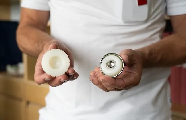
The new implant (to the right) consists of a small ball encased in a larger ball, which gives better stability. Photo: Sandra Gunnarsson. Credit: Queen Mary University of London

A major international clinical trial had found that an emerging type of hip replacement implant dramatically reduces complications in people with pelvic fracture. The study, published in The Lancet, involved 1600 patients across 44 hospitals in the UK and Sweden. The DUALITY trial is the largest clinical trial to compare dual mobility total hip replacements (DM-THR) with standard total hip replacements (THR). The team found that people treated with DM-THR were 70 per cent less likely to experience a dislocation post-surgery – the most common complication after hip replacement for fracture.

A broken hip in older people is one of the most common serious injuries worldwide, affecting more than 14 million individuals each year and accounting for 1.4% of total direct healthcare expenditure in established market economies. The type of hip fracture studied in the DUALITY trial represents approximately half of all hip fractures experienced globally.

A total hip replacement where both the ball and socket of the hip are replaced, is recommended for older, active individuals with some types of hip fracture. For most patients, the procedure improves mobility and quality of life, but dislocation post-surgery can be common and can have serious consequences for those affected. When dislocation occurs, patients often require emergency hospital admission, procedures to reset the joint, and sometimes further surgery. This can lead to longer recovery times, added distress, and increased risk of further health problems.

Dual mobility-THR uses a small ball encased in a much larger plastic ball, and was developed specifically to reduce the risk of dislocation. In the DUALITY trial, the team aimed to establish if DM-THR reduced the risk of dislocation compared to THR. The results showed that the DM-THR implant does improve stability in the hip joint, making it less likely to dislocate after surgery. They found that within one year of surgery, just 1.3 per cent of patients receiving DM-THR experienced a dislocation, compared with 4.2 per cent of those given a standard THR. Importantly, the study found no increase in other risks such as infection or death, and overall complication rates were lower in patients receiving DM-THR.

The researchers conclude that dual mobility implants should be considered the preferred option for suitable older patients undergoing total hip replacement after a broken hip. Crucially, DM-THR requires no new technology or training. Surgeons are already familiar with both implant types, meaning the change could be implemented immediately within existing practice.

Professor Xavier Griffin, study author and Chair of the Centre of Bone and Joint Health at Queen Mary University of London and Honorary Consultant Orthopaedic Surgeon at Barts Health NHS Trust, said:

“Dislocation is the most common major complication following hip replacement for a broken hip. People that experience this painful complication often require further surgery and the recovery following this is usually long, slow and painful. So, it has been an area that I, along with our surgeons around the world, have been wondering if there is any benefit to using these type of hip replacements in people with a broken hip. The fantastic news from DUALITY is that we can make a really substantial reduction in the risk. I hope that this research will make a real difference to many future patents who might avoid this catastrophic problem.

I’ve tried to run similar studies before but never been able to deliver one that is big enough to give us a reliable answer to the question. Meeting the team in Uppsala and making this international collaboration a reality has been a game changer for accelerating how quickly we can discover the answers to these sorts of questions.”

Professor Nils Hailer, study author, Chair of Orthopaedics at Uppsala University and Consultant Orthopaedic Surgeon at Uppsala University Hospital, said:

”After many years of analysing registry data and seeing both advantages and limitations of dual mobility hip replacements, I was eager to obtain solid evidence for or against the concept. Together with colleagues at Queen Mary, we succeeded in delivering a large, pragmatic, orthopaedic randomized trial involving both smaller hospitals and major referral centres across two countries.

“The results provide robust support for the use of dual mobility constructs in hip fracture patients in need of a total hip replacement. Beyond the findings themselves, I believe this collaboration sets a new benchmark for future randomized trials in European orthopaedic research, and we will jointly continue working towards that goal.”

Researchers say that beyond improving outcomes for patients, reducing dislocations could have a significant impact on healthcare systems. Complications such as post-surgery dislocations increase hospital stays, require additional operating time, and drive unplanned readmissions. Although dual mobility implants are currently more expensive than standard implants, researchers believe the reduction in complications could offset the higher upfront cost. A full economic analysis is underway.

Source: Queen Mary University of London

Neural Stimulation and Exoskeletons Improve Hand Function in Neurological Injuries

Study demonstrates the potential of personalised assistive technologies for people with impaired hand function

Researchers at the Medical University of Vienna, in collaboration with ETH Zurich, the Technical University of Munich and Medical Faculty Belgrade, have developed a wearable neurorobotic system that combines electrical neurostimulation with hand exoskeletons. In a clinical trial involving 14 patients with hand impairments caused by neurological injury, the technology supported finger mobility, tactile perception and grip control. The results demonstrate the potential of personalised assistive systems for people living with the consequences of spinal cord or brain injury. The study has recently been published in the journal Science Advances.

Hand movements and the sense of touch are essential for everyday activities such as grasping, eating, dressing or personal hygiene. However, after damage to the central nervous system, motor and sensory impairments of the hand often persist. Conventional rehabilitation can achieve improvements, but does not always lead to sufficient restoration of hand function. There is therefore a great need for assistive technologies suitable for everyday use.

A research team led by study director Stanisa Raspopovic from the Center for Medical Physics and Biomedical Engineering at MedUni Vienna has developed the “SensoExo” system for assisting people with hand sensorimotor impairements. It combines a wearable hand exoskeleton with a custom-fitted neurostimulation sleeve. The sleeve stimulates specific nerves and muscles in the forearm through the skin. Sensors on the fingers detect touch and gripping forces and translate this information into electrical stimulation, providing users with tactile feedback. In addition, functional electrical stimulation can assist users open and close their fingers more easily.

“Our aim was not only to provide mechanical support for movement, but also to restore their sense of touch,” says Stanisa Raspopovic. “The interplay of strength, movement and the sense of touch is crucial, particularly when gripping. Without feedback on how firmly an object is being held, hand function remains significantly limited in everyday life.”

Individualised support depending on the impairment

The system was tested on 14 patients with neurological hand impairments. All study participants exhibited sensory deficits and therefore received tactile feedback via transcutaneous electrical nerve stimulation. In seven individuals with particularly severe motor impairments, functional electrical muscle stimulation was also used to support hand opening and grip strength.

The study compared three conditions: no support, support from an exoskeleton alone, and the combined use of an exoskeleton and neurostimulation. Eight of the 14 participants also completed functional grasping and releasing tasks with bulky and fragile objects. This investigation revealed that the combination of exoskeleton and neurostimulation provided additional benefits compared to an exoskeleton alone. In patients with severe motor impairment, SensoExo improved finger mobility to a greater extent than the exoskeleton alone. The artificially mediated tactile feedback also increased the areas of the hand where touch sensations could be perceived.

“The results show that motor assistance and sensory feedback must be considered together,” explains lead author Andrea Cimolato from the Center for Medical Physics and Biomedical Engineering at MedUni Vienna. “The system can be adapted depending on the individual’s impairment profile. People with more severe motor impairments benefited particularly from additional motor support, while those with pronounced sensory loss used the sensory feedback to grasp fragile objects more precisely.”

Improved grasping of everyday objects

In the functional tests, participants using SensoExo achieved the highest success rates when grasping and carrying objects. With bulky objects, muscle stimulation supported grip strength. With fragile objects, sensory feedback helped to avoid applying too much pressure.

“The technology is currently a prototype and not a fully developed medical device for everyday use,” emphasises Raspopovic. “However, the study provides early clinical evidence that non-invasive neurostimulation combined with wearable robotics can form a realistic basis for future personalised assistance systems.”

Source: Medical University of Vienna

Why Antibiotics Fail Against a Common Dental Implant Disease

By Alexmit artOwn work, CC BY-SA 4.0, Link

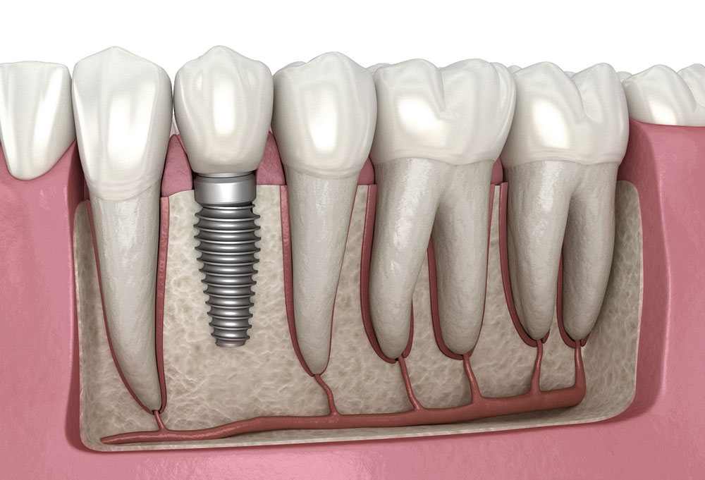
Dental implants have given tens of millions of people something dentures never could: a full set of fixed and fully functioning teeth. Unfortunately, 10% to 20% of implant patients eventually experience an aggressive jawbone infection called peri-implantitis. 

Antibiotics usually fail to stop the infection for reasons that researchers have never understood – until now.

A new study in PNAS Nexus by researchers with the Rutgers School of Dental Medicine found that bacteria corrode implants, causing them to shed microscopic titanium particles into the surrounding tissue. Those particles hijack the immune cells sent to clear the infection and lock them into a state of inflammation that destroys the jawbone they are supposed to protect.

Working with human tissue samples, cultured human immune cells and a genetically engineered mouse model, the team pinpointed a specific calcium channel in the body’s bacteria-eating macrophages that the titanium particles activate. Switching that channel off in mice prevented the disease. The result is the first credible drug target for a condition that affects up to one in five implant recipients and costs the global health system more than a billion dollars a year.

“For the first time, we show why all the antibiotic treatments that work around teeth do not work around implants,” said Georgios Kotsakis, the study’s senior author and the assistant dean for clinical research at the dental school. “Now that we know the cause, we can start developing therapeutics.”

Peri-implantitis has long been a puzzle because it initially looks like its counterpart in natural teeth, which is called periodontitis and begins with the same oral bacteria. In patients with natural teeth, antibiotics and routine cleaning resolve the infection. In patients with implants, the same drugs against the same bacteria succeed less than half the time, while the bone underneath continues to disappear.

Most research over the past 20 years has focused on the bacteria. Members of Kotsakis’ lab took a different approach and began looking at the implants. Bacteria living on the implant surface produce acidic biofilms that slowly corrode the titanium, releasing billions of particles smaller than a red blood cell. The same shedding can occur during routine cleaning, especially with instruments that dentists typically use on natural teeth.

Inside the gum, those particles get coated with a bacterial toxin called lipopolysaccharide. To the immune system, they suddenly look like enormous, indigestible bacteria – but macrophages cannot digest metal. The cells become trapped in a hyperinflammatory state, pumping out signalling molecules including interleukin-1 beta, an inflammatory protein also implicated in rheumatoid arthritis and Alzheimer’s disease. 

That inflammation eats away at bone. Worse, the immune cells lose their ability to deal with the original infection. In the lab, macrophages exposed to titanium particles took up less than half as many bacteria as unexposed cells. 

“These particles are little magnets that attract the bacterial toxin, and they hijack the immune system, preventing it from clearing bacteria,” said Kotsakis. “You have a perfect storm that defies antibiotics.”

Team members traced the cascade to a calcium channel (a specialised, pore-forming protein structure within cell membranes) called TRPC1. In mice engineered without it, the immune cells handled the same titanium-plus-bacteria challenge normally: abscesses were dramatically smaller, inflammatory cytokines dropped, and bacterial clearance was restored. 

Funded by the National Institutes of Health, members of Kotsakis’ group are testing drug candidates that target the same pathway in human cells.

For people who already have implants, the most useful finding may be a quieter one. The strongest known protective factor is regular professional cleaning, but the kind of cleaning matters. Until roughly a decade ago, many dentists scraped implants with the metal scalers used on teeth, a method the Rutgers lab and others have shown can itself corrode the implant and accelerate the disease. Nonabrasive techniques are now standard. 

By Andrew Smith

Source: Rutgers University

Clinical Trials Are Part of UP Professor’s Dream of ‘Making Deafness History’

University of Pretoria’s Professor Mashudu Tshifularo is leading a groundbreaking clinical trial for 3D-printed ossicles,

Forty-five patients with conductive hearing loss from middle-ear damage are eagerly awaiting the start of clinical trials, led by University of Pretoria’s Professor Mashudu Tshifularo, on a ground-breaking procedure to restore hearing.

The trials, due to begin within weeks at Steve Biko Academic Hospital in Pretoria, come seven long years after Prof Tshifularo successfully performed the world’s first middle-ear transplant using 3D-printed bones made from titanium. The patient was Thabo ***, whose middle-ear bones – the ossicles – had been injured in an accident, causing almost total hearing loss until he had the transplant in March 2019.

In a video screened at a UP Roundtable event held in April this year to announce the launch of the upcoming clinical trials, a beaming Thabo *** confirmed he had regained his hearing after the transplant and had continued to enjoy excellent hearing ever since.

While this procedure was hailed as a surgical breakthrough at the time, its full acceptance by the South African and global ear, nose and throat (ENT) community depends on formal clinical validation. Despite the many obstacles he knew would lie ahead, Prof Tshifularo, joint head of UP’s Department of Otorhinolaryngology, was determined to take his innovation through clinical trials.

“Today is a culmination that I never thought would come. There was a time when I cried, there was a time when I was very depressed, there was a time when I nearly gave up, but something inside me told me to remain steady,” he said at the Roundtable, where the announcement was made that clinical trials could finally move ahead now that all the necessary regulatory, ethical and licensing clearances have been obtained, including from the South African Health Products Regulatory Authority (SAHPRA).

The years since Prof Tshifularo performed that first transplant have also been spent developing, perfecting, testing and patenting a prototype of the titanium material and implants for the clinical trials. UP’s engineering partner on this project, the Council for Scientific and Industrial Research (CSIR), was responsible for the materials and prototype development, while HH Industries manufactures the implants, using 3D printing technology, and Marcus Medical is providing the robotic technology to be used during the surgery. Seed funding was provided by the Motsepe Foundation.

Ready to transform lives

Now that it is all-systems-go for this all-South African collaborative effort, Prof Tshifularo and his team aim to perform this life-changing surgery in the next 12 months on all 45 patients who have expressed interest in participating in the clinical trials.

The team will then focus on publishing their research and training future researchers and medical teams so that the work can be amplified far and wide for the benefit of humanity.

“My dream is to make deafness history,” Prof Tshifularo said, noting that an estimated two billion youth are living with undiagnosed, mostly noise-induced hearing loss, which would become a severe problem in the next 20 to 25 years. Hearing loss is also common among the world’s rapidly ageing population.

The procedure itself takes about three hours and uses advanced robotics to remove the damaged middle-ear bones and replace them with the 3D printed titanium bones. This is minimally invasive, carries significantly less risk than conventional procedures and leaves minimal scarring. The titanium used to manufacture the ossicles is biocompatible, meaning it can be introduced into the ear without causing harmful reactions.

“This innovation will ultimately transform the lives of many people, including newborn babies born with congenital middle-ear defects,” said Prof Themba Mosia, Vice-Principal: Student Life, who gave the opening address at the Roundtable. “It exemplifies the spirit of innovation and collaboration at the university, combined with the deep medical expertise needed to restore hearing.”

UP at the forefront of healthcare advancements

Prof Flavia Senkubuge, Dean of UP’s Faculty of Health Sciences, said innovations such as Prof Tshifularo’s middleear surgical procedure place the university “front and centre” of healthcare advancements on the African continent and the world stage.

She reiterated UP’s commitment to deploying its innovations for the benefit of local communities, such as by establishing a “one-stop shop” – most likely in the form of a private day hospital – where community members could benefit from advanced clinical technologies and the expertise of its researchers.

Paying tribute to Prof Tshifularo, long-time colleague Dr Christian Quitter thanked Prof Tshifularo “for having the guts” to persevere with his innovation, even when it was not always supported by the ENT establishment.

He also thanked the university for supporting researchers who “think out of the box in seeking to improve the lives of all humanity”.

Provided by the University of Pretoria

New Magnesium Coating Could Improve Safety of Medical Implants

Photo by DanR. CC BY-NC-SA-2.0

A team of scientists from the University of the Sunshine Coast in Australia and around the world has developed a promising way to reduce the risks from biodegradable medical implants. 

Scientists from UniSC’s Centre for Bioinnovation and advanced materials and manufacturing scientists from the School of Science, Technology and Engineering collaborated with Foshan’s First People’s Hospital in China and the University of Tokyo to develop a new coating for medical magnesium implants. 

The implants are coated in bioactive peptides, which are small, naturally occurring protein fragments that can support health and wellbeing. 

The new coating combines advanced metal processing with biomolecular science to improve compatibility with the body, reduce inflammation and boost antibacterial activity, enabling the implant to degrade safely as the bone heals. 

It has shown promising results, published recently in Biomaterials Research

Associate Professor Tianfang Wang said the technology could be most beneficial for orthopaedic implants such as plates, screws and pins used to repair fractures, as well as certain dental implants. 

“Our ultimate goal is to create self-absorbing implants that support healing then naturally disappear once no longer needed. This would reduce the physical and emotional burden associated with implant removal, giving patients greater confidence and comfort in their recovery. 

“It may also be suitable for cardiovascular stents or other devices where antibacterial protection and immune compatibility is essential in the critical early stages after implant,” he said.  

Traditionally, metallic implants are made from stainless steel or titanium and remain permanently in the body or sometimes require surgical removal after healing, which can cause pain, anxiety, and added costs. 

Magnesium alloys are among a new generation of degradable implant materials currently being developed based on naturally occurring trace elements in the body and designed to degrade naturally over time in unison with healing, so that they don’t require removal.  

 “While the magnesium alloys are biodegradable, these implants may still need to be removed if they degrade too rapidly, or cause infection,” said Professor Xiaosong Liu, the lead Chinese collaborator from the First People’s Hospital of Foshan. 

“Degradable, biocompatible magnesium implants with built-in antibacterial activity could eliminate these issues, reducing patient distress, surgical risks, and healthcare costs while promoting more sustainable medical practices,” said AMM materials scientist Dr Hejie Li.  

Associate Professor Damon Kent, leader of AMM, said the next step is to move the alloy to production for early pre-clinical trials, while exploring partnerships with biomedical companies to support the scale-up. 

“We are also exploring use of the coatings on other suitable metals and 3D metal printing options. There are a lot of possibilities,” he said. 

UniSC Deputy Vice-Chancellor (Research and Innovation) Professor Ross Young said the innovation was the latest in a growing body of world-class translational research and impact at the University, particularly in the health and medical space. 

“Expert researchers at UniSC continue to deliver new insights into cancer, chronic diseases, mental wellbeing such as PTSD and youth mental health, healthy ageing, nutrition and sports science,” Professor Young said. 

“Coupled with our commitment to introducing a Medical Program, UniSC is truly establishing its position on the world stage for its leadership and expertise in human health.” 

Source: University of the Sunshine Coast

Is it Safe to Have an MRI After Hip or Knee Replacement Surgery?

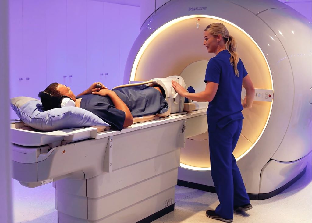
A patient with a knee replacement undergoing an MRI where modern technology reduces the distortions in the images.

It is a common concern for patients that metal implants, such as hip or knee replacements, may prevent them from having an MRI scan. In most cases, this is not true. Patients with modern joint replacements can safely undergo MRI, depending on the materials used in the implant. It is important to inform the radiology team about the implant before your scan.

Dr Jean de Villiers, a radiologist and director of SCP Radiology, answers some of the questions most frequently asked by patients, specifically around the process from referral to reporting in radiology imaging.

What is Magnetic Resonance Imaging (MRI)?

MRI is a non‑invasive imaging technique that uses powerful magnets and radio waves to create detailed images of the body’s internal structures. Unlike X‑rays or CT scans, MRI does not involve ionising radiation and is used extensively to diagnose a wide range of conditions.

Because MRI uses strong magnetic fields, many patients ask whether it is safe to have an MRI after a hip or knee replacement.

Can you have an MRI after a hip or knee replacement?

Yes, you can have an MRI scan on other parts of the body, as well as on the knee or hip where the implant is. Although some older MRI scanners may not be compatible with certain prostheses, the vast majority of MRI equipment in use today is safe and compatible with modern hip and knee implants.

How safe is MRI if the implant is made of metal?

Most implants are made from titanium or cobalt‑chromium alloys. Although these materials are metallic, they are not significantly affected by the magnetic field of an MRI scanner, nor do they heat up during the scan. Many implants also contain hard plastic components, all of which are designed to be compatible with MRI scanners. They are not attracted to the powerful magnet in the same way as older or highly magnetic materials.

Dr de Villiers explains, “The vast majority of joint replacements used today are MRI‑safe. The key is that we know about them in advance, so we can adjust the scan if needed.”

What is the main challenge with MRI and an implant?

The main challenge is image quality. Metal can sometimes cause image distortion, known as artefact, on MRI images. This may make it more difficult to assess structures close to the implant. However, modern MRI techniques have improved significantly and can often minimise these effects, allowing radiologists to assess surrounding tissues such as muscles and ligaments, and to detect complications such as infection or loosening. MRI is often the best imaging method for evaluating pain or complications after joint replacement surgery.

What happens if MRI does not produce clear diagnostic images?

In some cases, alternative imaging techniques such as CT or ultrasound may be recommended, depending on the clinical question. However, MRI remains safe and highly valuable for many patients with joint prostheses.

Are there implants that prevent you from having an MRI?

Certain implants and devices may be unsafe or require special precautions during MRI, including:

  • Implanted pacemakers
  • Intracranial aneurysm clips
  • Cochlear implants
  • Certain prosthetic devices
  • Implanted drug‑infusion pumps
  • Neurostimulators
  • Bone‑growth stimulators
  • Any other iron‑based metal implants

MRI is also contraindicated in the presence of some internal metallic objects such as bullets or shrapnel, as well as certain surgical clips, pins, plates, screws, metal sutures or wire mesh.

Having a hip or knee replacement does not automatically exclude you from having an MRI scan. With modern implants and appropriate planning, MRI is both a safe and important diagnostic tool. As technology continues to evolve, future developments are expected to further enhance MRI compatibility with hip and knee implants, making it an even more reliable tool for ongoing patient care.

It is crucial for patients to inform their healthcare providers about their joint replacement before undergoing an MRI. This allows the medical team to adjust the MRI settings and take appropriate precautions to ensure both safety and diagnostic accuracy.

New Global Study Estimates that Modern Hip Replacements Could Last at Least 30 Years

New global study using data from the National Joint Registry, estimates that modern hip replacements could last at least 30 years

Photo by DanR. CC BY-NC-SA-2.0

A major international study led by researchers who have used extensive data from the NJR estimates that modern total hip replacements, those using today’s more advanced bearing surfaces, are likely to last over 30 years in 92% of patients. This new finding marks a significant improvement in long term implant longevity and durability, compared with previous generations of medical implant devices.

Published on 26 February 2026, the research represents the largest and most contemporary analysis of hip replacement conducted to date. The study was a global collaboration including data contribution from eight joint registries. The data of just under two million hip replacement procedures were analysed, with the NJR accounting for almost two-thirds of that data. Registry data were combined with evidence from 29 long term clinical studies, across 18 countries.

Data was included from adult patients undergoing primary hip replacement with contemporary bearing surfaces: highly cross‑linked polyethylene (XLPE), ceramic‑on‑XLPE, or third‑ and fourth‑generation ceramic‑on‑ceramic articulations. Only implants that are still in routine clinical use were included, ensuring the study reflects modern practice, rather than historic device performance. Across all registries, cases were followed for a minimum of 10 years, with implant survival tracked until first all‑cause revision. All three material types demonstrated similarly high survivorship.

The results of the study provide patients with reassurance in consideration of the commonly asked question “How long will my hip replacement last?”  It is encouraging to know that modern hip replacements could last decades.

With regard to previous research on implant longevity, a 2019 study into hip replacement longevity which was supported by the NJR, suggested that over half, ie. 58% of hip replacements lasted 25 years, but those estimates were based on some implants made of materials that are no longer widely used. In 2022, another review of NJR data was conducted to enable further understanding of implant longevity, which produced the paper: ‘How long revised and multiply-revised hip replacements last?’ You can read more on that here.

You can read the recent Lancet paper here: Survivorship of modern total hip replacement to 30 years: systematic review, meta-analysis, and extrapolation of global joint registry data – The Lancet

Source: National Joint Registry

‘What’s Your Epic?’ Gathers Momentum as Amputee Riders Prepare for the 2026 Cape Epic

Movement is a Right, not a Privilege

Since launching late last year, Össur South Africa’s ‘What’s Your Epic?’ campaign has gained strong traction, with six amputee athletes now deep into training for the 2026 Cape Epic (15–22 March). As preparations intensify, the campaign continues to rally South Africans around a powerful belief: that access to mobility is fundamental to dignity, independence, and opportunity.

Three amputee teams will line up at one of the world’s most demanding mountain biking events, not only to test their physical limits, but to raise awareness and funds for three South African non-profit organisations restoring mobility and independence to people living with limb loss or disability: Jumping Kids, Rejuvenate SA, and Zimele.

Over the past few weeks, the riders have been balancing rigorous training schedules with advocacy, fundraising, and community engagement, using the build-up to the Cape Epic to shine a spotlight on the everyday barriers faced by thousands of South Africans who lack access to basic mobility solutions.

“Since launching ‘What’s Your Epic?’, the response has been incredibly encouraging,” says Blignaut Knoetze, Managing Director of Össur South Africa. “What’s been most powerful is seeing how this campaign has resonated beyond sport. It’s sparked conversations about access, inclusion, and what mobility truly means in people’s lives.”

For the six riders, the road to the Cape Epic is as much mental as it is physical. Long training rides, strength work, and recovery sessions are all undertaken with a deeper purpose in mind.

“Training for the Cape Epic is intense, but every ride reminds me why this matters,” says Rentia Retief, artist and amputee athlete. “With the right prosthetic and support, I’ve been able to reclaim the life I knew before losing my leg. Through this campaign, we’re trying to help make that same freedom possible for others.”

Mhlengi Gwala, international para-triathlete and African champion, adds, “This race is about more than endurance. It’s about representation and showing what’s possible when people are given the tools and support to move forward.”

Representing the third team, Brian Style, a passionate cyclist who rebuilt his life through mountain biking, says, “Preparing for the Cape Epic is both challenging and incredibly rewarding. Being part of this campaign gives real meaning to the training, knowing that every kilometre ridden helps create opportunities for others to regain their independence and confidence.”

Funds raised through the campaign will support:

  • Jumping Kids, which provides prosthetic limbs, education access, and sport opportunities to children with limb loss.
  • Rejuvenate SA, which supplies mobility aids to adults who cannot afford them, restoring dignity and independence.
  • Zimele, which supports adults with physical disabilities to regain independence, reintegrate into society, and build economic self-sufficiency.

Together, these organisations are changing lives every day, from helping a child take their first steps to enabling adults to return to work and participate fully in their communities.

“The riders may be the face of the campaign, but the real heroes are the organisations working on the ground,” says Knoetze. “Our role is to amplify their impact and encourage South Africans to get involved in any way they can.”

As race day draws closer, Össur South Africa is calling on individuals, corporates, and communities to support the campaign through donations, fundraising initiatives, partnerships, or simply by sharing the message.

“‘What’s Your Epic?’ asks a simple but powerful question,” says Knoetze. “How can each of us help remove barriers and create access? When we support mobility, we support inclusion, opportunity, and futures.”

Donate, fundraise, or get involved as an individual or company. Your support can help someone stand, walk, work, play, or believe in possibility again.

Donations: Össur Donations, ABSA Bank, Account number: 4123 215 542, Branch code: 632005, Reference: Company name and contact number. For more information or Section 18A certificates, please contact Amelda Potgieter at apotgieter@ossur.com.

This is more than a race. It’s a movement.
What’s your Epic?

Could a Living Implant End Daily Insulin Injections?

The development of a self-regulating, implantable living technology that could offer hope for millions with diabetes and other chronic diseases

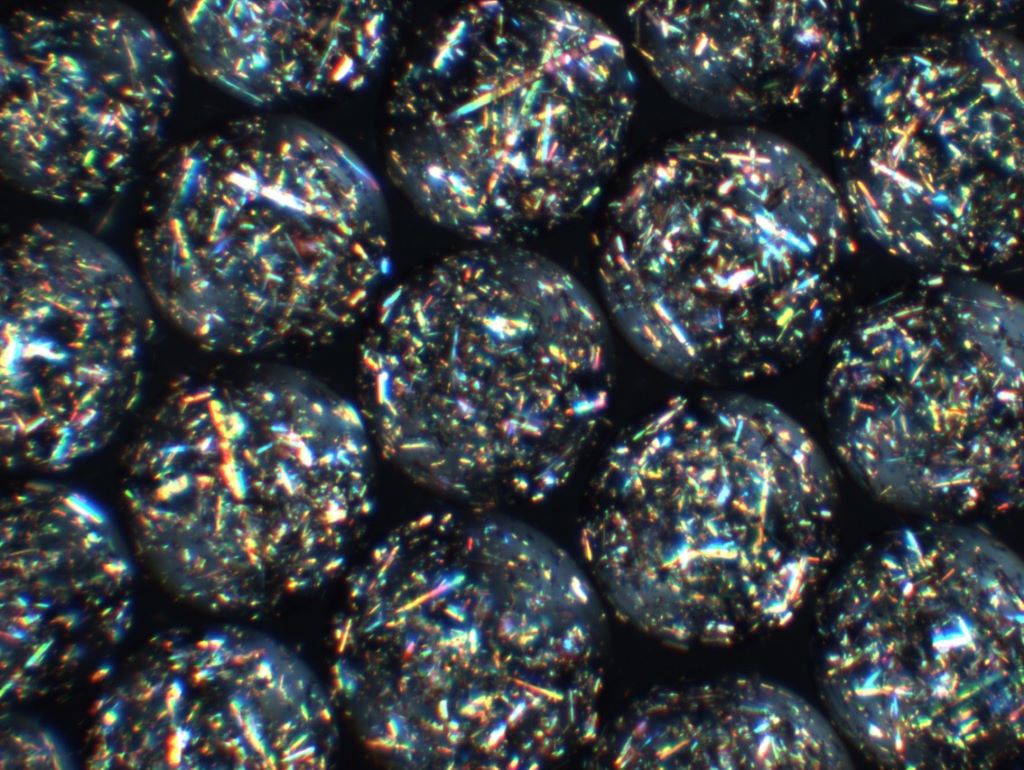
The crystal capsules developed by the researchers. They made the cover of Science Translational Medicine.

A pioneering study marks a major step toward eliminating the need for daily insulin injections for people with diabetes. The research introduces a living, cell-based implant that can function as an autonomous artificial pancreas, essentially a living drug that is long-term, thanks to a novel crystalline shield technology.

Once implanted, the system operates entirely on its own: it continuously senses blood-glucose levels, produces insulin within the implant itself, and releases the exact amount needed – precisely when it is needed. In effect, the implant becomes a self-regulating, drug-manufacturing organ inside the body, requiring no external pumps, injections, or patient intervention.

One of the study’s most significant breakthroughs addresses the longstanding challenge of immune rejection, which has limited the success of cell-based therapies for decades. The researchers developed engineered therapeutic crystals that shield the implant from the immune system, preventing it from being recognised as a foreign object. This protective strategy enables the implant to function reliably and continuously for several years.

The technology has already been successfully tested in a mouse model for effective and long-term regulation of glucose levels and in non-human primates for cell viability and functionality. These results represent a critical milestone and strongly support the potential for future translation to human patients.

From Postdoctoral Insight to Global Collaboration

The study was led by Assistant Professor Shady Farah of the Faculty of Chemical Engineering at the Technion – Israel Institute of Technology, in co-correspondence with MIT, and in collaboration with Harvard University, Johns Hopkins University, and the University of Massachusetts. Asst Prof Farah began developing the concept with colleagues in 2018 during his postdoctoral fellowship at MIT and Boston Children’s Hospital/Harvard Medical School, under the supervision of Prof Daniel Anderson and Prof Robert (Bob) Langer, a world leader in tissue engineering and co-founder of Moderna.

Today, the research continues in Asst Prof Farah’s laboratory at the Technion, in close collaboration with leading US institutions, including MIT, Harvard, the University of Massachusetts, Boston Children’s Hospital, and the Johns Hopkins University School of Medicine.

A Platform with Far-Reaching Potential

While the immediate focus is diabetes, the researchers emphasise that this implantable, closed-loop platform could be adapted to treat a wide range of chronic conditions requiring continuous delivery of biological therapeutics – including haemophilia and other metabolic or genetic diseases.

If successfully translated to the clinic, this technology could redefine how chronic diseases are treated, shifting from repeated drug administration to living, self-regulating therapies that work seamlessly from within.

To read the full article, click here

Source: TECHNION Israel Institute of Technology

Common Eye Ointment can Damage Glaucoma Implants, Study Warns

Research shows that petrolatum-based eye ointments can cause the device to swell and potentially rupture, prompting an urgent update to clinical guidance.

Photo by Tima Miroshnichenko


Widely-used eye ointments can cause glaucoma implants to swell and potentially rupture, according to new research from Nagoya University in Japan. This study is the first to show, using clinical and experimental evidence, that petrolatum-based eye ointments can compromise the PRESERFLO® MicroShunt, an implant used in over 60 countries to treat glaucoma.

Glaucoma is an eye disease that damages the optic nerve and can lead to vision loss. It often results from increased intraocular pressure caused by blocked drainage of eye fluid. A recent study estimated that 76 million people globally are affected by glaucoma.

Progression of visual field loss (from left to right) due to glaucoma
(Credit: Ryo Tomita)

MicroShunt is a small filtration device implanted in the eye to improve fluid drainage in glaucoma patients. Compared to traditional surgeries, it lowers post-operative complications and reduces reliance on additional medications.

MicroShunt is made from a styrenic thermoplastic elastomer based on a polystyrene-block-polyisobutylene-block-polystyrene (SIBS) block polymer, which is highly biocompatible, flexible, and less likely to cause inflammation or scarring. However, this material is vulnerable when it comes into contact with hydrocarbon- and oil-based materials. Due to its high oil affinity, exposure to petrolatum-based eye ointments may allow oil components to penetrate the device, causing swelling and potential changes in its shape and flexibility.

The MicroShunt manufacturer’s instructions state that “the MicroShunt should not be subjected to direct contact with petrolatum-based (ie, petrolatum jelly) materials, such as ointments and dispersions.” But this precaution is not widely recognised or consistently followed in clinical practice.

“Swollen MicroShunts can be structurally fragile,” said ophthalmologist and Assistant Professor Ryo Tomita of Nagoya University Graduate School of Medicine, the study’s first author. “During surgery, I observed a rupture in a swollen MicroShunt. If more clinicians are aware of this risk, they will be able to prevent similar problems.”

Tomita and colleagues, including Assistant Professor Taiga Inooka and Associate Professor Kenya Yuki from Nagoya University Hospital and the Graduate School of Medicine collaborated with Dr. Takato Kajita and Junior Associate Professor Atsushi Noro from the Graduate School of Engineering to examine changes in the MicroShunt after exposure to a petrolatum-based eye ointment.

The medical team reviewed clinical cases, while the engineering team conducted laboratory analyses. The findings were published in Graefe’s Archive for Clinical and Experimental Ophthalmology.

Clinical evidence

The clinical study examined seven glaucoma patients whose MicroShunt implants were later removed for different reasons. The results revealed a clear pattern. In three cases, the MicroShunt was exposed outside the conjunctiva, and patients received a petrolatum-based eye ointment. All three explanted devices showed significant swelling, and two of them ruptured.

In three other cases, the MicroShunt remained covered by the conjunctiva, and no ointment was administered. These devices retained their original structure. Crucially, in one additional case, the MicroShunt was exposed outside the conjunctiva, but no ointment was applied. The device did not swell. This indicates that direct contact with the ointment, rather than conjunctival rupture alone, is the primary cause of swelling.

Photographic comparison of MicroShunt illustrating size changes
Top: MicroShunt explanted from a patient, exhibiting diffuse swelling with fracture and loss of one fin
Middle: MicroShunt explanted from another patient, showing localized swelling around the fin
Bottom: Unused MicroShunt (control)

Scale: 1 division = 1 mm   
(Credit: Ryo Tomita)

Laboratory confirmation

Laboratory experiments confirmed the clinical findings. The team immersed unused MicroShunts in petrolatum-based eye ointment to reproduce the swelling seen in clinical cases. Microscopic measurements showed significant changes. After 24 hours in the ointment, the MicroShunt’s outer diameter increased to 1.44 times its original size, and the fin-like portion widened to 1.29 times its initial value.

Chemical analysis identified the cause of this change. After 24 hours of immersion, oil components made up approximately 45% of the MicroShunt’s total weight, rising to 73% after three months. These results confirmed the primary cause of swelling to be the absorption of oil-based ointment constituents into the material.

Clinical implications

The research team emphasises that clinicians should avoid using petrolatum-based ointments on patients with MicroShunt implants, particularly when the device is exposed outside the conjunctiva. Alternative post-operative treatments should be considered, while further research is needed to assess whether swelling impacts MicroShunt performance even when rupture does not occur.

“Our study found that commonly used medical materials can cause unexpected complications if their chemical properties and usage environments are not fully understood,” Noro stated. “From both medical and engineering perspectives, we emphasise the importance of understanding the chemical properties of medical materials and appropriately managing their usage environments.”

Paper information:

Ryo Tomita, Taiga Inooka, Takato Kajita, Hideyuki Shimizu, Ayana Suzumura, Jun Takeuchi, Tsuyoshi Matsuno, Hidekazu Inami, Koji M. Nishiguchi, Atsushi Noro, and Kenya Yuki. (2026) Petrolatum-based ointment application induces swelling of the PRESERFLO MicroShunt. Graefe’s Archive for Clinical and Experimental Ophthalmology
DOI: 10.1007/s00417-025-07075-2