Category: Emergency Medicine

Does Longer Therapeutic Hypothermia Improve Outcomes for Out-of-Hospital Cardiac Arrests?

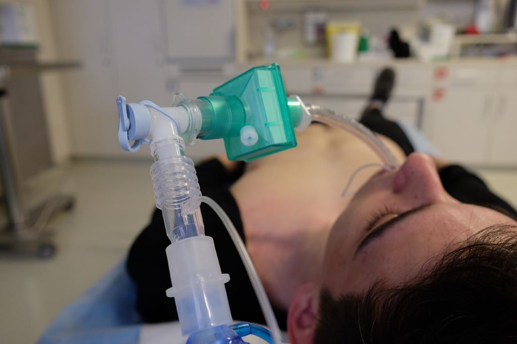
Photo by Navy Medicine on Unsplash

For the past two decades, doctors have been using therapeutic hypothermia to prevent or treat brain injury in comatose survivors of cardiac arrest. The process involves cooling down the body, which slows the body’s metabolism and triggers other protective mechanisms that reduce brain damage. However, clinical trials of therapeutic hypothermia for out-of-hospital cardiac arrests have not consistently shown that the process works.

To get a clearer understanding, University of Michigan doctors and researchers led the Influence of Cooling duration on Efficacy in Cardiac Arrest Patient, or ICECAP, trial to determine if longer durations of cooling are more effective than those previously used. The largest US trial focusing on this question, ICECAP included 71 hospitals in the US between 2020-2025 and investigated adults who underwent cooling durations between six and 72 hours.

“We knew that cooling reduces brain injury, but we didn’t know how to make that work in a clinical environment,” said Robert Silbergleit, MD, Professor of Emergency Medicine.

“Most of the previous trials used limited durations of cooling, but longer durations were better in laboratory experiments. We wanted to study whether longer durations were more beneficial for adults with cardiac arrest.”

The study, published in JAMA, focused on 1158 patients, across different genders and races, whose heartbeats were restored with or without a defibrillator. It also included a range of injury severity seen in emergency departments. Patients who suffer cardiac arrests fall into two categories: those who have a sudden arrest, usually from a heart attack and those whose bodies have slowed down over time.

“The two situations created a lot of uncertainty in previous studies because those who have a sudden arrest are usually less sick and their hearts can be quickly restarted with defibrillators,” said William Meurer, MD, Professor of Emergency Medicine and Neurology.

“It was important to study both populations because sometimes doctors can give up hope for patients who had a longer period of CPR and never received a defibrillator shock.”

The study found that outcomes did not change when they underwent cooling for longer durations. Future analyses will explore whether duration of cooling was more important for some types of patients but not others. Additionally, the survival rate in the non-shockable patients was higher than in previous studies with cooling.

“Our study showed that some patients undergoing therapeutic hypothermia can wake up even after two weeks,” Meurer said. “Although it can be a difficult process to go through, it gives families and doctors hope.”

The team is conducting a similar study for paediatric cardiac arrest patients where they are looking at the benefits of cooling for up to four days.

Source: Michigan Medicine

Closing the Gap Between Suspecting a Heart Attack and Confirming it

A highly sensitive cardiac test brings traditional lab diagnostics to the field, when it’s most needed

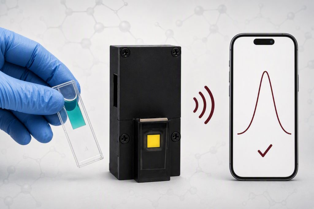
Illustration of a portable cardiac troponin I testing system that wirelessly transmits results to a smartphone.Credit: AI-generated image

A person having a heart attack may be only feet from medical care but miles from the laboratory testing needed to confirm it. That gap matters, especially for rural patients, those in nursing homes, ambulances and other places where access to specialised diagnostics is limited.

In a paper published in Biosensors and Bioelectronics, an international team of researchers details a highly sensitive test designed to bring that molecular evidence closer to the patient.

“A heart attack doesn’t wait for laboratory results,” said lead author Sayantan Tripathy, assistant research scientist in Texas A&M University’s Optical and Bio-Sensing Laboratory in the College of Engineering. “By reducing the time needed to detect heart muscle damage, we’re helping clinicians move more quickly from uncertainty to action, not only in hospitals, but also in ambulatory and resource-constrained settings where rapid diagnostic tools are often unavailable.”

Clinicians may suspect a heart attack based on symptoms or an electrocardiogram, but confirming heart muscle damage has occurred often requires testing for cardiac troponin I, a protein released into the bloodstream when the heart is injured. The most sensitive versions of those tests typically rely on centralised laboratory equipment, creating a potentially dangerous delay between suspicion and certainty.

“Our goal is for emergency medical technicians in an ambulance to be able to take someone’s fingertip sample – just a small finger prick – put that sample into a cartridge that contains all of our components, plug this cartridge into our device, and then be able to run the test in five minutes,” said Dr Samuel Mabbott, associate professor of biomedical engineering and study co-author.

Faster heart attack diagnosis

Many of today’s troponin tests depend on expensive laboratory instruments and trained personnel. As a result, obtaining the information needed to confirm a heart attack can take time, especially when advanced laboratory resources are not immediately available.

“Our vision is to bring affordable and sensitive advanced molecular diagnostics closer to where care is delivered,” said Dr Gerard Coté, professor of biomedical engineering and director of Texas A&M’s Center for Remote Health Technologies and Systems. “The less distance between the patient and the information clinicians need, the faster critical decisions can be made.”

To create the test, the researchers designed a specific structured DNA molecule that remains inactive until it encounters cardiac troponin I. Once it detects the biomarker, it triggers a series of molecular reactions that amplify the signal, making it easier to detect even very small amounts of heart damage. The amplified signal is then measured using portable optical sensing technology.

The researchers say the technology requires additional development before it could be used in clinical practice. The original work takes approximately two hours to complete, but the current version of the technology has reduced the testing time to less than 25 minutes, and further work is underway to shorten it to under 15 minutes, improving its user applicability.

Still, the study points toward a future in which life-saving diagnostic information is no longer tied to a centralized laboratory and can instead move closer to the patients who need it most.

Source: Texas A&M University

Simple Triage Protocol Cuts ER Wait Times Without Adding Beds or Staff, New Study Finds

Mayo Clinic field trial shows hospitals can reduce emergency department overcrowding using a simple patient-routing checklist based on information already collected at triage.

Photo by Camilo Jimenez on Unsplash

Emergency departments face a common challenge: how to reduce long wait times without adding more beds, hiring more staff or expanding facilities. New research suggests one answer may be hiding in plain sight – making better decisions about where patients receive care.

A new study published in the INFORMS journal Management Science, entitled, “Vertical Patient Streaming in Emergency Departments,” found that a simple, data-driven triage protocol reduced emergency department length of stay by 11 minutes without compromising patient safety or requiring additional resources.

Researchers from Harvard University, Oxford University and Mayo Clinic developed an evidence-based protocol to identify patients who could safely receive care in a seated treatment area – known as a vertical processing pathway – instead of occupying a traditional emergency department bed.

Although many emergency departments already have these areas, decisions about who should be treated there are often made on an ad hoc basis. The researchers sought to replace that variability with a standardised, easy-to-use protocol.

The team first analysed nearly 50 000 emergency department visits at Mayo Clinic Arizona to develop a machine learning model that predicts, using only information collected during triage, whether a patient will ultimately require an emergency department bed. They then combined those predictions with mathematical models of patient flow to determine the most efficient routing strategy before translating the results into a straightforward decision tree that clinicians could implement without new software or changes to hospital IT systems.

To evaluate the approach in practice, the researchers conducted a 13-week prospective field trial involving 11 015 patients at Mayo Clinic Arizona’s new emergency department.

The results demonstrated measurable improvements in efficiency:

  • 11-minute (4.2%) reduction in total emergency department length of stay.
  • Eight-minute (4.5%) reduction in time from arrival to clinical disposition.
  • No increase in 72-hour return visits, indicating patient care quality was maintained.

“Our goal wasn’t to add technology to the emergency department,” said Arshya Feizi, lead author of the study and a researcher at Harvard University. “It was to give clinicians a practical, evidence-based way to decide which patients can safely receive care without occupying one of the department’s limited beds.”

The protocol relies only on information hospitals already collect during triage, including a patient’s Emergency Severity Index score, presenting complaint and whether the department is operating over capacity. Because it requires no additional staff, equipment or software integration, the researchers say it could be implemented quickly in many emergency departments.

“Our findings show that improving patient flow doesn’t always require expanding capacity,” said Soroush Saghafian, co-author of the study and professor at Harvard University. “Sometimes the greatest opportunity comes from using existing resources more intelligently.”

The researchers estimate that a medium-sized emergency department treating approximately 40 000 patients annually could recover nearly 6800 bed-hours each year – enough capacity to care for roughly 2000 additional patients while potentially generating approximately $3 million in additional reimbursement, all without expanding facilities or increasing staffing.

Emergency department overcrowding has challenged hospitals for decades, contributing to treatment delays, patient dissatisfaction, clinician burnout and higher healthcare costs. The researchers believe their findings demonstrate that operational improvements grounded in analytics and implemented through simple clinical protocols can produce meaningful gains without sacrificing quality of care.

Read the full study here.

Source: INFORMS

Naloxone Use During Cardiac Arrest Linked to Improved Survival

Study shows benefits to drug often used for opioid overdose reversal

Photo by Mikhail Nilov

A new study by emergency medicine researchers at UC Davis Health set out to assess the effects of naloxone administration by first responders treating patients with out-of-hospital cardiac arrest (OA-OHCA).

The study, published in Jama Open Network, found naloxone administration during resuscitation by emergency medical service (EMS) personnel was associated with improved outcomes in patients with suspected OA-OHCA.

“This study provides important real-world evidence that naloxone may offer benefit even after cardiac arrest has occurred.”

David Dillon, study author

What the data shows

For this retrospective cohort study (looking back at existing patient records), researchers collected data from the California Resuscitation Outcomes Consortium between 2021 and 2022. In total, 3811 patients with suspected OHCA were treated by EMS.

Researchers found that people who received naloxone, a medication better known for reversing opioid overdoses, had higher rates of survival from the time they were treated by EMS to the time they were discharged from the hospital. The patients also benefitted from return of spontaneous circulation (ROSC) and favourable neurological outcomes compared to those who did not receive the drug.

The key findings included:

  • Survival to hospital discharge was higher among those receiving naloxone (8.1%) compared to those who did not (4.4%). 
  • Naloxone use was associated with a 2.8% absolute increase in survival, after accounting for patient and clinical factors. 
  • People treated with naloxone had improved neurologic outcomes (+3.2%) and ROSC (+3.3%).
  • Benefits were even greater among those with EMS-suspected drug-related cardiac arrest, with survival improvements approaching 8–9%.

The study also found that the association between naloxone and improved outcomes was weakened in certain situations – particularly among patients who required epinephrine during resuscitation. This suggests that timing, patient condition or resuscitation complexity may influence effectiveness.

Addressing a critical gap

Opioid overdose deaths in the United States have surged over the past two decades, contributing to a growing number of cardiac arrests outside the hospital. While naloxone is widely used to reverse opioid overdoses, its role during cardiac arrest has remained unclear and is identified by the American Heart Association as a key evidence gap.

“This study provides important real-world evidence that naloxone may offer benefit even after cardiac arrest has occurred,” said David Dillon, assistant professor of emergency medicine at UC Davis Health and one of the study’s authors. “While these findings are promising, randomised controlled trials are needed to determine whether naloxone directly improves survival in opioid-associated cardiac arrest.”

By Liam Connolly

Source: UC Davis Health

Whole Blood and Components Equally Effective in Prehospital Trauma Care

Photo by Mat Napo on Unsplash

Giving whole blood or the component parts of blood are equally effective options for paramedics and emergency medical technicians to use in treating patients with severe, traumatic bleeding before arriving at the hospital, according to a large, nationwide trial directed by University of Pittsburgh and UPMC clinicians and scientists.  

The results, published in the New England Journal of Medicine, provide flexibility to prehospital emergency care providers and could increase the odds that traumatically injured patients receive blood as soon as possible.  

“Traumatic bleeding is the leading cause of trauma death and is the most time-sensitive injury a person could suffer æ more time sensitive than a stroke or heart attack,” said co-lead author Jason Sperry,  professor of surgery in Pitt’s School of Medicine, and chief of trauma surgery at UPMC. “But it is preventable – and that starts with giving blood back to the injured person before they even arrive at the hospital.”  

Donated blood is usually separated into parts – red cells, plasma and platelets – for storage and so the parts can be used individually as needed. When someone is bleeding, emergency clinicians will often give all or some of these parts to the patient at once. Giving either whole blood or its component parts had long been considered safe options. 

But which is better for treating severe bleeding: Giving never-separated whole blood or giving the components? The answer matters for blood bank and emergency care logistics.  

Sperry and fellow principal investigators Francis Guyette, professor of emergency medicine in Pitt’s School of Medicine, and Stephen Wisniewski, professor of epidemiology and associate vice chancellor for clinical trials coordination at Pitt, launched the “Type O Whole Blood and Assessment of Age During Prehospital Resuscitation (TOWAR) Trial” to find out.  

The multicentre trial, which ran from May 2022 to June 2025, enrolled and included 1020 severely bleeding patients who were transferred to a trauma centre by medical helicopter. The patients were randomised 2-to-1 to receive either whole blood or blood components.   

The research team found no statistically significant difference between the two study arms. In both cases about a fifth to a quarter of the patients died within 30 days, compared to a third of traumatically bleeding patients who do not receive blood before arriving at the hospital. 

“This is good news,” said co-lead author Guyette, who is also medical director of STAT MedEvac, which is directed by a consortium of UPMC hospitals and is the nation’s largest academic, nonprofit critical care transport group. “It means that emergency responders can use whatever form of blood is most accessible to them. In U.S. civilian emergencies that may be component blood because that is how most blood banks package it, but in military settings whole blood is often all that is available. We’ve shown that both are equally great options.” 

In March, a European group announced the results of a similar, slightly smaller trial conducted in England, also published in the New England Journal of Medicine. Like the Pittsburgh team, they also found that giving whole blood or blood components was equally effective. The clinician-researchers believe that the combined findings will be reflected in guidelines set by various societies that oversee trauma care, surgery and blood handling.  

Whole blood is good for 21 days after donation, so the clinician-scientists were also curious if patient outcomes were any different if they were given new blood or blood closer to the expiration date. They learned that it made no difference—outcomes were the same for patients receiving newer blood within 14 days of donation compared to those receiving older blood within seven days of expiration. 

“Our thoughtful approach to the study design allowed us to not only answer the important question of the efficacy of whole blood compared to component therapy, but also to evaluate the health impact of an important public health question, the age of whole blood,” said senior author Wisniewski, who is also codirector of the Epidemiology Data Center at Pitt’s School of Public Health. “Our trial provides reassurance by verifying current standards that support the use of whole blood units throughout their entire shelf life.” 

The team also noted that the findings wouldn’t have been possible without the generosity of blood donors, study participants agreeing to share their data and the hard work of emergency care providers.  

“We’re very grateful to everyone involved, particularly the paramedics, emergency medical technicians and flight nurses,” Guyette said. “We are hopeful that this study and future research will give them better tools to save lives.” 

Source: University of Pittsburgh

Intubation Before Hospital Admission for Major Trauma Saves Lives

Photo by Mikhail Nilov

Trauma patients urgently requiring a breathing tube are more likely to survive if the tube is inserted before arriving at hospital compared to insertion afterwards, suggests a modelling study led by researchers at University College London and the Severn Major Trauma Network.

The researchers found that prehospital emergency intubation of high-risk trauma patients could improve 30-day survival by 10.3%, and could save 170 lives each year in the UK.

The findings of the new artificial intelligence (AI)-supported analysis, published in The Lancet Respiratory Medicine, provide\s the strongest evidence yet that prehospital emergency anaesthesia with intubation saves lives when delivered to those who need it most.

Trauma is a leading cause of death worldwide, with rates in South Africa 5–9 times higher than the global average. But there is a lack of high-quality evidence on the best time to start certain types of care for major trauma patients, such as the insertion of breathing tubes.

Prehospital intubation needs to be administered by an advanced critical care team, specially trained and equipped to administer the anaesthesia required to facilitate the insertion of breathing tubes. In the UK, that is currently provided only by the air ambulance services.

The researchers say their findings could inform policy discussions on funding specialist prehospital critical care teams, which could include public funding for air ambulances or funding additional training for ground ambulance teams, so that more high-risk major trauma patients can have breathing tubes inserted before arrival at hospital.

Joint first author Dr Amy Nelson (UCL Queen Square Institute of Neurology and King’s College London) said: “The airway is a top priority in major trauma, but the question of whether we should intubate before hospital arrival is unsettled because we cannot ethically conduct a randomised trial.

“Emergency care decisions made before hospital admission depend on the combination of many measurements taken under pressure. We used these measurements to answer the question in steps: we first built a machine learning model to identify high-risk patients, then we modelled the impact of early intubation in this group, which showed us that prehospital intubation saves lives.”

For the study, researchers analysed data from 6467 trauma patients treated at Southmead Hospital Major Trauma Centre, Bristol.

The researchers used AI-assisted modelling to predict both who would need intubation and who would likely survive – to isolate the impact that intubation had from other factors such as the injury severity. To facilitate their analysis, they developed a new machine learning model, called ‘Intub-8’, which predicted outcomes based on eight routinely collected prehospital measurements.

The researchers found that among high-risk patients who were identified by the model as needing intubation (229 patients), those who received it before arriving at hospital were 10.3% more likely to survive (within a 30-day period) compared with those who did not.

By scaling up their findings relative to national trauma incidence, the researchers estimate that if every trauma patient who needed prehospital intubation was given it, 170 lives could be saved each year in the UK – roughly one life saved every other day. 

Additionally, they conducted a cost-effectiveness analysis, finding that cost savings would be in the range of £101 million annually for the UK, due to reduced costs of further care and lives saved.

Professor Parashkev Nachev (UCL Queen Square Institute of Neurology), joint senior author, said: “In medicine, action and inaction are not morally asymmetric. When we cannot have randomised controlled trial evidence for an intervention, we must use the best available alternative: causal inference from real-world data, assisted by artificial intelligence, the only technology with the power to address the complexity of biological systems.”

Associate Professor Julian Thompson, joint senior author and Clinical Director of the Severn Major Trauma Network, said: “Until now, advanced air ambulance services across the world who respond to critically injured patients have struggled to conduct studies that assess the benefit and cost effectiveness of their life-saving interventions. The use of AI in this study has allowed us to analyse existing data in a totally new way. This reveals the huge impact that advanced care provides when delivered before arrival in hospital.

“These findings may have a huge impact on how UK and international health services look after the most severely injured patients in our societies.”

The authors note that the findings are specific to a mixed rural-urban UK setting where highly trained physician-paramedic teams perform all prehospital intubation. The survival benefit may differ in other healthcare systems or national contexts, and further research is needed to examine long-term outcomes and potential complications.

Source: University College London

Potential New Treatment for Sepsis Shows Promise in Trial

Griffith University researchers may have unlocked the secret to treating sepsis, with a Phase II clinical trial in China successfully concluding with promising results.

Photo by Alex Fedini on Pixabay

Griffith University researchers may have unlocked the secret to treating sepsis, with a Phase II clinical trial in China successfully concluding with promising results. The sepsis drug candidate, a carbohydrate-based drug called STC3141, was co-developed by Distinguished Professor Mark von Itzstein AO and his team from Griffith’s Institute for Biomedicine and Glycomics, and Professor Christopher Parish and his team at The Australian National University.

“The trial met the key endpoints to indicate the drug candidate was successful in reducing sepsis in humans,” Professor von Itzstein said.

STC3141 was administered as an infusion via a cannula and counteracted a significant biological molecule release phenomenon which occurred in the body during the course of sepsis.

The small-molecule experimental drug was a carbohydrate-based molecule and could treat sepsis by reversing organ damage.

Sepsis was known to affect millions of hospitalised patients across the world each year and occurred when the body’s immune response to an infection attacked and injured its own tissues and organs.

“When sepsis is not recognised early and managed promptly, it can lead to septic shock, multiple organ failure and death,” Distinguished Professor von Itzstein said.

The trial, conducted by Grand Pharmaceutical Group Limited (Grand Pharma), involved 180 patients with sepsis, one of the leading causes of death and long-term disability worldwide.

Currently, there is no specific anti-sepsis therapy available, and sepsis is considered a clinical unmet need.

Professor von Itzstein said Grand Pharma would now look to progress to a Phase III trial to continue testing the efficacy of the novel treatment.

“It’s hoped we could see the treatment reach the market in a handful years, potentially saving millions of lives,” he said.

Executive Director of the Institute for Biomedicine and Glycomics, Professor Paul Clarke, said: “I am thrilled to see the results of the trial which ultimately aims to save lives.”

“The Institute and its researchers collectively work on translational research to deliver real and immediate impacts both in Australia, and globally to transform lives.”

Source: Griffith University

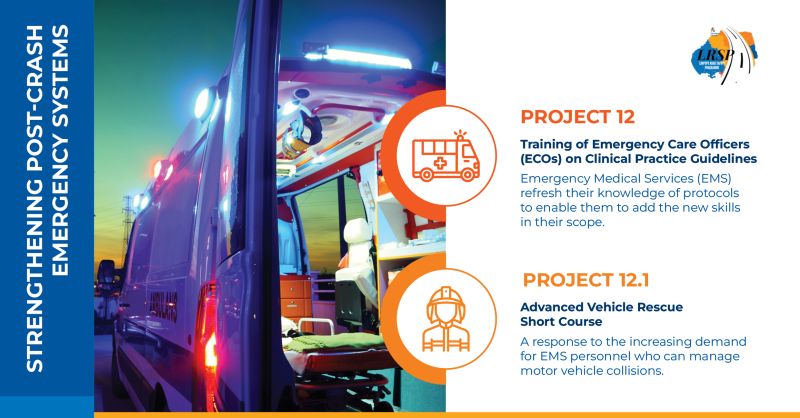
Case Study: Building a Stronger Emergency Response System in Limpopo

Strengthening Limpopo’s post-crash emergency response has been one of the most powerful achievements of the Limpopo Road Safety Programme (LRSP). Through a combined focus on updated clinical training, advanced rescue skills and improved operational systems, Projects 12 and 12.1 have reshaped how Emergency Medical Services (EMS) teams respond in the critical minutes after a crash – from the first emergency call to hospital handover.

Updating clinical skills to strengthen frontline emergency care

Across South Africa, the Clinical Practice Guidelines (CPGs) for emergency care have been substantially updated, including a major revision in 2018. These updates incorporated new evidence, improved patient outcomes, and standardised practice across the health system, shifting toward more user-friendly formats such as clinical decision-support tools. For Limpopo’s EMS, this presented both an opportunity and a challenge: although the guidelines were available, many personnel had not yet received training to apply them consistently in the realities of roadside emergencies. Project 12 addressed this need directly, rolling out comprehensive CPG training across all five districts. EMS practitioners were equipped with updated algorithms for trauma, medical, paediatric and obstetric emergencies, along with enhanced assessment, triage and stabilisation skills.

This clinical uplift aligned perfectly with major system improvements. In the 2023/2024 financial year, the Limpopo Department of Health procured more than 500 new, modern ambulances, significantly expanding the provincial fleet. The LRSP ensured this investment translated into real-world impact: EMS personnel were trained not only on updated CPGs but also to use the new vehicles and onboard equipment to their full potential; optimising monitoring, patient loading, scene workflow and en-route care. Modern ambulances combined with modern knowledge dramatically strengthened the quality of emergency care.

By 2025, the system advanced even further with the introduction of a Computer-Aided Dispatch (CAD) system, enabling more efficient call-taking, improved dispatch decision-making, clearer communication and better tracking of EMS resources across districts. The CAD system, together with updated CPGs and a modern ambulance fleet, created a tightly integrated platform for faster, smarter and more coordinated EMS response. For the first time, Limpopo could align clinical best practice, operational intelligence and fleet capacity into one cohesive system.

Introducing advanced rescue skills for high-severity crash scenes

Yet, while clinical updates and dispatch improvements strengthened core EMS response, Limpopo still faced a critical need for specialised capacity at high-severity crash scenes, especially those involving vehicle entrapment. Project 12.1 filled this gap by introducing the province’s first Advanced Vehicle Rescue Short Course, delivered by EPIC EM and the University of Johannesburg. Over seven intensive days, participants trained in vehicle stabilisation, extrication techniques, hydraulic tool use, and multi-casualty scene management, blending theory with realistic, high-pressure simulations. Many described the training as transformative, giving them the competence and confidence to manage complex incidents on Limpopo’s regional and mining routes.

Together, these interventions have created a step change in Limpopo’s post-crash care system. Today, EMS teams arrive at crash scenes equipped with modern ambulances, updated clinical guidance, advanced rescue skills and a CAD-supported operational network that ensures faster and more coordinated response. Patients benefit from safer extrication, quicker stabilisation and better continuity of care during the “golden hour”. Beyond improving skills, the programme has strengthened morale, professionalism and a culture of excellence within EMS.

Projects 12 and 12.1 have left a lasting legacy: a provincial emergency response system that is smarter, faster and better prepared to save lives on Limpopo’s roads.

Is There a Doctor on Board? New Research Reveals the Frequency of In-Flight Medical Emergencies

The study is the largest global analysis of in-flight medical events

Photo by Daniel Eledut on Unsplash

 With nearly five billion people flying each year, medical emergencies in the air may be more common than most realise and they can be deadly.

A new study led by Duke Health researchers analysed more than 77, 00 in-flight medical events reported to the world’s busiest airline medical support centre. The findings show that while most incidents are minor, thousands of passengers required hospital care after landing, and hundreds died or triggered aircraft diversions.

The study, published in JAMA Network Open, was conducted in partnership with MedAire, an aviation and maritime health and safety solutions company, which also provided the data for analysis. The paper offers a rare look into how airlines respond to medical crises and why some flights are forced to divert.

“This is the largest and most comprehensive study of in-flight medical emergencies ever conducted,” said Alexandre Rotta, MD, senior and corresponding author of the paper and chief of the Division of Pediatric Critical Care Medicine with the Department of Pediatrics at Duke University School of Medicine.

“It gives us a real-world snapshot of what happens when someone gets sick in the sky and how starkly the options differ from those in a hospital,” Rotta said.

Researchers reviewed medical calls from 84 airlines across six continents, covering over 3.1 billion passenger boardings between January 2022 and December 2023.

They found that one in every 212 flights involved a medical emergency. Of those flights, about 8% of passengers were taken to the hospital after landing, and 1.7% of the total medical events were so serious they caused the plane to divert.

The most common reasons for diversion were suspected strokes, seizures, chest pain, and altered mental status. Cardiac arrest occurred in 293 cases, with survival rates far lower than on land.

Medical volunteers (often physicians) assisted in nearly one-third of emergencies. Their involvement was linked to a higher likelihood of diversion, likely because they were called upon during more serious events.

“It’s humbling to practice medicine in the air,” said Rotta, who became interested in the topic after being called upon as medical volunteer during several flights. “You’re working with limited equipment, no lab tests and no backup. Even minor issues can become major challenges.”

Rotta emphasised that airlines are generally well-prepared, especially in the US, where regulations require defibrillators and basic medical kits. However, he noted that not all airlines partner with ground-based medical support centres, an approach he believes is essential.

“Airplanes aren’t hospitals, and we shouldn’t expect them to be,” he said. “But having expert guidance from the ground can make all the difference when someone’s life is at risk.”

The findings could help shape airline policies, improve crew training and inform passengers with chronic conditions about how to prepare for travel.

Source: Duke University

Innovative UK Project to Test Drones for Cardiac Arrest Response

Credit: University of Surrey

A project to test how drones can be integrated into the UK’s 999 emergency response system to rapidly deliver defibrillators to patients experiencing out-of-hospital cardiac arrest (OHCA) has been launched by the University of Surrey, Air Ambulance Charity Kent Surrey Sussex, South East Coast Ambulance Service NHS Foundation Trust. 

With survival rates for OHCA in the UK currently below 10%, a key challenge is the delay in delivering life-saving defibrillation. While public Automated External Defibrillators (AEDs) are widely available, getting them to a patient in time is often difficult. This 16-month project will explore using drones to rapidly deliver AEDs to the scene of an emergency. 

This research is the first step towards integrating drone technology into our emergency response systems. Our ultimate goal is to develop and test the procedures needed to seamlessly introduce drone delivery of AEDs into the 999-emergency system 

Dr Scott Munro, Lecturer in Paramedic Practice at the University of Surrey and co-lead on the project

The initiative, which has been funded by the National Institute for Health and Care Research (NIHR), will be divided into two sections: in the first, researchers will develop and refine the drone delivery process through a series of simulations, coordinating 999 call taking, Air Traffic Control, ambulance dispatch and drone operators.  

In the second part, interviews will be conducted with a diverse group of people -including OHCA survivors, family members, responders and members of the public – to understand the public’s perception of drone technology, including any barriers or concerns, and to ensure ease of use for responders. 

This project is a great example of how NIHR’s RfPB programme supports life-saving innovation. Using drones to deliver defibrillators could help emergency teams reach patients faster, improve survival after cardiac arrest, and bring cutting-edge technology directly to the NHS frontline, while working with the public to ensure it’s used safely and effectively. 

Professor Kevin Munro, Director of the NIHR Research for Patient Benefit (RfPB) Programme

Rapid intervention is vital in managing out-of-hospital cardiac arrests. As demand continues to grow, the opportunity to integrate this technology into future healthcare systems represents real progress in ensuring ambulance services can work with the communities they serve to strengthen the chain of survival and give patients the best chance of a positive outcome Being a partner in this research, we are eager to explore how this new initiative could strengthen our cardiac arrest care pathway. 

Dr Craig Mortimer, Research Manager at South East Coast Ambulance Service NHS Foundation Trust (SECAmb)

Source: University of Surrey