Category: Pain Management

Exercise Helps Hip Arthritis Pain, but Perhaps Less than We Thought

Photo by RDNE Stock project


A new Cochrane review finds that exercise may improve pain and function, but the benefits may fall short of what patients would notice in daily life.

Hip osteoarthritis is a condition affecting millions of people worldwide and a leading cause of chronic pain and disability. Exercise is widely recommended as a first-line treatment for managing hip arthritis pain.

Led by researchers from University of Sydney and University of Melbourne, the findings show exercise produces small improvements in pain and physical function for people with hip osteoarthritis, but those improvements may not be large enough to make a meaningful difference to patients.

The review included 18 clinical trials involving 1368 people. Participants were mostly women (63%) and were aged between 53 and 74 years, meaning findings may not apply to younger people. Exercise programmes in the included studies varied widely, lasting between two and 52 weeks and covering a range of types including strengthening, aerobic, and mind-body approaches.

Compared with no treatment or usual care, exercise probably reduces pain by around 7 points on a 100-point scale. However, experts generally consider an improvement of at least 12 points necessary for patients to notice a meaningful difference in daily life. Physical function showed a similar pattern. The authors note, however, that these thresholds were derived largely from knee and mixed osteoarthritis populations, and may not entirely reflect the hip osteoarthritis experience.

Quality of life, arguably the outcome patients care about most, showed little to no improvement with exercise regardless of the comparison used.
 

“Exercise is recommended as a primary treatment for hip osteoarthritis, and this review doesn’t overturn that, but it does suggest we should be honest with patients that the average benefit may be modest, and that we need better-designed trials to understand who benefits most and from which type of exercise.”

— Michelle Hall, co-lead author from the University of Sydney. 


This update analysed results differently from its 2014 predecessor, which pooled all available data together and found high-quality evidence that exercise slightly reduced pain. The new review separated trials according to what exercise was being compared against.

When exercise was tested against a placebo or sham treatment, the evidence for pain relief weakened considerably. And adding exercise on top of another treatment made little difference either. While no single type of exercise came out on top, the authors caution that the evidence to answer that question properly simply isn’t there yet.

Not a recommendation away from exercise

The review stops short of saying exercise is ineffective or should be abandoned as a recommendation. Exercise carries broad health benefits beyond arthritis, is low-cost, and is unlikely to cause harm. 

Most studies included in this review were small and unblinded, which may have influenced outcomes. Because pain and function were largely self-reported, and participants knew whether they were exercising, exercise may appear more effective than it truly is.

The authors call for larger, better-designed trials to give patients and clinicians a clearer picture of what exercise can realistically achieve for hip osteoarthritis specifically.
 

“There just isn’t a huge body of evidence out there. For some people struggling with hip pain, exercise can really be their only hope, but I also don’t want to give patients false hope. It’s important future research is done with larger, better-quality trials, examining what types of exercise work specifically for different people.” 

— Belinda Lawford, co-lead author from the University of Melbourne. 

Read the review

By Mia Parkinson

Source: Cochrane

Researchers Demystify the Molecular Mechanisms of General Anaesthesia

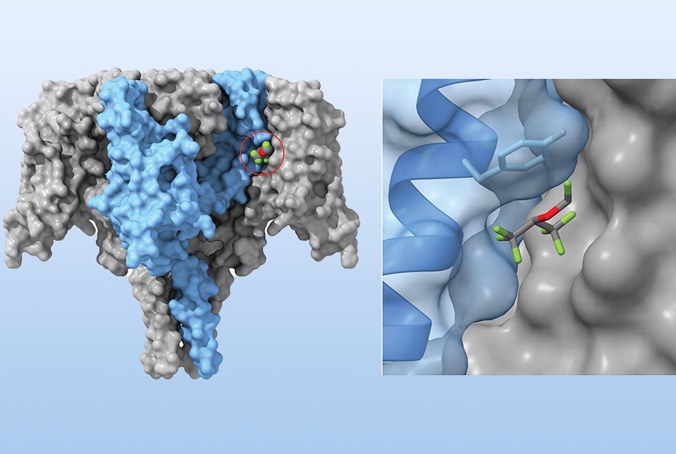
Left: Surface model of the bacterial sodium channel NavMs with one of the four subunits coloured light blue and the others grey. The anaesthetic sevoflurane (coloured spheres) sits in a pocket at the interface of two adjacent subunits. Right: Close-up of the binding pocket. The semi-transparent surface exposes the underlying ribbon and a tyrosine amino acid (light blue), which is key to binding sevoflurane (coloured sticks). Credit: Dr Karl Herold

Researchers at Weill Cornell Medicine and Birkbeck, University of London, have identified a site where a commonly used anaesthetic binds to sodium ion channels, revealing a molecular mechanism that may explain how these drugs dampen communication between neurons. Ion channels are proteins that regulate the flow of charged particles across cell membranes, enabling neurons to generate electrical signals. By reducing this signalling, inhaled anaesthetics help suppress brain activity, producing unconsciousness and immobility during surgery.

The findings, published June 19 in Nature Communications, shed light on a longstanding mystery: For 175 years, doctors have safely used inhaled anaesthetics to render patients unconscious, but didn’t fully understand how these drugs work.

“Sodium channels are critical for communication between neurons in the brain, and anaesthesia breaks down that communication,” said Dr Hugh Hemmings, senior associate dean for research and chair of the Department of Anesthesiology at Weill Cornell, who co-led the research. “So, there’s good reason to believe that the unconsciousness produced by volatile anaesthetics is related to their effects on sodium channels.” 

The study provides the first atomic-level view of how the anaesthetic sevoflurane binds to sodium channels and stabilises them in an inactive state. “The insights we gain from this study may enable us to design safer, more selective anaesthetics, with fewer side effects,” said Dr Karl Herold, co-first author and senior research associate at Weill Cornell.

Anaesthetising a bacterial counterpart

As far back as the 1970s, scientists suspected that inhaled, volatile anaesthetics could interact with ion channels – in particular, the voltage-gated sodium channels that play a crucial role in cell-to-cell communication throughout the nervous system. But determining how this interaction inhibits neuronal activity has been challenging because mammalian sodium channels were too large and complex for detailed structural analysis.

The researchers turned to a marine bacterium, Magnetococcus marinus, that uses voltage-gated sodium channels to swim toward nutrients and oxygen. Although structurally simpler than their mammalian counterparts, the bacterial channels operate similarly and share the same sensitivity to anaesthetics. “Volatile anaesthetics bind through weak, low-affinity interactions that are very hard to capture structurally,” Dr Herold said. “A bacterial channel that behaves like ours but is small enough to crystallise lets us finally see where sevoflurane sits and how it holds the channel inactive.”

Discovering a binding pocket

The Weill Cornell team joined forces with Birkbeck researchers, co-senior author Dr Bonnie Ann Wallace and co-first author David Hollingworth. The UK-based researchers have extensive expertise in structural analysis of these bacterial channels bound to a variety of drugs, including those that affect neuronal activity.

Using high-resolution X-ray crystallography, the researchers captured detailed snapshots of sevoflurane bound to the channel. They discovered that the anaesthetic tucks into a small pocket at the edge of the channel’s pore-forming region, but away from the pathway through which sodium ions flow. Binding in this pocket stabilises the channel in an inactive state, making it less likely to open and allow sodium ions through, thereby reducing a neuron’s ability to transmit electrical signals.

This interaction is key to the drug’s molecular effects. When the researchers altered a single amino acid in the binding pocket, sevoflurane could no longer bind effectively and lost its ability to keep the channel in its inactivated state.

The researchers are now working on translating their findings to the mammalian system. “The bacterial channel is just a testing ground,” Dr Hemmings explained. “If naturally occurring mutations affecting anaesthetic binding exist in humans, studying them could help explain why some people respond differently to anaesthesia and may provide new insights into the biology of consciousness.”

“As anaesthesiologists, it’s our responsibility to understand how these drugs work, so we can resolve issues when people don’t react well to anaesthesia,” said Dr Hemmings, who is also anaesthesiologist-in-chief at NewYork-Presbyterian/Weill Cornell Medical Center.

Source: Weill Cornell Medical Center

Epidurals Not Linked to Increased Harm for Newborns or Children

Study provides strong evidence that epidural analgesia in labour is safe for newborns, say researchers

Photo by Duda Oliveira

Having an epidural during labour is not associated with clinically significant increased risks of harm to newborn babies, including brain injury, severe breathing problems, sepsis and death, or cerebral palsy later in childhood.

The researchers say these findings “support widening availability and equitable access to epidural analgesia as a safe component of intrapartum care.”

Epidural analgesia in labour provides effective pain relief and may help reduce complications in mothers after giving birth, but evidence of its effect on newborn and child health is limited.

To address this, researchers analysed data for 495 695 births in Scotland over a 13 year period (2007-2019) to examine whether epidural analgesia during labour was associated with serious neurological conditions occurring within 28 days of birth.

Only women who delivered a single baby vaginally or via unplanned caesarean section between 24 and 42 weeks of pregnancy were included in the analysis.

Further measures included other severe newborn illness, sepsis, low Apgar score (a routine test of a baby’s health) five minutes after birth, death within 28 days of birth, and cerebral palsy diagnosed at any point during childhood.

Factors such as mother’s age, ethnicity, weight, existing pre-eclampsia or diabetes, smoking history, birth location and gestational age at birth, were also taken into account.

Of nearly 500 000 women included in the study, around one in four had an epidural during labour. Overall, serious neurological conditions were rare, affecting fewer than 1 in 1000 babies. These conditions occurred at the expected rate and were no more common among babies whose mothers had an epidural compared with those who did not.

No association was found between epidural analgesia in labour and serious neurological conditions, other severe newborn illness, sepsis, low Apgar score at five minutes, death at 28 days, or cerebral palsy in childhood.

This is an observational study so no firm conclusions can be drawn about cause and effect, and the authors acknowledge that the study was limited to women delivering in Scotland, a predominantly white population, so the findings may not apply to more ethnically diverse populations or other healthcare settings.

However, this was a large study with long term follow-up of newborn and childhood outcomes, and results were consistent after additional analyses across various groups including women considered to have high risk pregnancies and preterm births, supporting the reliability of the findings.

As such, the authors conclude: “These results should reassure parents and clinicians that epidural analgesia use in labour is safe for babies and support informed, evidence based decision making about analgesic options in labour.”

Source: The BMJ Group

Melatonin May Help Ease Chronic Muscle and Joint Pain, New Study Suggests

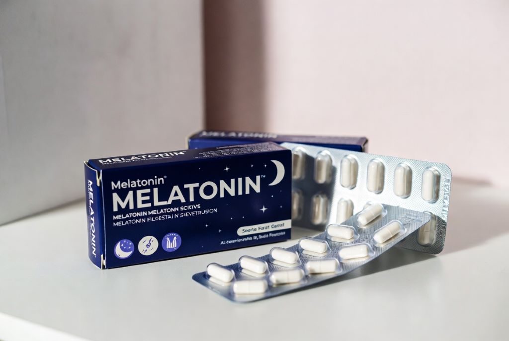
Made with AI

Dipa Kamdar, Kingston University

Melatonin is best known for helping us sleep. But a new study suggests it might have another surprising use: easing muscle and joint pain.

Produced naturally by the pineal gland in the brain at night, melatonin helps regulate the body’s sleep-wake cycle. That’s why this hormone is widely used as a treatment for insomnia and jet lag.

Sleep and pain are closely linked (poor sleep can make pain feel worse, and pain can make sleep harder). But melatonin may also reduce pain directly. Researchers believe it dampens pain signals in the brain and spinal cord, reduces inflammation, calms overactive nerves and protects cells from oxidative stress – the cellular wear and tear caused when harmful molecules build up.

In the new study, researchers combined the results of 23 clinical trials involving over 2,000 participants to identify overall patterns. These trials looked at melatonin for long-term muscle and joint pain as well as pain after surgery.

Overall, melatonin reduced both pain and sleep problems in people with chronic muscle and joint pain. But the improvements were modest. On average, pain scores fell by about nine points on a 100-point scale. That’s within the range reported for some anti-inflammatory drugs in similar studies, although the two treatments haven’t been directly compared.

Melatonin’s effects may also depend on whether people already have sleep problems or other long-term health conditions. Most of the chronic pain studies included people who already had poor sleep to begin with, but none of the trials looked at results separately for people with and without sleep issues. Because of this, it’s unclear whether melatonin works better for people who struggle with sleep or whether the effects are similar for everyone.

The findings were much less convincing for pain after surgery. Melatonin did not make a noticeable difference to pain or sleep. One analysis found a tiny improvement (about 2.5 points on a 100-point pain scale), but this is far below what would matter to patients recovering from surgery.

The benefits in chronic muscle and joint pain are modest. Based on the current evidence, melatonin should be seen as a possible add-on treatment rather than a replacement for established therapies. The evidence suggests melatonin could complement treatments such as physiotherapy, exercise and anti-inflammatory medicines rather than replace them.

What we still don’t know

There is also still a lot we don’t know. The trials in this study used a wide range of doses, from 1mg to 10mg, and the researchers couldn’t determine which dose worked best.

There were hints that longer treatment helped more in chronic pain, but this was based on only a few studies. There is also very little evidence on the effectiveness of higher doses, even though they appear safe in other studies.

Melatonin is widely used and generally considered safe for short-term use, but it can cause side-effects, such as daytime sleepiness, dizziness, headaches and nausea.

People with liver or kidney conditions, or those with autoimmune conditions like rheumatoid arthritis, should speak with a doctor or pharmacist before taking it.

It is also worth noting that melatonin is regulated very differently around the world. In the US, melatonin is sold as a dietary supplement, meaning people can buy it easily in supermarkets and online without medical advice. But in the UK, melatonin is a prescription-only medicine and is only licensed for short-term sleep problems and jet lag.

For now, the findings suggest melatonin may offer modest relief for some people with chronic muscle and joint pain, particularly if poor sleep is part of the problem. It’s unlikely to replace established treatments, but it could eventually earn a place alongside them. Larger, well-designed trials will be needed before doctors can say with confidence who is most likely to benefit.

Dipa Kamdar, Senior Lecturer in Pharmacy Practice, Kingston University

This article is republished from The Conversation under a Creative Commons license. Read the original article.

Supermarket Receipts Show Trends in Menstrual Pain Relief

An analysis of 211 million supermarket transactions found that more than a quarter of customers buying menstrual products bought pain relief at the same time.

Photo by Sora Shimazaki on Pexels

More than a quarter of women buying menstrual products also purchase pain relief at the same time – and those in lower-income areas are significantly less likely to do so – according to a new study published this week in the open-access journal PLOS Digital Health by Dr. Victoria Sivill of the University of Bristol, UK, and colleagues, which used supermarket loyalty card data to map menstrual pain disparities across England.

Menstrual pain is a common concern affecting many individuals globally. Existing research highlights its negative impact on daily activities, including school and work attendance.

In the new study, researchers analysed anonymised loyalty card data from a major UK health and beauty retailer, encompassing 211 million transactions by 3.4 million individuals between 2006 and 2015. They analysed how often shoppers purchased menstrual products at the same time as pain relief, and how that compared to a customer’s baseline rate of buying pain relief.

The analysis found that 26.7% of customers who purchased menstrual products also bought pain relief in the same transaction. These customers were nearly four times more likely to buy pain relief while buying menstrual products compared to other shopping trips. As a validation of the approach, the most common interval between consecutive menstrual purchases across the dataset was exactly 28 days – consistent with the average menstrual cycle.

Regional income emerged as the strongest predictor of menstrual pain purchases: customers in the lowest-income areas were 32% less likely to purchase pain relief at the same time as menstrual products compared to those in the highest-income areas. The authors note that lower rates of pain relief purchases in deprived areas likely reflect an inability to afford over-the-counter medication rather than lower rates of menstrual pain itself

“The study highlights the need for greater awareness and policy interventions to address the high prevalence of menstrual pain as well as socioeconomic dimensions of menstrual pain,” the authors say. “Public health initiatives should incorporate menstrual pain relief as part of broader efforts to improve health equity.”

 Co-author Dr James Goulding notes: “It is wonderful that smart data research in the UK is able to bring issues which may have once been overlooked in scientific settings – such as the sheer scale and impact of menstrual pain – to light. This is well overdue.”

Co-author Dr Anya Skatova adds: “Like many women, I was aware of how common menstrual pain is, but the scale of painkiller purchases alongside menstrual products was still striking. Using shopping data, we can see just how widespread the need for pain relief really is. This kind of evidence helps make menstrual pain visible at a population level and provides a strong foundation for systemic change in how it is recognised, treated, and prioritised in public health.” 

Provided by PLOS

Press Preview: https://plos.io/42wSl1W

In your coverage please use this URL to provide access to the freely available article in PLOS Digital Health: https://plos.io/4wzrwbh

Contact: Anya Skatova, anya.skatova@bristol.ac.uk; James Goulding, james.goulding@nottingham.ac.uk 

Image Caption: Fig 1. Average (mean) individual summary statistics for Menstrual, Pain and Menstrual Pain customer sets via analysis of transactional logs between 30th April 2006 to 16th April 2015. 

Image Credit: Sivill et al, PLOS Digital Health, 2026

High-Resolution Image Link: https://plos.io/4ujYPxl

Citation: Sivill V, Ljevar V, Goulding J, Skatova A (2026) What can shopping transactional data reveal about relative prevalence of menstrual pain and period poverty in England? PLOS Digit Health 5(5): e0001308. https://doi.org/10.1371/journal.pdig.0001308 

TENS Shown to Improve Pain and Fatigue in Fibromyalgia

Real-world trial finds long-lasting benefit for TENS with physical therapy in reducing movement pain, fatigue

Credit: University of Iowa

Adding TENS (transcutaneous electrical nerve stimulation) to outpatient physical therapy reduced movement-based pain and fatigue in patients with fibromyalgia, and the effects lasted for at least six months, according to a new study led by researchers at University of Iowa Health Care.  

The study, led by Kathleen Sluka, PT, PhD, is the first real-world trial of TENS for fibromyalgia. The findings, published on March 27 in the journal JAMA Network Open, show that TENS is a safe, effective, inexpensive, and readily available treatment for fibromyalgia, a chronic condition that causes pain, tenderness, and fatigue throughout the body. 

“It is one of the few treatments that specifically targets movement-evoked pain and fatigue, which are major barriers to participation in daily activities,” says Sluka, UI professor of physical therapy and rehabilitation science. 

TENS uses a small device with adhesive electrodes to send mild electrical pulses through the skin to block or reduce pain. The study found that the effect of TENS for reducing pain was similar, if not better, than current FDA-approved medications for fibromyalgia. 

“We were excited to see that patients also had less fatigue,” Sluka added. “Right now, there are no good treatments for fatigue. So, the fact that we had anything that touched the fatigue was pretty powerful.”

Fibromyalgia: complicated, misunderstood, and hard to treat 

Fibromyalgia affects about 4% to 7% of the population. It significantly impacts a person’s physical function, cognitive abilities, and sleep. In addition to chronic pain, a key feature of the condition is whole-body fatigue, which interferes with day-to-day life and contributes to patients’ inability to concentrate and perform functional activities. 

Exercise is often the first line of treatment recommended to people with fibromyalgia, and research has shown that it can be beneficial. However, fibromyalgia causes fatigue and pain, which is a key reason why the research team focused on alleviating pain with movement. 

“Pain with movement hinders a person’s ability to participate in an effective exercise program and do their day-to-day activities.” Sluka says.  

Science translated to real-world benefit 

Sluka and her colleagues have spent decades studying the biological mechanisms affected by TENS, developing the ideal parameters of TENS stimulation and testing the efficacy of TENS for treating chronic pain and fatigue in human trials. 

They have previously shown that under the ideal conditions of a randomized, controlled clinical trial, TENS in conjunction with physical therapy can significantly decrease movement pain. 

The new Fibromyalgia TENS in Physical Therapy (FM-TIPS) study was designed to test the effect of TENS under real-world conditions. The study was conducted in 28 outpatient physical therapy clinics across six health care systems in the Midwest, and included 384 people of different ages, education levels, and socioeconomic backgrounds. Almost 50% of the participants were from rural areas.  

“It was a challenge to recruit participants for this study, but the clinics and the physical therapists we worked with were great. This would never have happened without them,” Sluka says.

The clinics were randomised to provide either physical therapy (PT) with TENS or physical therapy alone. In the PT-TENS group, participants were asked to use TENS for two hours a day for six months. That time could be split into short periods or done all at once. The TENS electrodes were placed on the upper and lower back and delivered a mixed frequency signal at an intensity as strong as the participant could tolerate. 

After 60 days, movement-evoked pain during TENS treatment was significantly improved in the PT-TENS group. Adding TENS also significantly reduced resting pain and resting and movement-fatigue. In contrast, participants who received only physical therapy had no change in their movement-evoked pain. 

When we gave the PT-only patients the TENS unit and they started using it, we also saw the same improvements as the PT with TENS patients, which is powerful. – Kathleen Sluka, PT, PhD

The response also was dose-dependent, with people who used TENS daily for 60 days having the best outcomes. 

Unlike many pain-relieving drugs that can become less effective over time as the body develops a tolerance for the medication, the study shows that over time, TENS maintained its ability to improve pain and fatigue at a significant level. 

After the primary endpoint at day 60, the PT-only group was also given TENS, and all the participants continued in the study for another four months. 

“When we gave the PT-only patients the TENS unit and they started using it, we also saw the same improvements as the PT with TENS patients, which is powerful,” Sluka says. 

Overall, the study showed that 80% of patients found TENS helpful. At six months, 80% were still using TENS once a week, and over 70% reported they felt better after using TENS. 

TENS adds benefit

Dana Dailey, PT, PhD, UI assistant research scientist and the first author of the study, notes that it’s important for people to realize that the benefit of TENS comes from using it as a part of a total treatment plan that includes physical therapy.  

“Using TENS on its own will not give the same benefits,” Dailey says. “However, the study shows that TENS provides an added benefit on top of any relief from other treatments. All the study participants were also using pain medications and receiving physical therapy, yet TENS still provided additional relief.” 

Fibromyalgia often needs multiple interventions to help patients feel less pain and fatigue and improve their overall function. The new findings suggest that TENS could be particularly helpful as a part of a multipronged approach because it can be safely and easily used as a self-management tool that uniquely targets movement-associated pain and fatigue. 

“Often, when you move a randomised, controlled clinical trial into a real-world setting, it doesn’t work because there are too many confounding factors. But this intervention still works,” Sluka says. “Not only did the treatment reduce movement pain and fatigue during the testing period, but patients continued to use it at six months.” 

By Jennifer Brown

Source: University of Iowa

Pain Neurons Protect Nerve Health and Offer New Therapeutic Targets

A healthy neuron. Credit: NIH

Researchers at Karolinska Institutet, have uncovered a previously unknown mechanism that helps pain sensing nerve cells stay healthy and respond to injury. The findings, published in Nature Communications, may improve understanding of chronic pain and nerve damage and maintenance of myelin integrity.

A new study shows that a molecule called RNase4, is produced by specialised pain-sensing neurons. It plays a key role in maintaining their normal function and influences both these neurons and the structure of nearby nerve fibres, positioning pain-sensing neurons not only as sensory transducers but also as sentinels of nerve integrity.

The researchers showed that RNase4 is expressed in unmyelinated sensory neurons, including neurons that innervate the auditory organ, and in the pain-sensing neurons that innervate the face, head, dura mater, and the rest of the body. By combining multiple experimental approaches on mice, they demonstrated that loss of RNase4 alters mechanical pain responses and disrupts the myelin structure surrounding neighbouring nerve fibres. They also found that RNase4 levels increase after nerve injury, both during the pain phase and the subsequent recovery period.

“Our results point to RNase4 as part of a regulatory pathway that supports nerve integrity. This molecule has not previously been linked to pain sensing neurons, so its presence and role came as a surprise,” says corresponding author Saida Hadjab, head of the Neurobiology of pain & Therapeutics laboratory at the Department of Neuroscience.

Chronic pain is often difficult to treat, partly because the underlying biology is still not fully understood. The findings suggest that pain sensing neurons may take on a more active role in maintaining the health of surrounding nerve tissue.

“This work has enabled us to identify a novel mechanism and position RNase4 as a regulator of afferent neuron integrity and local microenvironment. The localisation of RNase4 and its function in sensory neurons made it directly relevant to hearing dysfunction, headache, and chronic pain,” says Saida Hadjab.

RNase4 shows a comparable expression pattern in human pain-sensing neurons, supporting its potential relevance in humans. While further research is required to develop therapies targeting the RNase4 pathway, these findings provide a strong foundation for advancing the study of myelin integrity and long-lasting pain in humans.

Source: Karolinska Institutet

Sugar Comforts Newborn Babies During Painful Procedures

Researchers have found that sucrose can relieve newborn babies’ pain during common hospital procedures

Photo by Christian Bowen on Unsplash

A new Cochrane review has found that sucrose can help with pain relief in newborn babies during common hospital procedures, such as venepuncture. This involves drawing blood with a needle, typically for testing. 

Newborns, especially preterm infants in neonatal intensive care units (NICUs), undergo numerous painful procedures. Because of their immature pain regulation, they can experience these procedures intensely. Preventing and treating procedural pain in hospitalized newborns is important, as repeated untreated pain has been associated with poorer physical growth and potential effects on brain development.

Accessible, low-cost solutions such as sucrose – a sweet sugar solution placed in a baby’s mouth shortly before needle procedures – have been used for decades. However, evidence specific to some procedures, such as venepuncture, has been limited.

Despite sucrose being recommended in multiple guidelines for procedural pain relief in infants, its use in clinical settings remains inconsistent.

Low-cost, safe intervention

The new review examined 29 clinical trials involving more than 2700 preterm and full-term babies undergoing venepuncture in hospital. It found that sucrose probably reduces pain during and immediately after the needle procedure when compared to no treatment, water or standard care. The findings also suggest that sucrose works especially well when combined with non-nutritive sucking, such as a pacifier or dummy. 
 

“Newborn babies undergo frequent needle procedures in hospital without any pain relief or comforting measures, even though older children and adults rarely have these procedures done without pain care.

The evidence shows that a small amount of sucrose given just before the procedure is a simple, fast and effective way to reduce that pain. Our review helps clinicians use this evidence more confidently and consistently in practice.”

 Mariana Bueno, University of Toronto


None of the studies included in the review reported immediate side effects from sucrose when used in the small amounts required for pain relief. However, the studies focused on short-term effects, and more research is needed to understand any potential long-term effects of repeated use in babies who spend extended time in neonatal care.
 

“Parents may be surprised to learn that something as simple as a few drops of sugar solution can make a real difference to their baby’s comfort during blood tests. 

This is a low-cost, safe intervention that works within minutes, and it can be especially helpful when other comforting methods like skin-to-skin contact or breastfeeding aren’t possible.”

 Ligyana Candido, University of Ottawa

Treated like other medications

Although sucrose is already widely used in neonatal units, the researchers found considerable variation in how it is given, including differences in dose and timing. 

Bueno added:
 

“What stood out to me when doing this review was the wide variation in how sucrose was given to newborns.”


The authors suggest the findings can help inform clearer clinical protocols and more consistent practice.

They also highlight that sucrose should be used purposefully for painful procedures and documented appropriately, rather than being given routinely to settle a crying baby. 


“To ensure safety and clinical consistency, sucrose must be administered under formal medication protocols that define specific timing and dosage for painful procedures.”

— Jiale Hu, Virginia Commonwealth University


The review authors say future research should focus on comparing effective comfort measures such as skin-to-skin contact, breastfeeding and sucrose with each other, rather than continuing to compare them to no treatment, and on understanding any potential long-term effects of repeated use in babies who spend extended time in neonatal care. 

By Mia Parkinson

Read the review

Source: Cochrane

Why is Migraine More Common in Women than Men?

Photo by Andrea Piacquadio

Lakshini Gunasekera, Monash University; Caroline Gurvich, Monash University; Eveline Mu, Monash University, and Jayashri Kulkarni, Monash University

We’ve known for a long time that women are more likely than men to have migraine attacks.

As children, girls and boys experience migraine equally. But after puberty, women are two to three times more likely to experience this potentially debilitating condition.

Recently, an Australian study showed it may be even more common than we previously thought – as many as one in three women live with migraine.

For comparison, migraine affects roughly one in 15 men in Australia.

So, what’s behind the difference? Here’s what we know.

More than a headache

Migraine is not just a bad headache – it is a complex disorder that causes the brain to process sensory information abnormally.

This means “migraine brains” can have difficulty processing information from any of the five senses:

  • sight (leading to problems with light sensitivity and glare)
  • sound (leading to noise sensitivity)
  • smell (certain smells can trigger headaches)
  • touch (leading to face or scalp tenderness)
  • taste (causing distorted taste, nausea and vomiting).

Migraine attacks typically last anywhere from four hours to three days – but can be longer.

In addition to the symptoms above, attacks can include throbbing head pain, dizziness, fatigue and difficulty concentrating. It is these extra symptoms that help diagnose migraine – not the location of head pain or pain severity.

Why are attacks more frequent in women?

Puberty is when the difference between men and women emerges. This is when our bodies massively increase the production of sex hormones.

People are often surprised to learn that both men and women produce oestrogen, progesterone and testosterone. Testosterone levels are higher in men, whereas women have higher levels of oestrogen and progesterone.

However, it is not just the type of hormone that makes a difference, but the way they fluctuate over time.

For many women, there are certain “milestone moments” when their migraine tends to worsen due to hormonal fluctuations – puberty, menstruation, pregnancy and perimenopause (the lead-up to your final period).

For example, some women notice migraine flare-ups every month, linked to phases in their monthly menstrual cycle when oestrogen levels drop.

They might even be able to predict when their period will start, as migraine attacks typically start a few days before the bleeding.

How hormones affect the brain

Women with migraine can be more sensitive to hormonal changes. This is particularly the case for sudden decreases in oestrogen. But even more subtle changes to hormone levels can cause migraine attacks.

These hormonal changes can activate brain processes that trigger migraine, such as cortical spreading depression. This is a very slow wave of electrical activity that spreads in the brain, causing some areas to function more slowly than others after it passes.

Decrease in oestrogen can also affect how we receive and process information through the trigeminal nerve. This plays a key role in the onset and maintenance of migraine pain.

Diagram showing the trigeminal nerve in the head.
Oestrogen can affect how we process information through the trigeminal nerve. ttsz/Getty

All kinds of fluctuations can be a trigger

Pregnancy can often destabilise migraine again and make attacks more likely, even when someone has previously enjoyed a period of good migraine control.

Migraine symptoms often become uncontrolled in the first trimester in particular, due to rapid hormonal changes needed to sustain a pregnancy. This usually settles in the second and third trimesters, when hormonal changes stabilise.

However, giving birth is yet another change.

Towards the end of pregnancy, oestrogen levels can be 30 times higher than pre-pregnancy levels, and progesterone can be 20 times higher. When these hormones plummet back to normal after giving birth, migraine attacks can often sharply worsen again.

Perimenopause can also involve random surges of oestrogen from the dwindling supplies of eggs within the ovaries – which previously produced these hormones cyclically and in abundance. This irregular hormone production can cause random spikes in migraine attacks. It can be extra challenging when combined with other symptoms of menopause such as hot flushes or mood changes.

Hormonal contraceptives and menopause hormone therapy can also affect migraine control. Sometimes, supplementing hormones at a regular, steady daily dose can help manage the hormone-sensitive headaches and other symptoms. However, for others, adding extra hormones can cause head pain to flare up.

Does migraine run in the family?

Genes also play a role. It’s not a coincidence that migraine is passed down in families through the maternal side.

This is because mothers pass on mitochondria to children (while fathers do not). Mitochondria are parts inside the cell that control energy.

People with migraine have fewer functional enzymes within their mitochondria, meaning their brains are in an energy-deficient state. This worsens with migraine attacks as there is even more stress to the system.

This is also why extra stress (such as sleep deprivation, missed meals, or emotional stress) can trigger a migraine and worsen pain.

There is also a strong link between migraine in women and anxiety and depression – conditions women are more likely to develop in response to stressful life events.

Knowing your own patterns

If you suspect hormones may be affecting your migraine attacks, it is helpful to keep a diary of symptoms, including headaches. Mark each day per month where you get migraine symptoms, as well as your period, to find patterns.

Identifying patterns in pain flares helps doctors guide you to a personalised medication plan, which may include hormone therapies or non-hormonal therapies.

Lakshini Gunasekera, PhD Candidate in Neurology, Monash University; Caroline Gurvich, Associate Professor and Clinical Neuropsychologist, Monash University; Eveline Mu, Research Fellow in Women’s Mental Health, Monash University, and Jayashri Kulkarni, Professor of Psychiatry, Monash University

This article is republished from The Conversation under a Creative Commons license. Read the original article.

Scientists Find Hidden Trigger Behind Achilles Pain and Tennis Elbow

Achilles tendon injury. Credit: Scientific Animations CC0

Complaints such as pain in the Achilles tendon, tennis elbow, swimmer’s shoulder and jumper’s knee are familiar to many young sportspeople, as well as to older individuals. These conditions are all caused by overloading of tendons and are generally very painful.

“Tendons are fundamentally susceptible to overuse,” explains Jess Snedeker, a professor of orthopaedic biomechanics at ETH Zurich and Balgrist University Hospital in Zurich. “They must withstand powerful loads, with all the forces of our muscles being concentrated to the relatively thin tendons that transmit these forces into movement of our skeleton.”

In medical terms, the aforementioned conditions are known as tendinopathies. They are some of the most frequent conditions seen by orthopaedic specialists, but treatment options are extremely limited. Although physiotherapy can help, there are many serious cases for which this treatment does not achieve much. Scientists are therefore keen to research these tendon problems in greater depth with a view to developing effective treatments.

Not just correlation – causation

Now, a team of researchers led by Snedeker and by Katrien De Bock, professor of exercise and health at ETH Zurich, has reached a new milestone. In the HIF1 protein, they have identified a central molecular driver of tendon problems of this kind. A part of HIF1 acts as a transcription factor, which controls the activity of genes in cells.

This protein was already known to be present at elevated levels in diseased tendons. However, it was unclear whether the increase was simply a concomitant phenomenon or whether the conditions are actually triggered by the protein. In experiments in mice and with tendon tissue from humans, the team of researchers has now shown the latter to be the case.

Treatment before it is too late

In mouse experiments, the researchers either activated the HIF1 protein permanently or switched it off completely. Whereas they observed tendon disease even without overloading in the mice with permanently activated HIF1, no tendon disease occurred in the mice if HIF1 was deactivated in tendons, even in the case of overloading.

Both in the mice and in the experiments with human tendon cells, which the researchers obtained from tendon surgeries at the hospital, they were able to show that elevated HIF1 levels in the tissue leads to a pathogenic remodelling of the tendons: More crosslinks form within the collagen fibres that make up the basic structure of the tendons.

“This makes the tendons more brittle and impairs their mechanical function,” explains Greta Moschini, a doctoral student in De Bock and Snedeker’s groups and lead author of the study. In addition, blood vessels and nerves growth into the tendon tissue. “This could be the explanation for the pain commonly observed in tendinopathy,” says Moschini.

“Our study not only provides new insight into how the disease develops. It also shows that it’s important to treat tendon problems early,” says Snedeker. He is thinking particularly of young athletes, who frequently struggle with tendinopathies. In these cases, it is often still possible to treat the problems. “However, the damage caused by HIF1 in tendon tissue can accumulate and become irreversible over time. Physiotherapy then no longer helps, and the only treatment at this moment is to surgically remove the diseased tendon.”

A starting point to search for treatments

The fact that HIF1 has now been identified as a molecular driver raises the question whether it is possible to develop medicines that deactivate HIF1 and therefore can prevent or cure tendon disease. It is not quite that easy, explains ETH Professor De Bock. In many organs of the body, HIF1 is responsible for detecting hypoxia and activating a physiological adaptation. “Switching HIF1 off throughout the body would likely lead to side effects,” she says.

It may be possible to look for methods that specifically deactivate HIF1 only in the tendon tissue. In De Bock’s view, however, the more promising approach would be to explore the biochemical processes around HIF1 in the cells in greater detail. This could help to identify other molecules that are influenced or controlled by HIF1 and that could be more suitable targets for the treatment of tendinopathy. The researchers will now embark on precisely that search.

Source: ETH Zurich