Beyond Straight Teeth: Why Orthodontic Health Matters More than You Think
Angelo Maura, General Manager Africa and Middle East at Align Technology
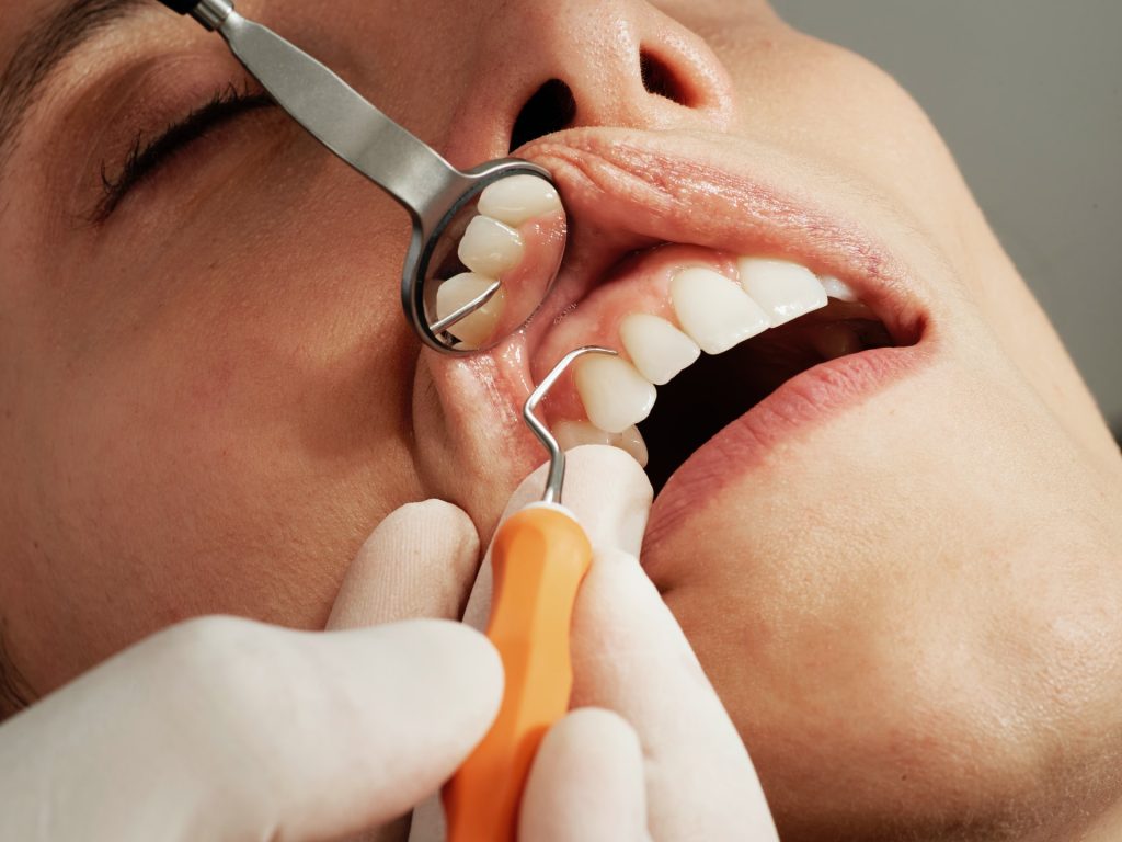
Orthodontic treatment goes beyond getting a better smile; it can also support important oral functions such as chewing and speaking, as well as help patients maintain good oral health over a lifetime. Angelo Maura, General Manager for Africa and Middle East at Align Technology, discusses World Orthodontic Health Day 2026 (WOHD 2026), what “Beyond Straight Teeth” means, and how digital innovation is reshaping orthodontic care for South African patients and practitioners.
Q1: What does “Beyond Straight Teeth” mean to orthodontics?
“Beyond Straight Teeth” strongly reflects how we have always approached orthodontic care. For nearly 30 years, our focus has been on improving the journey to a healthy, confident smile, but that journey has never been limited to aesthetics.
Orthodontic treatment plays an important role in oral function, including how patients chew and speak. It can also support long-term oral health by helping create tooth positions that are easier to clean and maintain. For general dentists and orthodontists, the theme is a reminder that case assessment and treatment goals extend beyond alignment to include function, hygiene access and long-term stability.
Q2: Oral health is said to have an impact on overall health. What are the health benefits of orthodontics?
Misaligned teeth and bite issues can be associated with uneven wear and may make oral hygiene more difficult, which can contribute to plaque build-up and gingival inflammation. In some people, bite problems may also be linked to jaw discomfort. Depending on the individual case, misalignment can also affect everyday functions such as eating and speaking.
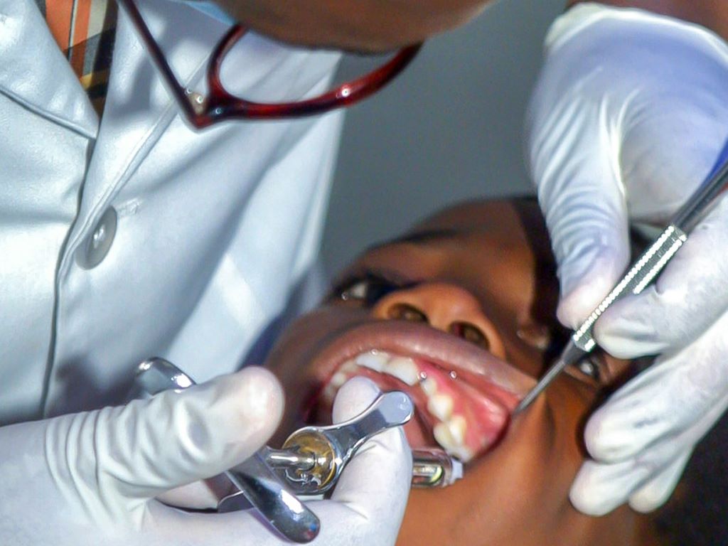
At Align Technology, we design solutions that help clinicians address a wide range of malocclusions through modern, evidence-based orthodontic care. The Invisalign® System is designed to treat a wide range of malocclusions, and starting the conversation early can help patients understand their options and plan the right care with their doctor.
Q3: Orthodontic treatment is often associated with teens and young adults. How does it benefit patients at different life stages?
Our aim at Align Technology is to ensure patients of all ages have access to treatment that fits into their daily lives while supporting overall oral health.
For children, early orthodontic assessment can help identify developing issues such as crowding and spacing. Invisalign First™ aligners are designed for growing patients and are removable, which can support oral hygiene when used as directed and supervised appropriately.
For teens, adherence and day-to-day practicality are important. Removable aligners can help many teens maintain normal activities and oral hygiene routines, while clinicians can use digital planning and monitoring to support progress throughout treatment.
For adults, treatment often needs to fit around work and family commitments, and many patients benefit from an interdisciplinary approach. Clear aligner therapy can be an option that balances aesthetics with planned tooth movement, particularly when coordinated with periodontal maintenance and restorative goals where needed.
To date, approximately 22.8 million patients worldwide have been treated with the Invisalign® System, including more than 6.5 million teens and kids.*
*Data on file at Align Technology, as of December 31, 2025.
Q4: How is Align Technology equipping clinicians to raise the standard of care beyond straightening teeth?
We support clinicians through comprehensive education programmes and tools. There are currently approximately 299,500 Invisalign-trained doctors globally. In South Africa, this includes bringing international specialists to work directly with local clinicians through in-market education sessions and academic engagements, ensuring global best practice is shared in a way that is locally relevant.
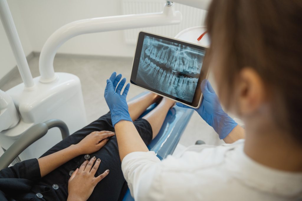
Q5: Digital technology and AI are changing healthcare. How is the Align™ Digital Platform reshaping what happens in dentistry?
The Align™ Digital Platform connects diagnosis, treatment planning, manufacturing, and monitoring into a single workflow.
One key development is ClinCheck® Live Plan, which automates the generation of an initial doctor-ready ClinCheck® treatment plan within about 15 minutes after an eligible case is submitted with Flex Rx, so the doctor can review and approve the plan faster.
The Align™ Oral Health Suite offers a comprehensive set of digital tools that assist clinicians in evaluating, monitoring, and managing patients’ oral health. By integrating advanced assessment capabilities and patient education resources, the suite supports effective communication and engagement, helping doctors deliver personalised care and promote long-term oral wellness.
These technologies are designed to support clinical expertise, with the doctors central to every treatment decision.
Q6: WOHD 2026 calls for global unity in prioritising orthodontic health. What does this commitment look like in South Africa?
South Africa is an important market for us. There is strong engagement from clinicians, and patient awareness continues to grow. We are also seeing increased adoption of digital dentistry.
At the same time, practitioners are at different stages of their digital journey. A high-volume practice in Johannesburg will have different needs from a smaller practice beginning with aligner therapy.
Our approach is to support clinicians at every stage and grow with them.
Our focus is on expanding access to innovation, strengthening engagement with the dental community, and ensuring clinicians have the tools and support needed to deliver strong patient outcomes.
Ultimately, a healthy smile contributes to overall health.