Students and dermatologists are determined to find how rosemary and rosemary extract can repair damaged skin without leaving scars
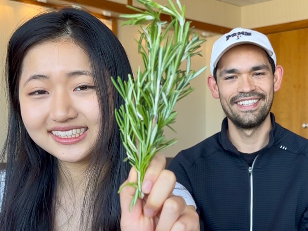
Penn undergraduate student Jiayi Pang (left) and Penn PhD candidate Emmanuel Rapp Reyes (right) found that rosemary can help skin wounds heal without causing scars.
The social media trend touting rosemary and rosemary extract as part of skincare routines is now backed by science. A compound found in rosemary leaves may significantly improve the healing of skin wounds and reduce scarring, according to new research published in JCI Insight from the Perelman School of Medicine at the University of Pennsylvania.
“Many skin injuries end in scars, and in some people, it can lead to long-term cosmetic and even functional issues,” said senior author Thomas Leung, MD, PhD, an associate professor of Dermatology at Penn. “Our findings suggest that rosemary extract, and specifically the antioxidant, carnosic acid, can shift the healing process from scarring to healthy skin regeneration. We don’t have proven ways to consistently do that in humans.”
The hypothesis behind the hype
The inspiration for this study stemmed from an unusual place: TikTok and Instagram. After seeing beauty influencers and other social-media users touting the skin-healing benefits of homemade rosemary extract serums and products with rosemary, Penn undergraduate student Jiayi Pang and Penn PhD candidate Emmanuel Rapp Reyes turned to Leung for expertise. Then, they did what all good scientists do: they went to the lab and ran their own tests.
“We hypothesized there was likely something real behind the hype because rosemary contains many antioxidants,” said Pang, co-lead author of the study. “But we knew in order to really uncover its potential, we needed to prove its healing properties and uncover how exactly it was facilitating healing.”
Conducting the research in mice, the researchers made cream with carnosic acid, a naturally occurring antioxidant mostly existing in rosemary, to accelerate wound closure and restore hair follicles, oil glands, and cartilage. They also found that a particular nerve sensor in the skin previously identified as essential to scarless healing, TRPA1, was critical for stimulating the healing in this instance, too. When tested in mice without the TRPA1 sensor, which previous research from Leung showed is responsible for scarless healing, carnosic cream lost its impact.
“We also identified other herbs, such as thyme and oregano, that may activate TRPA1. But rosemary stood out for its potency and safety,” said Rapp Reyes, co-lead author of the study. “Other natural ingredients, such as mustard oil, or the topical medication imiquimod are known to also stimulate the TRPA1 receptor, but unlike rosemary, those can cause irritation and inflammation,”
The researchers also found a localised effect from rosemary; scarless healing only occurred when carnosic acid cream was applied to the site of the injury but not when it was applied to skin far from the wound.
The team at Penn, however, notes that individuals should speak with their doctors before incorporating rosemary skincare products in their daily regimens or mixing up their own rosemary-based concoctions. Nevertheless, given rosemary’s accessibility and low cost, the researchers hope their findings will inspire further investigation into its use in human wound care, especially for patients at risk of excessive scarring.
“If rosemary is part of your skincare regimen and you think it’s working, it likely is,” said Leung. “I’m proud that the young scientists that led this research sought answers to questions in their everyday lives.”
QIMR Berghofer scientists have uncovered hundreds of genes that play a role in the growth of both moles and melanoma, in a discovery that could lead to new ways of preventing and treating the deadliest form of skin cancer.
The world’s largest genetics study of ‘moliness’, published in Nature Communications, is unravelling the complex causes of both moles and melanomas that are not related to well-known risks caused by sun exposure, skin colour, and pigmentation.
The team found risk genes linked to biological pathways that could lead to the development of a mole or melanoma. These include an immune response pathway that may be failing to control cell growth, and genes implicated in harmful cell proliferation in other types of cancer, such as breast cancer, prostate, and brain cancers.
Working out how to stop these risk pathways could lead to new melanoma drug targets and prevention strategies that go beyond sun protection.
“We know how to reduce sun exposure and risk through SunSmart behaviours, and new immunotherapies have greatly improved survival rates. But people still get melanoma and people still die from melanoma,” A/Prof Law said.
“Existing immunotherapies fail to work for half of all patients with late-stage melanoma, so we need to find other ways to target the disease. By studying moles, we’re learning more about the biology of melanoma so we can find new ways of controlling it.”
Moles and melanomas share the same cellular origin, forming from a pigment-producing cell called a melanocyte that gives skin its colour. In moles, the cell multiplies to form a cluster then stops growing, leaving a harmless spot. In melanoma, the cell growth continues aggressively.
Moliness is strongly influenced by your genes and having a high mole count is a major risk factor for melanoma. Around a third of melanomas develop from a mole.
The QIMR Berghofer research analysed data from more than 85000 participants of European ancestry discovering 24 new genetic regions that determine the number of moles someone has. This is a five-fold increase on the five areas found in an earlier 2018 study also led by QIMR Berghofer researchers.
All but one of the genetic regions for mole count also play a role in melanoma. The team pinpointed more than 250 key genes in these regions to prioritise for further research.
One of the new genes, SIKE1, regulates immune responses to viral infections. The researchers think it could enable the development of melanomas by malfunctioning and affecting the immune system’s ability to detect and destroy melanocytes that are multiplying abnormally. This could be a promising target for a potential immunotherapy that could possibly prevent early stage melanoma growth.
“I’m really proud to be continuing this long legacy of research. Our study increases understanding of why some people have a lot of moles and why some people develop melanoma so we can better treat and prevent this skin cancer,” Ms Jayasinghe said.
The researchers used the study insights to create a Polygenic Risk Score (PRS) for moliness to predict those who are genetically more likely to have a large number of moles, which could be integrated into melanoma screening tools in future to improve their accuracy in finding those at high risk so they can receive extra monitoring.
The next step is to analyse even larger data sets to find more genetic regions involved in moliness and melanoma. The researchers are also searching for existing drugs that could potentially target the newly identified biological pathways.
New findings identify keratinocytes as replication hubs and immune responders, contributing to the risk of rabies infection from superficial scratches or minor bites
Skin cell (keratinocyte) This normal human skin cell was treated with a growth factor that triggered the formation of specialised protein structures that enable the cell to move. We depend on cell movement for such basic functions as wound healing and launching an immune response. Credit: Torsten Wittmann, University of California, San Francisco
While it was previously thought that keratinocytes (skin cells) were only passive conductors that allow the rabies virus to pass through, novel research reveals that these cells play a much more active role. The findings of a new study in the Journal of Investigative Dermatology (JID), published by Elsevier, provide direct evidence that keratinocytes can support viral replication and transmit the rabies virus to neurons. The investigators offer a mechanistic explanation for how superficial skin exposures from scratches or minor bites by dogs and bats can lead to neuroinvasion, contributing to the risk of infection.
Rabies is a fatal zoonotic infection caused by rabies virus (RABV), responsible for at least 59 000 human deaths per year. The virus is transmitted through the saliva of infected animals. While most cases are caused by dog bites, superficial exposures such as bat bites or scratches can also lead to infection, although the underlying mechanisms remain poorly understood.
“In our previous work, we discovered that keratinocytes – cells that form the epidermis, the outermost layer of the skin – were infected at the site of entry of the rabies virus, both in natural and experimental infections. This was unexpected, as rabies pathogenesis has traditionally focused on muscle cells and motor neurons,” explains lead investigator Corine H. Geurts van Kessel, MD, PhD, Department of Viroscience, Erasmus Medical Centre, Rotterdam, The Netherlands. “Given the strategic position of keratinocytes at the skin barrier and their close proximity to sensory nerve endings, we wanted to understand whether these cells are simply bystanders or active participants in early rabies infection and neuroinvasion.”
The investigators used primary human keratinocyte cultures to investigate susceptibility to rabies virus infection and characterise the resulting antiviral immune responses. Three viral strains were tested: a vaccine strain and two wild-type (“street”) strains derived from fatal human cases associated with bat and dog exposures. The dog-associated strain caused only minimal infection and limited keratinocyte immune activation, whereas the other two strains infected keratinocytes more readily and triggered a pronounced antiviral response.
To simulate the close contact between keratinocytes and intra-epidermal nerve endings, a co-culture model of keratinocytes and neurons was developed. In this model, virus produced in infected keratinocytes was successfully transmitted to adjacent neurons, giving the virus a direct route into the nervous system. Once the virus has established infection in the central nervous system, it is almost inevitably fatal.
“Our study demonstrates that the skin might play a more important role in rabies infection than previously recognised. We were particularly surprised by the strong antiviral response mounted by keratinocytes to the bat-related rabies virus strain,“ notes co-investigator Keshia Kroh, PhD candidate, Department of Viroscience, Erasmus Medical Centre, Rotterdam, The Netherlands. “Wild-type rabies viruses are known for their immunosuppressive capacities, and we expected an immune evasive effect in keratinocytes. Instead, we observed the opposite. This raises new questions about how keratinocyte-derived immune responses influence overall disease progression in rabies and other viral infections of the skin.”
This in vitro co-culture model is the first to study rabies virus entry to the nervous system across a cell barrier. Future in-depth studies should be performed to provide mechanistic insight into the differential strain tropism, the interactions of infected keratinocytes with immune cells, and the mechanisms of neuroinvasion from superficial skin contact.
According to the World Health Organization (WHO), any transdermal exposure (including small scratches or abrasions) should be assessed as a potential rabies risk and managed appropriately based on exposure category and clinical context.
“Our study provides a biological rationale for these recommendations,” says co-investigator Carmen W.E. Embregts, PhD, Department of Viroscience, Erasmus Medical Centre, Rotterdam, The Netherlands. “At the same time, it is important to emphasise that the risk of rabies virus infection via superficial exposures depends on multiple factors, including the nature of the exposure and the epidemiological setting. Rather than causing alarm, our findings support informed decision-making. Awareness that superficial skin exposures can represent a route of neuroinvasion helps ensure that potential risks are recognised and evaluated appropriately, while treatment decisions remain guided by established public health criteria.”
“The data in this study support the increasingly recognised concept that cells in the skin are in snug communication with the nervous system. That a scratch or bite is needed for the transmission of rabies is further evidence of the importance of an intact skin barrier in health,” observes JID Associate Editor Ethan Lerner, MD, PhD, Associate Professor of Dermatology, Harvard Medical School, and Massachusetts General Hospital, Boston, MA, USA.
Study led by UTSW researchers defines how skin hormones influence bacteria and results in potential treatment for Staph infections
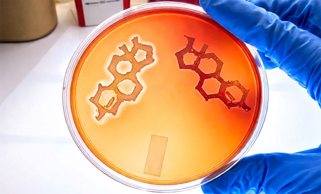
This laboratory image shows Staphylococcus aureus bacteria streaked in the shape of a sex steroid, like testosterone. The left shape is of wild-type S. aureus, with the lighter halo around the shape indicating haemolysis, or the breakdown of red blood cells, releasing their haemoglobin into the surrounding fluid. The right shape is a quorum-sensing mutant strain of S. aureus, which cannot damage blood cells.
Men are more susceptible than women to skin infections caused by Staphylococcus aureus bacteria, but the biological basis for this disparity has remained unclear. A new study led by UT Southwestern Medical Center researchers is the first to reveal that testosterone as a key driver of infection. The sex steroid activates a bacterial communication pathway known as quorum sensing, increasing skin cell death and promoting the destruction of red blood cells and white blood cells called neutrophils.
Published in Nature Microbiology, the study also reported that a mirror-image form of testosterone, known as an enantiomer (ent-T), blocks quorum sensing and prevents S. aureus from damaging tissue in mouse models.
Senior author Tamia Harris-Tryon, MD, PhD, Associate Professor of Dermatology and Immunology at UT Southwestern, and first author Maria S. John, PhD, a UTSW postdoctoral researcher, have a patent pending for an ent-T-based therapeutic along with collaborators at the University of Colorado.
“This research has important implications for treating Staph skin infections and conditions complicated by Staphylococcus, such as atopic dermatitis, pemphigus, abscesses, and wound infections, including the deadliest skin infections caused by methicillin-resistant Staphylococcus aureus [MRSA],” Dr Harris-Tryon said. “It also explains why men are more susceptible to Staph infections.”
S. aureus is the leading cause of skin infections. When it enters the bloodstream, it can cause septicaemia, a life-threatening infection that may lead to organ failure.
During infection, the bacteria use quorum sensing to detect neighbouring cells of the same species. As bacterial density rises, they produce short signalling molecules called auto-inducing peptides (AIP), which activate virulence programs and trigger toxin release, resulting in host-cell damage.
The research team found that male skin cells consistently secrete higher levels of testosterone than female skin cells. They also found the same is true for male mice, which were significantly more susceptible to S. aureus colonisation and skin damage than female mice when exposed to a strain of MRSA. However, mice engineered to secrete less testosterone displayed greater resistance to the bacteria, while applying testosterone to the skin of female mice increased MRSA’s severity.
In laboratory experiments, testosterone activated quorum sensing even in the absence of AIPs. Other sex steroids, including progesterone and oestrogen, had no measurable effect on quorum sensing.
While using ent-T as an experimental control, the researchers unexpectedly identified its therapeutic potential. In lab tests, ent-T inhibited quorum sensing and reduced the bacteria’s virulence. The molecule also inhibited quorum sensing on male and female mice when applied to their skin.
Dr Harris-Tryon won an Innovation Award from the UTSW Office for Technology Development in 2024 to fund development of an ent-T-based transdermal therapeutic for Staph.
“Our exciting finding suggests we can inhibit S. aureus virulence rather than killing the bacteria directly, an approach that prevents infection, preserves beneficial skin microbes, and reduces the selective pressure that drives antibiotic resistance while offering a potential new strategy to treat infections, including MRSA,” Dr. John said.
This work builds on Dr Harris-Tryon’s studies in 2023 and 2025 with Jeffrey McDonald, PhD, Professor in the Center for Human Nutrition and of Molecular Genetics at UTSW, which demonstrated sex-specific differences in skin hormone production. The team also has previously uncovered how the immune system stimulates testosterone production in skin cells. Dr Harris-Tryon said the current research builds on UT Southwestern’s longstanding leadership in steroid and skin hormone biology, a field in which the institution has been a global leader for decades.
A team of researchers at the Icahn School of Medicine at Mount Sinai, Weill Cornell Medicine, and other institutions have uncovered a key biological explanation for why atopic dermatitis (eczema) so often starts in childhood. The study, in young mice, found that some types of immune cells in early-life skin are more reactive than those in adults, a difference that may help explain why children are more vulnerable to inflammation and allergic skin disease.
The findings, reported in Nature, suggest that early childhood represents a critical window for immune-driven skin disease and may shed light on why atopic dermatitis is often the first condition in a broader pattern of allergic disease.
Atopic dermatitis (AD) affects nearly one in four children and often appears early in life. It can also precede other allergic conditions, including asthma and food allergies. Until now, scientists have not fully understood why the disease is so strongly linked to early childhood.
“We found that allergy risk is shaped very early in life, when the skin’s immune system is biologically programmed to overreact to allergens, with important consequences for understanding how immune-mediated diseases emerge and should be treated,” says senior study author Shruti Naik, PhD, Associate Professor of Immunology and Immunotherapy, and Dermatology at the Icahn School of Medicine. “By pinpointing the cells and hormonal signals that control this window of vulnerability, we open the door to strategies that could prevent allergic disease before it spreads from the skin to the lungs, gut, and beyond.”
The researchers discovered that a specific immune cell type, the dendritic cell, in young skin behaves differently than in adults. These cells do not overreact to everything – but when it comes to allergens, they respond faster and more strongly, setting the stage for inflammation and AD early in life. In adult skin, the same cells are far less reactive.
To understand why allergies often start in early childhood, researchers exposed infant mice to everyday allergens such as dust mites and mould. Unlike adult mice, the infants developed strong skin inflammation, revealing a brief early-life period when the skin’s immune system is especially sensitive.
The scientists traced this response to dendritic cells, which are unusually active shortly after birth and triggers allergic inflammation. When this pathway was blocked, the young mice did not develop skin allergies.
The team also found that infants lack normal levels of stress hormones that later help keep immune reactions in check, allowing these allergic responses to take hold. Importantly, signs of the same immune activity were found in skin samples from children with early-onset AD, but not in adults, suggesting this early-life window may also be important in humans.
“This work was only possible through a true clinic-to-lab collaboration – where insights from paediatric patients shaped the questions we asked in the lab,” says study co-author Emma Guttman-Yassky, MD, PhD, professor at the Icahn School of Medicine. “By studying allergic disease where it actually begins, in early life, and by modelling clinically relevant allergens and disease features, lead author Yue Xing, PhD, uncovered immune biology that simply doesn’t appear in adult models. By revealing what’s unique about the early-life immune system, this work explains why eczema so often begins in infancy.”
Next, the investigators plan to explore ways to block this early-life immune pathway to stopallergic disease before it spreads from the skin to other organs.
“Beyond eczema, this study reinforces a critical point for medicine,” says Dr Naik. “Children are not simply small adults when it comes to immunity. Their immune system follows a unique set of rules, and recognising that difference is essential for understanding – and ultimately preventing – allergic, immune-driven diseases that begin in childhood.”
From genetics to stress myths, researchers reveal what really drives greying and the breakthroughs pointing to natural colour restoration
Photo by Ravi Patel on Unsplash
Grey hair is more than a cosmetic concern – it drives a booming industry, influences how people are perceived, and can affect confidence. Globally, the hair colour market was valued at nearly USD 28 billion in 2025, with over half of purchases linked specifically to concealing greys. In South Africa, spending on hair colourants is projected to grow from roughly USD 172 million in 2021 to over USD 228 million by 2028, highlighting the demand for solutions that go beyond temporary cover-ups.
By age 50, roughly 50-70% of adults have visible grey hair, while premature greying can appear in some as early as the 20s. The psychological weight is clear: studies indicate grey hair can make people appear 20-30% older, influencing workplace perception, social interactions, and self-esteem. Studies show faces with grey hair are consistently perceived as more subdued than the same faces without greys, confirming that hair colour alone can shape social impressions.
“Many popular beliefs about greying hair are misleading,” says Dr Kashmal Kalan, Medical Director at Alvi Armani. “Stress does not turn hair grey overnight, plucking one strand won’t trigger several more, and no supplement or home remedy has been proven to restore pigment reliably. The reality is far more biological – genetics and pigment cell behaviour are the keys we are finally beginning to understand.”
At the heart of greying are melanocyte stem cells (McSCs) within hair follicles. In youth, these cells migrate and maintain melanin production, the pigment responsible for hair colour. With age, many become inactive or “trapped,” interrupting pigment delivery and causing grey strands. In mouse models, freeing these cells restored pigment production in roughly half of cases – a major step toward therapies that could reawaken natural colour without dyes.
Emerging research aims to tackle the root cause rather than just the appearance of grey hair. Scientists are exploring topical agents that target dormant pigment cells, metabolic modulators that influence follicle behaviour, and activation therapies designed to revive pigment production. These innovations could allow hair to regain its natural shade – not just cover it – while supporting overall follicle health.
“We are witnessing science that was once purely theoretical become reality,” says Dr Sunaina Paima, aesthetic and hair-restoration physician at Alvi Armani Johannesburg. “For patients, this could mean seeing grey strands regain their original shade naturally – a moment the hair science world has long dreamed of. The potential impact on confidence and self-esteem is enormous, because this isn’t just about covering colour, it’s about restoring it at a biological level.”
While most pigment-restoring therapies remain in development, advances in genetics, dermatology, and biotechnology are converging at unprecedented speed. “For decades, grey hair was seen as an irreversible hallmark of ageing,” adds Dr Kalan. “Today, that assumption is being seriously challenged. We’re on the brink of options that rejuvenate hair from the inside out, not just cosmetically.”
These breakthroughs signal a new era in hair science: ageing hair may no longer be inevitable or purely cosmetic, but a biological process that can be understood, guided, and ultimately restored.
Peptides have become one of the skincare industry’s most popular ingredients. It’s no wonder why, with evidence showing these powerful molecules hold the secret to healthier, firmer and more radiant skin.
But out of the many peptides that exist, one in particular has been gaining attention lately in the beauty industry: copper peptides.
It’s not surprising that copper peptides are garnering so much attention. This peptide is special because of its ability to multitask – with research showing that not only does it help make the skin firmer and more supple, it also protects the skin from damage.
The human body naturally produces many types of peptides. Each supports vital body functions, acting like tiny building blocks of life. Many help form the foundation of essential proteins – such as collagen and elastin, which help keep skin healthy and youthful.
The three main types of peptides in cosmetics are: carrier peptides, signal peptides and neurotransmitter-inhibiting peptides.
Carrier peptides aid in wound repair by physically transporting important minerals into the cells to initiate repair.
Signal peptides can prevent ageing by stimulating the activation of the skin’s fibroblasts – specialised skin cells that produce substances such as collagen, a protein which helps maintain the skin’s elasticity.
Neurotransmitter-inhibiting peptides act like botulinum toxin, relaxing facial muscles by blocking the signals that make them contract. This may reduce wrinkles.
Copper peptides are actually a type of carrier peptide. They’re produced naturally by your body. But as we age, the concentration of copper peptides in our bodies drops. Applying synthetic, lab-made versions – found in creams, serums and masks – can help replenish these molecules and help your skin.
Copper peptides were first discovered in 1973. Research found that these molecules aided wound healing, which is why the first commercialised carrier peptide in 1985 was designed to deliver copper into wounded tissue.
After gaining research attention for this role, further studies examined what other functions copper peptides had on the skin. Researchers found that they had anti-ageing, anti-inflammatory and renewing properties and also supported hair growth.
Copper peptides act as little helpers that tell your skin cells to repair and rebuild themselves. They do this by boosting collagen and elastin, key proteins that keep your skin feeling smooth and firm.
Copper peptides have been also found to reduce inflammation and calm skin redness, too. But perhaps most crucially, they have been found to act as antioxidants, fighting damage caused by pollution and the sun’s ultraviolet rays.
On top of that, copper peptides improve wound healing. This is why they’re often used after cosmetic treatments – such as face and neck lifts and micro-needling – that can damage the skin. Copper infused wound dressings are also used to help chronic wounds heal faster.
Overall, skin cell studies have shown that copper peptides increase collagen production, improve skin thickness and skin elasticity. Clinical trials and lab tests confirm these benefits, making copper peptides one of the most researched anti-ageing ingredients.
For best results, you might want to try applying it twice a day – first in the morning so it can act as a potent antioxidant, then in the evening so it can replenish collagen overnight.
Copper peptides can also penetrate the skin more effectively when delivered with microneedles, which makes them even more useful in advanced skincare products.
Copper peptides v other peptides
Other peptides do work well on the skin – such as palmitoyl-based peptides and acetyl hexapeptide-8 peptide – both of which fight wrinkles. But these both work differently to copper peptides.
Palmitoyl peptides signal the skin to make more collagen, while acetyl hexapeptide-8 relaxes facial muscles to reduce expression lines, acting like a less expensive version of botulinum toxin.
Copper peptides stand out among these other peptides because they can do the work of multiple peptides in one. Copper peptides boost collagen, improve skin healing and fight oxidative stress. This appears to make them better at preventing the signs of ageing.
Some skin cell studies show they work even better when combined with other well known skincare ingredients, such as hyaluronic acid (which boosts hydration).
Copper peptides themselves can also cause, in a few people, some skin irritation and mild allergic reactions. If you find you experience these symptoms after using copper peptides, stop use immediately.
Copper peptides are more than just a trend – they’re backed by science. They help keep skin healthy and speed up healing. They might even play a role in future cancer treatments.
Research has shown copper peptides turn on genes that tell damaged cancer cells to shut themselves down and stop replicating. They’ve also been shown to fix other genes that control cell growth and repair.
If you’re curious about skincare, copper peptides may be worth incorporating into your daily routine. Just remember that good, healthy skin also needs other measures – such as sunscreen, hydration and a healthy lifestyle.
The protein Dickkopf 3 plays a key role in the development of radiation-induced fibroses – and could be a promising target for novel therapies
Picture by Macrovector on Freepik
Radiotherapy is one of the main treatment forms for cancer. Among its most common side effects is skin damage, right up to chronic inflammations and fibroses. At present, such long-term damage can only be treated symptomatically and leads to thickened, painful, or sensitive skin for months to years after the radiation treatment. A team led by LMU immunologist Professor Peter Nelson (LMU University Hospital) and Roger Sandhoff and Peter E. Huber from the German Cancer Research Center (DKFZ) has identified a protein called Dickkopf 3 (DKK3) as a main cause of long-term skin damage after radiotherapy – a decisive step for the development of novel, more targeted therapy options.
By investigating mouse models and human cells and tissue samples, the researchers demonstrated that DKK3 is activated after radiotherapy in a certain group of skin cells that are responsible for skin renewal. This activity triggers a chain reaction which promotes inflammations and the formation of scar-like tissue and leads to chronic skin damage. The key findings were driven by the work of LMU students, Li Li and Khuram Shehzad. Their efforts were essential in identifying DKK3 as the critical molecular mediator and in establishing the mechanistic framework presented in the paper. “We also observed similar processes in the kidney,” says Nelson. “This indicates that the activation of DKK3 is a fundamental mechanism that promotes fibrosis in various tissues.”
According to the researchers, these findings underscore that DKK3 represents a promising new treatment target. “Drugs that block DKK3 could one day help prevent or reduce long-term skin damage after radiotherapy and thus improve the quality of life of cancer patients and survivors,” says Nelson. The researchers are currently investigating, moreover, whether this approach could also contribute to the prevention of scar formation in other organs.
The face is privileged when it comes to scarring after injury. A Stanford Medicine study in mice not only discovers why but also finds a drug that helps skin from other sites regenerate.
Tweaking a pattern of wound healing established millions of years ago may enable scar-free injury repair after surgery or trauma, Stanford Medicine researchers have found. If results from their study, which was conducted in mice, translate to humans, it may be possible to avoid or even treat the formation of scars anywhere on or within the body.
Scarring is more than a cosmetic problem. Scars can interfere with normal tissue function and cause chronic pain, disease and even death. It’s estimated that about 45% of deaths in the United States are due to some type of fibrosis – usually of vital organs like the lungs, liver or heart.
Scars on the skin’s surface, while rarely fatal, are stiffer and weaker than normal skin and they lack sweat glands or hair follicles, making it difficult to compensate for temperature changes.
Surgeons have known for decades that facial wounds heal with less scarring than injuries on other parts of the body. This phenomenon makes evolutionary sense: Rapid healing of body wounds prevents death from blood loss, infection or impaired mobility, but healing of the face requires that the skin maintain its ability to function well.
“The face is the prime real estate of the body,” said professor of surgery Michael Longaker, MD. “We need to see and hear and breathe and eat. In contrast, injuries on the body must heal quickly. The resulting scar may not look or function like normal tissue, but you will likely still survive to procreate.”
Exactly how this discrepancy happens has remained a mystery, although there were some clues.
“The face and scalp are developmentally unique,” said professor of surgery Derrick Wan, MD. “Tissue from the neck up is derived from a type of cell in the early embryo called a neural crest cell. In this study we identified specific healing pathways in scar-forming cells called fibroblasts that originate from the neural crest and found that they drive a more regenerative type of healing.”
Activating this pathway in even a subset of fibroblasts around small wounds on the abdomen or backs of mice caused them to heal with much less scarring – similar to untreated facial or scalp wounds.
Longaker, the Deane P. and Louise Mitchell Professor in the School of Medicine, and Wan, the Johnson & Johnson Distinguished Professor in Surgery II, are the senior authors of the study, which was published January 22 in Cell. Plastic surgery resident Michelle Griffin, MD, PhD, and clinical and postdoctoral scholar Dayan Li, MD, PhD, are the lead authors of the research.
“Many of the authors on this paper are fellow physician scientists,” said Li, who is board certified in dermatology. “This project was inspired by what we’ve observed in our patients – facial wounds in general heal with less scarring. We wanted to understand, mechanistically, why this is.”
Proteins determine scarring
Li and his colleagues used laboratory mice to investigate differences in wound healing at various sites on the animals’ bodies. They anesthetised the mice before creating small skin wounds on the face, scalp, back and abdomen. The wounds were stabilised by suturing small plastic rings around them to prevent differences in mechanical forces as the animals moved. Mice were given pain relief during the healing process.
After 14 days, the wounds on the face and scalp expressed lower levels of proteins known to be involved in scar formation as compared with those on the abdomen or back of the animals. The sizes of the scars were also smaller.
The researchers then transplanted skin from the face, scalp, back and abdomen of mice onto the backs of control mice. After the transplants had engrafted, they repeated the experiment on the transplanted skin. As before, wounds in the skin transplanted from the faces of the donor mice expressed lower levels of scarring-associated proteins.
Additionally, Li and his colleagues isolated fibroblasts from skin samples from the four body sites in the donor mice and injected them into the backs of control mice. They observed reduced levels of scarring-associated proteins on the recipient animals’ backs injected with fibroblasts from the donor animals’ faces as compared with fibroblasts from the scalp, back or abdomen.
Now that we understand this pathway and the implications of the differences among fibroblasts that arise from different types of stem cells, we may be able to improve wound healing after surgeries or trauma.”
–Derrick Wan
“We found you don’t need to change or manipulate all fibroblasts within the tissue to have a positive outcome,” Li said. “When we injected fibroblasts that we had genetically altered to more closely resemble facial fibroblasts, we saw that the back incisions healed very much like facial incisions, with reduced scarring, even when the transplanted fibroblasts made up only 10% to 15% of the total number of surrounding fibroblasts. Changing just a few cells can trigger a cascade of events that can cause big changes in healing.”
A less-fibrotic wound healing
Digging deeper, the researchers identified changes in gene expression between facial fibroblasts and those from other parts of the body and followed these clues to identify a signaling pathway involving a protein called ROBO2 that maintains facial fibroblasts in a less-fibrotic state. They also saw something interesting in the genomes of fibroblasts making ROBO2.
“In general, the DNA of the ROBO2-positive cells is less transcriptionally active, or less available for binding by proteins required for gene expression,” Li said. “These fibroblasts more closely resemble their progenitors, the neural crest cells, and they might be more able to become the many cell types required for skin regeneration.”
In contrast, the DNA in fibroblasts from other sites of the body allows free access to genes like collagen that are involved in the creation of scar tissue.
“It seems that, in order to scar, the cells must be able to express these pro-fibrotic genes,” Longaker said. “And this is the default pathway for much of the body.”
ROBO2 doesn’t act alone. It triggers a signalling pathway that results in the inhibition of another protein called EP300 that facilitates gene expression. EP300 plays an important role in some cancers, and clinical trials of a small drug molecule that can inhibit its activity are underway. Li and his colleagues found that using this pre-existing small molecule to block EP300 activity in fibroblasts prone to scarring caused back wounds to heal like facial wounds.
“Now that we understand this pathway and the implications of the differences among fibroblasts that arise from different types of stem cells, we may be able to improve wound healing after surgeries or trauma,” Wan said.
The findings are likely to extend to internal scarring as well, Longaker said. “There’s not a million ways to form a scar,” he said. “This and previous other findings in my lab suggest there are common mechanisms and culprits regardless of the tissue type, and they strongly suggest there is a unifying way to treat or prevent scarring.”
Your guide to safe, effective, and natural hair restoration in 2026
Hair restoration is one of the fastest-growing aesthetic procedures worldwide. The International Society of Hair Restoration Surgery (ISHRS) reports hundreds of thousands of procedures performed globally each year, with demand climbing steadily. As more people seek confidence-boosting solutions to start the new year, South African specialists warn that choosing the wrong clinic can turn a life-changing decision into lasting damage.
Dr Kashmal Kalan, Medical Director at Alvi Armani South Africa, explains: “January brings a sense of renewal. Many people reassess their goals, and hair restoration has become one of the most transformative ways to invest in yourself. It’s no longer just about fitness or weight loss – hair and skin now play a central role in personal confidence.”
A successful hair restoration journey begins long before the procedure and continues well beyond it. At Alvi Armani, every patient undergoes a thorough, personalised consultation. The team evaluates hair loss patterns, donor density, scalp condition, hair type, and personal goals. Advanced AI-assisted microscopic analysis helps ensure patients are suitable and that the procedure is planned for optimal, natural results.
On procedure day, patients enter a calm, controlled environment. Hairline design is finalised, Follicular Unit Extraction (FUE) is performed with precision, and grafts are implanted to follow the natural flow of hair. Recovery is gradual, with initial shedding giving way to new growth. Density and texture refine over 12-18 months, and ongoing check-ins ensure progress stays on track.
Hairline design is the most artistic aspect of the process. Age, facial symmetry, ethnicity, and donor capacity all influence the final outcome. “We aim for perfection within imperfection. The goal is a hairline that complements the face naturally. No one should be able to tell a transplant took place.”
Strategic density planning is equally critical. Every follicle in the donor area is finite, and poor planning can create gaps or thin patches. This can leave permanent aesthetic imbalance. Reputable clinics plan for decades, not just the first few months. Patients should also understand that growth is gradual, and progressive hair loss may require more than one procedure to achieve the desired result.
Alvi Armani ensures every procedure is doctor-led and supported with ongoing care, including stabilisation medications, regenerative therapies, and annual check-ups. Dr Kalan cautions against so-called “dark clinics” offering prices too good to be true, often operating in unhygienic or mobile facilities. “These clinics treat hair restoration as a commodity rather than medicine. They overharvest donor areas, produce unnatural results, and leave patients needing urgent repairs. Repair procedures now make up roughly a quarter of our cases.”
Beyond procedural excellence, Alvi Armani educates patients on lifestyle choices that support lasting results, from nutrition and scalp care to ongoing therapies. While the process requires patience, the rewards – confidence, natural appearance, and the security of a clinic that plans carefully for the future – make it worthwhile.
For anyone considering hair restoration in 2026, the advice is clear: invest in quality from the start. With the right clinic, personalised planning, and medical oversight, patients can achieve safe, natural results that endure for years to come.