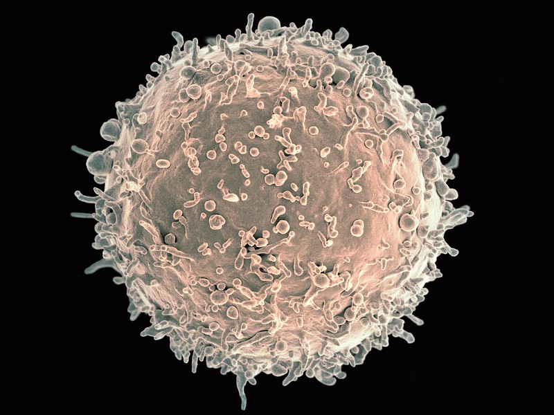
Scanning electron micrograph image of a human B cell. Credit: NIH/NIAID
New research in Nature Immunology has found that B cells play a surprising role in increasing or decreasing the bone marrow’s output of white blood cells. The findings may lead to new treatments for conditions that arise when white blood cell production goes out of balance.
Professor Matthias Nahrendorf, senior author of the study, explained that the nervous system plays a role in controlling blood cell production through neurotransmitters. “This is for instance important in people exposed to stress, where stress hormones — part of the ‘fight-or-flight’ response controlled by the sympathetic nervous system — may increase bone marrow activity and cardiovascular inflammation in response to the neurotransmitter noradrenaline,” he said. The parasympathetic nerves on the other hand, slow down responses and bring about a state of calm to the body, mainly through the neurotransmitter acetylcholine.
Because acetylcholine can have a protective effect against inflammation and heart disease, the researchers studied this neurotransmitter in the bone marrow. “When we looked into how acetylcholine acts on the production of blood cells, we found that it does the expected — it reduces white blood cells, as opposed to noradrenaline, which increases them,” said Prof Nahrendorf. “What was unexpected though was the source of the neurotransmitter acetylcholine.”
In the bone marrow, the typical nerve fibres that are known to release acetylcholine were not found. Instead, it was the antibody-producing B cells supplied the acetylcholine in the bone marrow. “Thus, B cells counter inflammation — even in the heart and the arteries — via dampening white blood cell production in the bone marrow. Surprisingly, they use a neurotransmitter to do so,” said Prof Nahrendorf.
Tapping into this process may help investigators develop strategies to block inflammation in cardiovascular conditions such as atherosclerosis. “Ultimately this may lead to new therapeutics that combat myocardial infarction, stroke, and heart failure,” said Prof Nahrendorf.
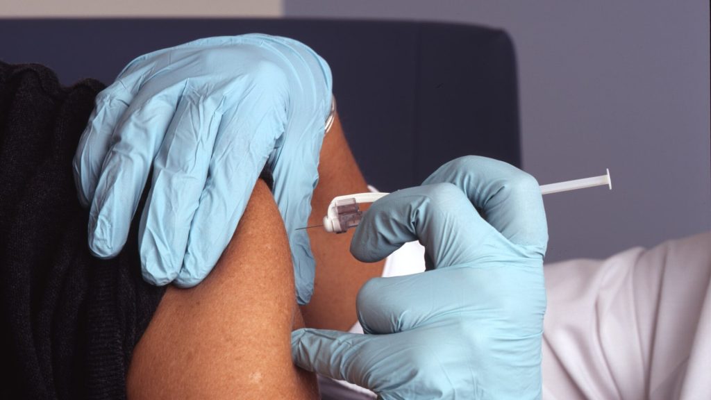
A small clinical trial published in Pediatrics has shown that intranasal flu vaccine is just as safe for children with asthma as the intramuscular vaccine. According to the researchers, within 42 days of vaccination, 10.8% of children who received the intranasal quadrivalent live attenuated influenza vaccine (LAIV4) had an asthma exacerbation compared with 14.7% of those who received the intramuscular quadrivalent inactivated influenza vaccine (IIV4).
According to the researchers, regardless of asthma severity, LAIV4 remained noninferior to IIV4. Among those with mild asthma, one of 25 kids who received the LAIV4 experienced an asthma exacerbation versus three of 16 in the IIV4 group, the researchers reported. In children with moderate to severe asthma, exacerbations occurred in seven of 49 in the LAIV4 group and seven of 52 in the IIV4 group.
“These data add to the compelling safety record of LAIV in children, including those with persistent asthma,” the researchers wrote.
The two groups also did not differ significantly in the frequency of asthma-related symptoms, including nighttime awakening, unscheduled albuterol use, cough, wheezing, or chest tightness, within 14 days of administration. Similarly, no differences were seen in peak expiratory flow rate, or changes in childhood asthma control test or asthma control test scores from baseline through 42 days.
At present, the CDC recommends against the nasal spray vaccine for children and people with asthma, citing an increased risk of exacerbations.
A previous study had suggested that the LAIV was linked increased asthma risk and reactive airway disease in children under 36 months of age, but more recent research has found no difference in risk between the LAIV and IIV, the researchers explained.
“Building off these previous studies, our prospective study suggests that LAIV may be appropriate for some children with asthma,” they noted.
“These data support reexamining precautions to using LAIV4 in children with asthma, which could be particularly important during influenza pandemics, at times when IIV4 supplies are limited, in situations of public/school mass vaccination clinics using LAIV, or for children with significant needle aversions,” they added.
The study was conducted over the 2018 and 2019 flu seasons with children aged five to 11 but expanded to include children ages 5 to 17 in its second year. The primary outcome of asthma exacerbation after 42 days was defined as an episode for which the participant sought medical care or a new prescription for corticosteroids.
The median age of the 151 enrolled participants was 9 years, and 58% were boys.
Systemic reactogenicity events in the 14 days after vaccination were not different between the LAIV4 and IIV4 groups, with the exceptions of myalgia and sore throat, which were more common in the IIV4 group.
Cedars-Sinai researchers have developed a clinical algorithm that is the first to be able to distinguish between treatable sudden cardiac arrest and untreatable forms of the condition. The findings, published in the Journal of the American College of Cardiology: Clinical Electrophysiology, may help prevent sudden cardiac arrest based on key risk factors identified in this study.
“All sudden cardiac arrest is not the same,” explained Professor Sumeet Chugh, MD, lead author of the study. “Until now, no prior research has distinguished between potentially treatable sudden cardiac arrest versus untreatable forms that cause death in almost all instances.”
In the US, 300 000 people die due to out-of-hospital sudden cardiac arrest each year. For those affected, 90% will die within 10 minutes of cardiac arrest.
Prevention could have an enormous impact for this largely fatal condition. The biggest challenge, however, lies in distinguishing between those who stand to benefit the most from an implantable cardioverter defibrillator and those who would not.
“Defibrillators are expensive and unnecessary for individuals with the type of sudden cardiac arrest that will not respond to an electrical shock,” said Prof Chugh. “However, for patients with treatable, or ‘shockable,’ forms of the disease, a defibrillator is lifesaving.”
Prof Chugh said that this new research provides a clinical risk assessment algorithm that can better identify patients at highest risk of treatable sudden cardiac arrest—and thus, a better understanding of those patients who would benefit from a defibrillator.
The risk assessment algorithm consists of 13 clinical, electrocardiogram, and echocardiographic variables that could put a patient at higher risk of treatable sudden cardiac arrest.
The risk factors include diabetes, myocardial infarction, atrial fibrillation, stroke, heart failure, chronic obstructive pulmonary disease, seizure disorders, syncope—a temporary loss of consciousness caused by a fall in blood pressure—and four separate indicators found with an electrocardiogram test, including heart rate.
New computational simulations of the behaviour of the SARS-CoV-1 and SARS-CoV-2 spike proteins before they fuse with human cell receptors show that SARS-CoV-2, is in fact more stable and slower changing than SARS-CoV-1 that caused the SARS epidemic in 2003.
Though severe acute respiratory syndrome coronaviruses 1 and 2 (SARS-CoV-1 and SARS-CoV-2) have striking similarities, why the latter is more transmissible remains unclear.
The spike proteins of each, which bind to host cell angiotensin converting enzyme 2 (ACE-2), otherwise known as the human cell receptor, have been proposed as the reason for their difference in transmissibility. A more detailed understanding of the spike proteins prior to binding could lead to the development of better vaccines and medications.
The new finding, which appears in the Journal of Biological Chemistry, does not necessarily mean that SARS-CoV-2 is more likely to bind to cell receptors, but it does mean that its spike protein has a better chance of effective binding.
“Once it finds the cell receptor and binds to it, the SARS-CoV-2 spike is more likely to stay bound until the rest of the necessary steps are completed for full attachment to the cell and initiation of cell entry,” explained Associate Professor Mahmoud Moradi, of the Fulbright College of Arts and Sciences.
To determine differences in conformational behaviour between the two versions of the virus, the researchers performed equilibrium and nonequilibrium simulations of the molecular dynamics of SARS-CoV-1 and SARS-CoV-2 spike proteins, leading up to binding with cell angiotensin converting enzyme 2.
Equilibrium simulations allow the models to evolve spontaneously on their own time, while nonequilibrium simulations change according to external input. The former is less biased, but the latter is faster and allows for many more simulations to run. Both methodological approaches provided a consistent picture, independently demonstrating the same conclusion that the SARS-CoV-2 spike proteins were more stable.
The models revealed other important findings, namely that the energy barrier associated with activation of SARS-CoV-2 was higher, meaning the binding process happened slowly. Slow activation allows the spike protein to evade human immune response more efficiently, because remaining in an inactive state longer means the virus cannot be attacked by antibodies that target the receptor binding domain.
Researchers understand the importance of the receptor-binding domain (RBD), which viruses use to gain entry to human cells. The team’s modelling confirms the importance of the RBD but also suggest that other domains, such as the N-terminal domain, could play a crucial role in the different binding behaviour of SARS-CoV-1 and -2 spike proteins.
N-terminal domain of a protein is a domain located at the N-terminus or simply the start of the polypeptide chain, as opposed to the C-terminus, which is the end of the chain. Though it is near the receptor-binding domain and is known to be targeted by some antibodies, the function of the N-terminal domain in SARS-CoV-1 and -2 spike proteins is not fully understood. Moradi’s team is the first to find evidence for potential interaction of the N-terminal domain and the receptor binding domain.
“Our study sheds light on the conformational dynamics of the SARS-CoV-1 and SARS-CoV-2 spike proteins,” Moradi said. “Differences in the dynamic behaviour of these spike proteins almost certainly contribute to differences in transmissibility and infectivity.”
Girls and boys might be more vulnerable to the negative effects of social media use at different times during their adolescence, according to a study in Nature Communications. Girls were found to experience a negative link between social media use and life satisfaction when they are 11–13 years old and boys when they are 14–15 years old. Increased social media use again predicts lower life satisfaction at age 19 years. At other times the link was not statistically significant.
Since its rapid emergence over a decade ago, social media has prompted concern over its possible impacts on wellbeing, especially in younger people.
A team of researchers analysed two UK datasets which included longitudinal data on 17 400 young people aged 10–21 years old. The team looked for a connection between estimated social media use and reported life satisfaction and found key periods of adolescence where social media use was associated with a decrease in life satisfaction 12 months later. Working backwards, the researchers also found that teens who have lower than average life satisfaction use more social media one year later.
In girls, social media use between ages 11 and 13 was associated with a drop in life satisfaction one year later, whereas in boys this occurred between 14 and 15. This suggests that sensitivity to social media use could be linked to developmental changes, possibly changes in the structure of the brain, or to puberty, which occurs later in boys than in girls.
In both females and males, social media use at the age of 19 years was again associated with a decrease in life satisfaction a year later. The researchers suggest that that social changes at this age, such as leaving home, may make people particularly vulnerable.
At other times, the link between social media use and life satisfaction one year later was not statistically significant. Decreases in life satisfaction also predicted increases in social media use one year later; however this does not change across age and or differ between the sexes.
Dr. Amy Orben, the study leader, said: “The link between social media use and mental wellbeing is clearly very complex. Changes within our bodies, such as brain development and puberty, and in our social circumstances appear to make us vulnerable at particular times of our lives.”
Professor Sarah-Jayne Blakemore, a co-author of the study, said: “It’s not possible to pinpoint the precise processes that underlie this vulnerability. Adolescence is a time of cognitive, biological and social change, all of which are intertwined, making it difficult to disentangle one factor from another. For example, it is not yet clear what might be due to developmental changes in hormones or the brain and what might be down to how an individual interacts with their peers.”
Dr. Orben added: “With our findings, rather than debating whether or not the link exists, we can now focus on the periods of our adolescence where we now know we might be most at risk and use this as a springboard to explore some of the really interesting questions.”
A further complication is that social media use can negatively impact wellbeing, but also the reverse is true, previously reported and confirmed by this study.
The researchers stress that these population-level findings do not predict which individuals are most vulnerable.
Professor Rogier Kievit said: “Our statistical modeling examines averages. This means not every young person is going to experience a negative impact on their wellbeing from social media use. For some, it will often have a positive impact. Some might use social media to connect with friends, or cope with a certain problem or because they don’t have anyone to talk to about a particular problem or how they feel—for these individuals, social media can provide valuable support.”
Professor Andrew Przybylski said: “To pinpoint which individuals might be influenced by social media, more research is needed that combines objective behavioural data with biological and cognitive measurements of development. We therefore call on social media companies and other online platforms to do more to share their data with independent scientists, and, if they are unwilling, for governments to show they are serious about tackling online harms by introducing legislation to compel these companies to be more open.”
Researchers in Spain have found that caffeine may be beneficial in alleviating cognitive symptoms of ADHD, such as improving attention span and retention capacity. Their findings, published in Nutrients, may provide a less controversial addition to the therapeutic arsenal for this disorder.
Attention deficit hyperactivity disorder (ADHD) diagnoses have increased exponentially over the last 20 years. It is currently estimated that this disorder affects between 2% and 5% of children in Spain, an average of one or two children per classroom, and up to 4% of the adult population.
Despite these high incidence rates, controversy surrounds the treatment of this pathology and the therapeutic approach to it. This varies widely depending on each patient, the symptoms they present and their intensity. For this reason, experts are continuing to investigate different components and substances that may be capable of providing new treatment opportunities for patients diagnosed with ADHD.
A team of experts at the Universitat Oberta de Catalunya (UOC) has investigated caffeine to alleviate some of the symptoms of ADHD, given the controversy surrounding the use of some medicines derived from methylphenidate, among others. Their systematic review of animal studies concludes that a prescribed consumption of caffeine can increase attention and retention capacity in adolescents and adults with ADHD.
“The therapeutic arsenal for alleviating ADHD is limited, and there is a certain degree of controversy around the use of some types of medications and stimulants, especially during childhood and adolescence. That’s why it’s useful to study the efficacy of other substances, such as caffeine,” explained Javier Vázquez, one of the paper’s main authors.
This is the first systematic review with results linking caffeine consumption in different animal models of ADHD with an increased attention span, improved concentration, learning benefits, and improvements in some types of memory.
“This substance improves these types of cognitive procedures, and increases capacity and flexibility in both spatial attention and selective attention, as well as in working memory and short-term memory,” emphasised Vazquez, who added that controlled treatment with this substance “doesn’t alter blood pressure, and doesn’t lead to an increase or reduction in body weight.”
The researchers point out that while possibly effective for cognitive symptoms, the results are unclear for other characteristic symptoms of ADHD, such as hyperactivity and impulsivity. “The results are very positive, but we must be much more careful when prescribing a caffeine-based medical treatment for these symptoms. In diagnoses in which the problem is purely attentional, caffeine may be an appropriate therapy, but if there’s a symptomatological presence of hyperactivity or impulsivity, we must be more cautious,” said Vasquez.
However, the results show that caffeine has a clear benefit in ADHD’s cognitive symptoms. “Our results reinforce the hypothesis that the cognitive effects of caffeine found in animal models can be translated and applied in the treatment of ADHD in people, especially at young ages such as adolescence,” the authors concluded.
“We want to emphasise that we aren’t against medication for ADHD, but we’re open to investigating all possible alternatives for improving this type of disorder, and for being able to use caffeine from a therapeutic point of view with all the appropriate medical supervision, a prescribed treatment and follow-up,” said Vázquez.
For the first time, children with severe congenital myopathy may have a better chance at learning to walk thanks to a new therapeutic approach using enzyme-inhibiting cancer drugs, as reported in the journal eLife.
Professor Susan Treves remembers seeing one child affected by the condition at the age of six months. The boy seemed more like a newborn, she said. Today, several years later and thanks to intensive physiotherapy, he is at least able sit. “He made it,” she said. As yet there is no cure for children like this one. Their first priority is survival. Another child with mutations in the same gene as the boy mentioned above, did not survive. However, his genetic alterations now form the basis of a therapeutic approach presented by the research group led by Professors Susan Treves and Francesco Zorzato.
The affected gene is for the calcium channel RYR1 in skeletal muscle. The mutations render the gene useless, which severely impacts muscle function. The researchers used the gene alterations found in a patient, as a template to develop a mouse model for this type of congenital myopathy. “The mice don’t die, but their muscle system is severely impaired,” says Treves. “They’re smaller, and move much less.” With a combination of two drugs, however, the research team was able to significantly improve muscle function and movement of the mice.
The therapy is based on the observation that certain enzymes are produced in excessive quantities in the skeletal muscles of affected patients. These enzymes – histone deacetylases and DNA methyltransferases – affect how densely genes are packed, making them less accessible to the cellular machinery that reads them and translates them into instructions for protein production.
Prof Treves and her team used inhibitors against these enzymes, which are already approved as cancer drugs or are in clinical trials. The treatment significantly improved the mice’s movement ability, although they were still smaller. No adverse side effects were noted during the study period.
The approach is still far from being a clinical therapy, said Prof Treves. “But it’s a first step in the right direction.” The researchers aim to further optimise the treatment and test combinations of newly developed drugs targeting the same enzymes for even better effects. “We anticipate around about two more years of optimisation and testing before we can initiate a phase I clinical trial,” she said.
For Profs Treves and Zorzato, these first promising results are the culmination of 10 years of research – especially as Prof Zorzato was the one who first isolated the gene affected in these muscle disorders years ago. “We’ve now succeeded in bridging the gap from the isolation of the affected gene to a therapeutic approach,” said Prof Treves.
New research published in the journalJCI Insight shows that immune responses to the Pfizer-BioNTech mRNA vaccine differ significantly in individuals depending on whether or not they had a prior COVID infection. Notably, those who had COVID before vaccination produced a surge of antibodies after the first dose, with little or no increase seen after the second dose. The opposite pattern was observed in infection-naïve individuals.
“Our study shows that the presence of immune memory induced by prior infection alters the way in which individuals respond to SARS-CoV-2 mRNA vaccination,” explained first author Professor Steven G. Kelsen. “The lack of response after the second vaccine dose in previously infected individuals is especially relevant, because it could mean that some people may require only one dose or could potentially skip the booster shot.”
Prof Kelsen and colleagues carried out the study in health care workers, some having previously tested positive for SARS-CoV-2 infection and others never having been infected. The researchers measured levels of neutralising antibodies in blood samples taken at three different time points, including before vaccination and after each vaccine dose. They also performed qualitative assessment for local reactions and systemic symptoms, such as fever, headache, and fatigue, associated with vaccination.
While levels of neutralising antibodies hit their maximum in some people with prior COVID after the first vaccine dose, individuals with no history of infection showed massive responses after the second dose. Those high levels also plummeted quickly, while the COVID group retained longer lasting immunity, despite the lack of response to a second dose. However, prior infection was also linked to more frequent and longer-lasting adverse reactions to the vaccine.
“Previous studies had similarly reported long-lasting immunity and strong immune reactions in COVID patients,” Prof Kelsen said. “We now provide new information on how prior infection interacts with vaccination in terms of measurable immune response and how individuals react to mRNA vaccines based on infection history.”
The next steps for Prof Kelsen and collaborators are to modify their neutralising antibody assay to detect Omicron and other SARS-CoV-2 variants. “We also are interested in understanding how long protection from a booster dose of the vaccine lasts,” he said.
A new review paper, published in the journal Brain, has shown that a mysterious brain region called the claustrum may play an important role in the experience of pain. This densely interconnected, but difficult to access area of the brain may be the next frontier in improving outcomes for brain damage patients.
The claustrum is a brain region that has been investigated for over 200 years, yet its precise function remains unknown. A 2005 article suggested it to be critically linked to consciousness, which spurred a renewed interest in this region, with recent research revealing its high level of interconnectedness.
Credit: Oxford University
Oxford University researchers reviewed studies of patients with rare cases of lesions in the claustrum, which show cognitive impairments and seizures. There may be many more cases to be uncovered due to the lack of clinical focus on the claustrum.
They also uncovered an underappreciated link between the claustrum and pain. It is already known that there are links between the claustrum and perception, salience and the sleep-wake cycle, but this is the first time a research team has shown how the claustrum might be more involved in the debilitating experience of pain.
Dr. Adam Packer, the lead author of the study, says that “The problem with understanding how the claustrum works is that it is deep inside the brain, and damage that is specific to it is a very rare occurrence. What makes it more difficult to work out what the claustrum actually does is that these rare occurrences are also linked to such a broad range of symptoms.”
“Clearly, when the claustrum is damaged the effects are severe and better therapies are urgently needed. It is possible that claustrum damage is more common than we currently realise, and it may be a crucial component in many more brain damage cases.”
“This work is important because it gives us some insight into the cognitive and neurological processes in which the claustrum may be involved, and gives us targets to pursue in basic research in the lab.”
The researchers found several recorded instances of either infection, autoimmune, or other process that attacked the claustrum in particular, and by analysing the results of these studies and others the most common symptoms in patients were cognitive impairment and seizures.
Additional research is needed for a better understanding of the claustrum and the impact of damage to the claustrum, which could eventually change clinical guidelines.
A study interviewing patients who received total knee replacement for osteoarthritis find that, despite the welcome pain relief, some also experience less pleasant psychological impacts.
For their study published in Arthritis Care & Research, UK researchers sought to bridge a knowledge gap of where people were dissatisfied with their total knee replacements even though they reported less pain and better function.
Using semi-structured interviews, researchers elicited comments from 34 patients, meant to explore patients’ thoughts about their knee implants. They received a lot of feedback about non-pain discomfort and feelings of dissonance.
“My leg feels like it’s made of lead,” one patient told researchers.
“It feels like someone is holding your knees, when you move, it’s like someone is … putting pressure there,” said another.
A third said: “I know it’s not my knee. It’s an alien knee in there. I don’t really feel connected to it.”
“Typically, the assessment of patient-reported outcomes after joint replacement focuses on functional outcome and pain relief as the main determinant of satisfaction,” the researchers explained. “This narrow perspective is compounded by poor definitions of satisfaction after surgery, and there is little research on how and why some patients express dissatisfaction with joint replacement and what they are dissatisfied about.”
Citing a study of hand surgery patients in which patients “spoke about their hand as if it were an object separate from their self,” the researchers argued that a psychological concept called embodiment could help explain the patients’ feelings of dissonance.
They wrote: “Embodiment refers to the experience of the body as both subject and object, such that this idea impacts the way in which a person sees and interacts with the world, and vice versa. Embodiment provides a way of understanding how one experiences limits of possible action, a sense of control, and empowerment over physical action.”
Initially, the researchers weren’t planning to focus on embodiment, but, they explained, “by the third interview we noted that some participants described sensations of discomfort such as heaviness or numbness when discussing pain and some described their knee as ‘alien,’ ‘foreign,’ or ‘not part of’ themselves. In response to these findings, the interviewer sought to elicit views about any such sensations in subsequent interviews, if this topic was not broached first by the participant.”
Their study emerged from an earlier one focusing on reasons for avoiding healthcare encounters post-surgery and involved the same participants, who had lingering pain and discomfort. The semi-structured interview covered pain as well as how patients managed it. After a third interview, patients who reported feelings of alienation from their implant were asked about it in more detail.
Participants reflected the general knee-replacement population – most in their 60 and 70s, and just over half were women. Of the 34 patients, 24 were between 2 and 4 years out from their surgery.
Physical types of non-pain discomfort were commonly reported, such as feelings of numbness and/or heaviness, as well as sensations of pressure applied externally. One man said it felt like the skin over his knee was stretched tight. Separate from these sensations were reports that the limb no longer felt like a part of them but something foreign, like an external prosthesis. Some patients complained of a lack of control. “That knee just wouldn’t do what it’s told to do,” one told the interviewer.
Others said they hadn’t regained trust in the knee, with one man still using a cane for fear of falling.
Overall, the researchers found that the patients’ experience were quite similar to those of amputees getting used to their prosthetic limbs. This could be partly explained by often experiencing years of pain and loss of function before the joint replacement.
“Presurgical chronic pain, instability, and untrustworthiness might continue to influence [mental] incorporation of the prosthesis afterwards,” the researchers suggested.
And there is a potential clinical implication for the findings: “Our study suggests that the interest for rehabilitation becomes not only strengthening the joint and promoting full recovery to tasks, but also modifying a person’s relationship with the new joint to achieve full incorporation or re-embodiment.”
The researchers proposed that other programmes developed for other conditions could be helpful, such as external prosthesis use as well as complex regional pain syndromes.
“Our focus should not be on the absence or loss of embodiment,” the researchers added, “but on employing a multidisciplinary approach to using the concept to guide the development of pre-rehabilitative strategies and appropriate outcome measures.”