Urine Test Could Help Detect Lung Cancer Years Before Symptoms Occur
Cambridge scientists hunting tell-tale killer ‘zombie’ cells that signal early lung cancer have developed a world-first urine test that could transform diagnosis and survival for thousands of patients.
[The test] could one day be used easily in GP surgeries and hospitals to help detect recurrence in this hard-to-treat cancer much earlier.
Ljiljana Fruk
As published this week in Nature Aging, the team has shown that this simple and affordable test could detect the earliest signs of lung cancer months, or even years, before symptoms appear, as well as monitor whether treatment is working and identify potential relapse.
It works by identifying the presence of senescent cells in the lungs – so called ’zombie cells’ – that stop dividing but linger and release abnormal inflammatory signals that damage surrounding tissue and help create an environment that lowers the body’s ability to fight the cancer.
The study, funded by Cancer Research UK, marks a major leap towards more precise therapy and a test for early cancer and treatment efficiency that could be rolled out across the NHS one day.
Lung cancer is the UK’s most common cause of cancer death taking the lives of around 32,800 people every year. Thanks to huge strides in prevention, detection and treatment, in the UK, lung cancer has seen a 22% reduction in death rates in the last decade. And around two in three people (65%) with lung cancer in England survive their disease for five years or more when diagnosed at the earliest stage. But when diagnosed at the latest stage, this falls to 5 in 100 (5%).
This new test could save and improve thousands more lives in the future.
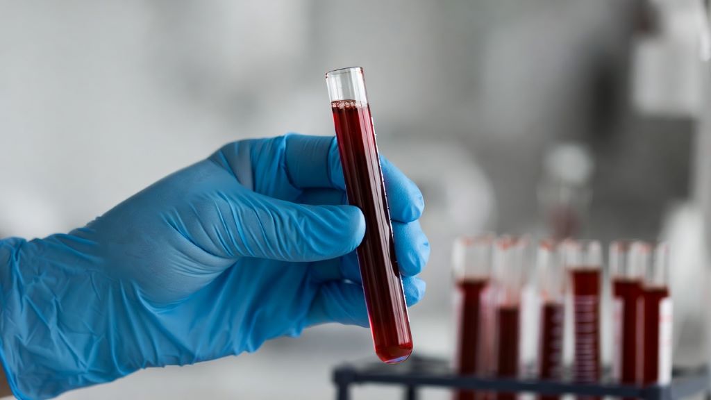
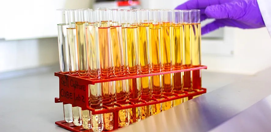
The researchers created an injectable sensor that interacts with proteins released by senescent cells. When these proteins are present, the sensor triggers the release of a detectable compound that appears in urine – signalling the earliest biological signs of therapy resistance and lung cancer development.
The researchers say that early identification is critical to saving more lives, as the disease often relapses silently with few or no symptoms until it has already spread. By detecting signs of lung cancer development and therapy resistance early, their simple urine test can spot lung cancer and treatment resistance early, helping doctors to tailor and adapt the treatment to the patient and start that treatment earlier when it works best.
The team confirmed their results using real patient samples and large genetic datasets.
Professor Ljiljana Fruk, from the Department of Chemical Engineering and Biotechnology at Cambridge, said: “The sensor has not yet been tested in humans, next is the clinical trials and it is likely it will take few years to bring it to patients, but it is a first big step and it could one day used easily in GP surgeries and hospitals to help detect recurrence in this hard-to-treat cancer much earlier.”
Nearly half (46%) of lung cancers in England are diagnosed at the latest stage.
Professor Daniel Munoz-Espin from the Early Cancer Institute and co-lead for the Cancer Research UK Cambridge Centre Thoracic Cancer Programme, said: “Our previous studies showed that senescent cells in response to chemotherapy can cause treatment resistance and an aggressive lung cancer relapse. We also found that senescent immune system cells promote lung cancer development by causing immunosuppression.
“Our urine nano sensor may allow primary care detection of therapy resistance and lung cancer early development in future clinical settings.”
Professor Robert Rintoul of the Department of Oncology, and co-lead for the Cancer Research UK Cambridge Centre Thoracic Cancer Programme said: “Novel approaches for lung cancer detection and response to treatment are urgently needed to improve patient outcomes. This work forms the basis for testing within clinical trials with a view to future use in the clinic.”
Cancer Research UK’s spokesperson for the East of England, Patrick Keely, said: “With new technologies opening doors to new discoveries, we’re living in a golden age of research, which is powerfully underlined by this innovative new urine test to detect early lung cancer.”
Adapted from a press release from Cancer Research UK
Reference
Hartono, M et al. Urinary detection of therapy-induced senescence and fibrosis using an injectable albumin-based nanoprobe. Nature Aging; 13 May 2026; DOI: s43587-026-01116-z
Republished from the University of Cambridge under a Creative Commons licence.
Read the original article.