A model using 71 proteins associated with retinal degradation could predict risk in diabetics
Plasma proteomic signatures for early risk stratification of diabetic retinal neurodegeneration. Credit: Wei Wang and Huangdong Li / Zhongshan Ophthalmic Center, Sun Yat-sen University (CC-BY 4.0, https://creativecommons.org/licenses/by/4.0/)
An AI-assisted model based on 71 different blood proteins could help doctors better predict retinal degeneration in diabetic patients before symptoms occur, according to a study published June 2nd in the open access journalPLOS Medicineby Huangdong Li from the Guangdong Provincial Clinical Research Center for Ocular Diseases in Guangzhou, China, and colleagues.
More than half a billion people around the world are now affected by diabetes. People with the disease are at risk of different neurodegenerative conditions, including the breakdown of the retina, the part of the eye that detects light, in a condition called diabetic retinal neurodegeneration (DRN). It can cause severe visual impairment and vision loss, and scientists believe that DRN is a “window” into the diabetic degeneration of other parts of the nervous system, including cognitive impairment and dementia, as well as degradation of nerves in peripheral areas like the fingers and toes.
Unfortunately, DRN is only detected after symptoms appear, when damage is already irreversible. To better predict who might suffer from DRN and when, the researchers sampled the blood plasma from 1492 patients in the Guangzhou Diabetic Eye Study with type 2 diabetes who did not yet have DRN, and examined the eyes of 1218 of them through scans over a six-year period. They compared their results with another 502 people with diabetes in the United Kingdom BioBank.
The researchers identified 71 different plasma proteins associated with DRN. The proteins were part of cell pathways for processes like inflammation and cellular maintenance. Using machine learning, the scientists used the protein levels in plasma to develop a predictive model called Pro-DRN which was able to improve on the best-performing model by 26 percent. The scientists have already put the model online to allow doctors to assess the risk. While Pro-DRN is based on plasma protein levels and relies on associations between protein levels and DRN and not direct causes, the authors hope that it could help doctors predict and potentially prevent neurodegeneration, using a simple blood test analysed by AI.
The authors add, “Our study suggests that early retinal nerve damage in diabetes leaves measurable signals in the blood. By combining plasma proteomics, longitudinal retinal imaging, and explainable AI, Pro-DRN may help move diabetic eye care from detecting established damage toward earlier, molecularly informed risk stratification, so that closer monitoring and future neuroprotective interventions can be directed to the people most likely to benefit.”
Groundbreaking research from the University of Houston shows that a single low-dose atropine eye drop can produce daylong effects in managing myopia, or nearsightedness.
Professor of Optometry Lisa Ostrin and postdoctoral researcher Barsha Lal are reporting that even one drop in the eye of low-dose atropine (0.01%–0.1%) produces clear changes in pupil size and focusing ability that persist for at least 24 hours. Importantly, they also found that the drop shows no short-term structural effects on the eye, with only temporary changes in blood flow inside the retina.
Ostrin’s latest research is published in the journal Eye and Vision. It adds to a growing body of vision research from David Berntsen, Golden-Golden Professor of Optometry at the University of Houston, who is co-leading a clinical trial to delay the development of myopia in children by using the atropine drops.
Low concentration atropine is widely prescribed to slow myopia progression in children, yet its short-term retinal and choroidal effects remain incompletely understood. Ostrin’s new study evaluated short-term effects of a range of low atropine concentrations on the length of the eye, the blood vessels in the retina and the thickness of the retina and choroid, which sits just behind the retina. These are important measurements because longer eye length is associated with myopia and as it gets longer, the retina and choroid are stretched.
“These findings indicate that a single instillation of atropine does not alter axial length or retinal or choroidal thickness over 24 hours but may transiently affect superficial retinal perfusion in a time-dependent manner,” said Ostrin.
In the double-masked, randomised study, twenty healthy adults received a single instillation of either a placebo or atropine in the right eye during five separate sessions. Researchers then checked the eye structure, thickness, and length in the central retina both one-hour and 24-hours later.
“Characterising these short-term effects is important for a better understanding of the physiological responses to atropine in clinical and research settings,” said Ostrin who previously published research results of a study investigating the short-term effects of a range of low-dose atropine concentrations on the pupils of young adults. In that study, she found similar results with a single drop of atropine inducing significant changes in the pupils.
Together, the studies indicate that atropine induces early functional and vascular effects in the eye, in the absence of structural change.
“By linking objective ocular responses with subjective visual experience, this work advances our understanding of how atropine works and supports more precise, evidence-based, and individualised approaches to myopia management,” said Ostrin.
Photoreceptor cells in the retina. Credit: Scientific Animations
A new Yale School of Medicine (YSM) study has uncovered surprising new details about how our eyes process what we see.
When we look at something, our visual system breaks down different aspects of the scene – such as colour, contrast, and motion – and processes those components separately. It’s called parallel visual processing and it’s what allows our brains to work out what we’re seeing so quickly.
This separation of information starts in the retina, and scientists have thought that separation is maintained as the information travels through the visual system. But in a study published in Neuron, researchers have found that information channels are more integrated than previously thought. This may help cells process weak visual signals, such as low-light conditions, the researchers say.
“We found that while different channels can deliver their own features, they’re also interconnected by underlying electrical circuitry,” says Yao Xue, PhD, a postdoctoral fellow in the department of ophthalmology and visual science at YSM and the study’s first author.
Untangling bipolar cell signals in the retina
The rods and cones in our retinas detect light and transmit signals to a type of neuron called bipolar cells. In these cells, visual components such as night, day, colour, shape, and contrast begin to separate into more than a dozen parallel channels.
But when researchers zoomed in on bipolar cell synapses, they found these information channels intermingle.
Neurons have two types of synapses: chemical and electrical. At chemical synapses, neurons release chemical messengers known as neurotransmitters that bind to the recipient cell. Electrical synapses, also known as gap junctions, facilitate communication with electric currents. Bipolar cells primarily communicate through chemical synapses.
The researchers found, however, that in the mouse and human retinas they studied, electric synapses were integrating most of those seemingly separate bipolar cell information channels. When the scientists electrically stimulated one bipolar cell, instead of seeing a localised release of neurotransmitters just within that cell’s channel, they observed cloud-like patterns of signalling – suggesting crosstalk among the different types of cells.
“When we stimulated one bipolar cell, many bipolar cells released neurotransmitters,” says Z. Jimmy Zhou, PhD, Professor of Ophthalmology and Visual Science and principal investigator.
“If the signal is already very weak and is divided into several channels, there isn’t much left for each channel to process. The integration is particularly useful for detecting low contrast signals or signals from very small objects.”
To their surprise, they also identified one type of bipolar cell, called BC6, that drove this signalling. These cells generated strong signals that travelled through the parallel channels in a hierarchical manner. “People had assumed that the different types of bipolar cells were more or less autonomous,” Zhou says. “But we found a driver among all these cell types that creates this network with a hierarchy.”
Having distinct parallel channels can help bipolar cells divide and conquer as they process different parts of a visual signal. The linkage of these channels through electrical synapses, on the other hand, could help the cells process weak visual signals, the researchers say.
“If the signal is already very weak and is divided into several channels, there isn’t much left for each channel to process,” says Seunghoon Lee, PhD, a research scientist in the department of ophthalmology and visual Science at YSM and co-corresponding author of the study. “The integration is particularly useful for detecting low contrast signals or signals from very small objects.”
“And the cells aren’t cooperating in a random way,” adds Xue. “There’s a commander within them – BC6 – that leads them in relaying signals to the downstream target.”
Recording from hard-to-reach cells
For the study, the researchers used several methods to study the synaptic circuitry of bipolar cells, including imaging to observe the cells’ activity and how they released and responded to neurotransmitters, as well as stimulating activity in bipolar cells and recording responses in recipient cells.
One challenge of studying signal transmission in bipolar cells is that they live in the middle of the retina. Previous studies have cut the retina into slices in order to access the cells, but that can disrupt the synaptic circuitry. In the new study, however, the researchers were able to apply the dual patch-clamp technique in fully intact mouse retinas. This method uses electrodes to stimulate activity in different types of bipolar cells and records the responses of recipient cells.
“No other lab in the world has been able to pull off these kinds of recordings systematically,” says Zhou. “It is a tour de force of Yao Xue’s PhD thesis work, pairing an innovative approach with exceptional electrophysiological skill.”
The team then repeated the experiment in human retinas, which they obtained from the department of pathology’s Legacy Tissue Donation Program. These are the first experiments of their kind in an intact human retina, the YSM researchers say.
Access to good eye care in South Africa remains uneven, resulting in many conditions being diagnosed too late. World Optometry Week, observed from 22 to 28 March, shines a light on this reality, where one in 10 South Africans suffers from some form of vision loss, highlighting the importance of eye health and the role early detection plays in preventing avoidable vision loss.
This challenge is exacerbated by the fact that, while there are approximately 4 200 registered optometrists in South Africa, only a small proportion practise in the public sector. This limits access to care for many communities and delays diagnosis, particularly in under-resourced areas. As a result, prevention remains one of the most important, yet underutilised, tools in protecting eye health.
“The reality is that many serious eye conditions develop without noticeable symptoms early on,” says Dr Themba Hadebe, Clinical Executive at Bonitas. “By the time vision is affected, the condition may already be advanced. Regular eye tests are critical in detecting issues early and preventing avoidable vision loss.”
This year’s World Optometry Week theme, “A Shared Vision: Collaboration in Global Eye Care”, underscores the need for a coordinated approach to improve access, strengthen prevention and enable early diagnosis. This is one way to ease pressure on the broader healthcare system, since identifying conditions earlier reduces the likelihood of more complex interventions later, benefiting both patients and providers.
Why early detection matters
Conditions linked to chronic illnesses, particularly diabetes, remain a significant contributor to vision loss in South Africa. Diabetic retinopathy is among the leading causes of blindness in working-age adults, yet it often develops without pain or early warning signs.
Advances in optometric technology are beginning to shift how the risks of permanent damage are identified and managed. Developments highlighted by the American Optometric Association point to a growing role for AI-assisted diagnostics and enhanced imaging in improving both the speed and accuracy of screening. These tools support clinicians by flagging potential abnormalities during routine eye tests, enabling earlier referral for further assessment where needed.
Within this context, collaboration between medical schemes and provider networks plays a role in strengthening preventative care. Through its partnership with PPN, Bonitas provides members with access to diabetic retinopathy screening as part of the eye testing process at participating network practices.
The screening process uses AI-assisted technology to evaluate retinal images in real time, flagging any irregularities that could indicate early-stage disease. This allows clinicians to identify potential issues ranging from diabetic retinopathy to glaucoma or macular degeneration before they progress to more serious stages. Patients who require further assessment are referred for secondary care, ensuring timely intervention and reducing the risk of irreversible vision loss.
“This approach extends the reach of early detection by combining advanced technology with coordinated care and helps make the most of the limited number of specialists available,” says Hadebe. “Spotting problems early dramatically improves outcomes while reducing pressure on our healthcare system. In practice, it means a member could walk into a routine check-up and leave with peace of mind, or if something is flagged, a clear path to treatment.”
As World Optometry Week highlights, awareness must translate into action. In a healthcare environment where access is not equal, regular eye tests, particularly for those at higher risk, remain essential to safeguarding vision and improving long-term health outcomes.
University of Pittsburgh School of Medicine researchers have developed an early-stage, experimental “living eye drop” that uses a naturally occurring eye bacterium to support corneal wound healing.
The proof-of-‑concept study, published in Cell Reports, demonstrates that the harmless eye-dwelling microbe Corynebacterium mastitidis can be genetically modified to secrete an anti-inflammatory therapeutic that promotes healing following corneal injury in a mouse model.
“This is the first demonstration that a microbe that lives on the ocular surface could be engineered to deliver a therapeutic that improves eye health,” said senior author Anthony St. Leger, associate professor of ophthalmology and of immunology and a faculty member of the UPMC Vision Institute. “It opens the door to the idea of ‘living medicine’ for the eye – something you apply once, and it stays, protects and helps the tissue heal.”
Because tears continually wash medications away, treating ocular surface disease often requires multiple daily applications of eye drops. This can limit the effectiveness of therapies for conditions such as corneal abrasions or dry eye disease.
To explore an alternative delivery method, the Pitt team engineered C. mastitidis, a benign bacterium that naturally resides under the eyelid, to continuously secrete cytokine interleukin10 (IL10). In mice, corneas that were gently scratched and treated with the engineered bacteria healed faster than those treated with regular bacteria or saline. When the IL10 receptor was blocked, this benefit disappeared – confirming the therapeutic effect was IL10-dependent.
The researchers also created a version of the microbe that releases human IL10, which improved wound closure in lab-grown cells that make up the outermost layer of human cornea and reduced inflammatory signaling in human immune cells. These studies offer an initial indication that the approach could eventually be adapted for use in people, though substantial development remains.
“What makes this exciting is that the system is modular,” St. Leger explained. “We built it so you can swap in different genes – different cytokines, growth factors or other proteins – to tailor the therapy to specific eye diseases.”
Though promising, the technology is still in early development. The researchers note that many steps must be completed before any clinical translation is possible, including developing built-in “off switches” to safely and reliably remove or deactivate the engineered bacteria after they are no longer needed.
Magnetic resonance imaging (MRI) is one of medicine’s most powerful diagnostic tools. But certain tissues deep inside the body – including brain regions and delicate structures of the eye and orbit that are of particular relevance for ophthalmology – are difficult to image clearly. The problem is not the scanner itself, but the hardware that sends and receives radio signals.
Now, researchers at the Max Delbrück Center have developed an advanced materials-based MRI antenna that overcomes these limitations – delivering enhanced images more quickly and that can be used in existing MRI machines. The research, led by Nandita Saha, a doctoral student in the Experimental Ultrahigh Field Magnetic Resonance lab of Professor Thoralf Niendorf, was published in Advanced Materials.
Niendorf and his team worked closely with researchers at Rostock University Medical Center, combining expertise in MRI physics with clinical ophthalmology and translational imaging. The Rostock team is also supporting clinical validation of the technology.
“By using concepts from metamaterials, we were able to guide radiofrequency fields more efficiently and demonstrate how advanced physics can directly improve medical imaging,” says Niendorf, senior author of the paper. “This work shows a pathway toward faster, clearer MRI scans that could benefit patients in many clinical areas.”
Rethinking MRI hardware with metamaterials
MRI works by sending radiofrequency (RF) signals into the body and detecting how tissues respond inside a strong magnetic field. The stronger the signal response, the better the image. Conventional MRI antennas – also called RF coils – often struggle to collect enough signal from deep or anatomically complex regions. This leads to images that lack detail and prolongs scan times.
The research team addressed this bottleneck by integrating metamaterials directly into the MRI antenna. Metamaterials are engineered structures that interact with electromagnetic waves in ways not found in natural materials. The engineered RF antenna increases signal strength from targeted tissues, improves spatial resolution and image sharpness and enables faster data acquisition. Crucially, the antenna fits into existing MRI systems, avoiding the need for new infrastructure. The team validated the technology by imaging the eye and orbit region in a group of volunteers at 7.0 Tesla.
“Our research demonstrates clear relevance for ophthalmological applications as it can facilitate anatomically detailed, high-spatial resolution MRI of the eye,” says Professor Oliver Stachs, a co-author of the paper at University Medicine Rostock. “It offers the potential to open a window into the eye and into (patho)physiological processes that in the past have been largely inaccessible.”
“Our goal was to rethink MRI hardware from the modern physics of antenna design,” adds Saha. This technology can also be tuned to protect sensitive areas of the body during MRI, for example, to reduce unwanted heating around medical implants, she adds. It could also be used to focus RF energy more effectively for MRI guided therapies for various cancer treatments, such as gentle heating of tumors (hyperthermia) or thermal ablation of tissue.
Better diagnostics
For patients, MRI scans can be uncomfortable and time-consuming – even more so when images need to be repeated because important details are hard to see. Faster scans mean patients spend less time inside scanners. Clearer images mean doctors can make diagnoses with greater confidence. And because the new antenna is lightweight and compact, it can also be designed to better fit specific parts of the body, improving comfort even further.
The technology could also be adapted to support MRI systems running at magnetic field strengths lower or higher than 7.0T, to image target anatomy other than the eye, orbit or the brain or to track metabolism or drug movement inside the body, says Niendorf. Special MRI scans that use other atoms, such as sodium or fluorine, could also benefit from this technology by producing clearer signals and better images, he adds.
“Innovations in imaging hardware have the potential to transform diagnostics, and this study is an important step toward next-generation MRI technology,” says Dr Ebba Beller, a co-author of the paper at Rostock University Medical Center.
The researchers are already planning larger studies at multiple hospitals and adapting the design for other organs, such as the heart and kidneys. The collaboration will continue to be strengthened by long-standing reciprocal visiting scientist appointments of Stachs and Niendorf.
Retina showing reticular pseudodrusen. Although they can infrequently appear in individuals with no other apparent pathology, their highest rates of occurrence are in association with age-related macular degeneration (AMD), for which they hold clinical significance by being highly correlated with end-stage disease sub-types, choroidal neovascularisation and geographic atrophy. Credit: National Eye Institute
Doctors have found further evidence that metformin is associated with less progression of age-related macular degeneration (AMD), the most common cause of blindness in Western countries. In a study of over 2000 people with diabetes, people over the age of 55 years taking metformin were 37% less likely to develop the intermediate stage of AMD over five years compared to those not taking metformin. The results were published in BMJ Open Ophthalmology.
AMD is a disease which affects the central retina or macular at the back of the eye. It eventually causes the light-sensitive tissue to die off (geographic atrophy, a form of ‘dry’ AMD) or be damaged by abnormal blood vessel growth (‘wet’ AMD). Intermediate and advanced AMD affects 10-15% of people over 65 years of age (1.1 to 1.8 million people in the UK), and is the commonest cause of blindness in high-income countries.
The annual cost of AMD is estimated to be £11.1billion in the UK. Geographic atrophy has no treatment in the UK and Europe, while treatments for wet AMD are expensive and unpleasant (repeated injections into the eye).
The research from the University of Liverpool used pictures taken of the eyes of 2000 people attending the routine diabetic eye disease screening programme in Liverpool over 5 years. The researchers assessed whether AMD was present on the photographs and how severe it was, and then compared those taking metformin and those who were not. They also adjusted for factors which might bias the result, such as age, sex, and duration of diabetes. The odds of developing intermediate AMD over 5 years in the metformin group was 0.63 compared to the no metformin group (95% confidence range 0.43 to 0.92).
A potential benefit from metformin in AMD has been suspected before, but this is the first study to grade AMD from eye photographs. Previous studies on metformin have used secondary information on AMD such as GP diagnostic codes, or insurance claims in the US.
Dr Nick Beare, an eye doctor who led this research, says: “Most people who suffer from AMD have no treatment, so this is a great breakthrough in our search for new treatments. What we need to do now is test metformin as a treatment for AMD in a clinical trial. Metformin has the potential to save many people’s sight.”
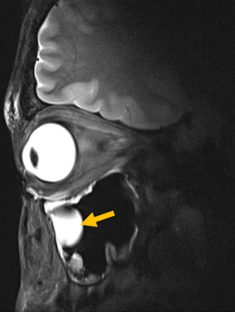
Research shows that petrolatum-based eye ointments can cause the device to swell and potentially rupture, prompting an urgent update to clinical guidance.
Widely-used eye ointments can cause glaucoma implants to swell and potentially rupture, according to new research from Nagoya University in Japan. This study is the first to show, using clinical and experimental evidence, that petrolatum-based eye ointments can compromise the PRESERFLO® MicroShunt, an implant used in over 60 countries to treat glaucoma.
Glaucoma is an eye disease that damages the optic nerve and can lead to vision loss. It often results from increased intraocular pressure caused by blocked drainage of eye fluid. A recent study estimated that 76 million people globally are affected by glaucoma.
Progression of visual field loss (from left to right) due to glaucoma (Credit: Ryo Tomita)
MicroShunt is a small filtration device implanted in the eye to improve fluid drainage in glaucoma patients. Compared to traditional surgeries, it lowers post-operative complications and reduces reliance on additional medications.
MicroShunt is made from a styrenic thermoplastic elastomer based on a polystyrene-block-polyisobutylene-block-polystyrene (SIBS) block polymer, which is highly biocompatible, flexible, and less likely to cause inflammation or scarring. However, this material is vulnerable when it comes into contact with hydrocarbon- and oil-based materials. Due to its high oil affinity, exposure to petrolatum-based eye ointments may allow oil components to penetrate the device, causing swelling and potential changes in its shape and flexibility.
The MicroShunt manufacturer’s instructions state that “the MicroShunt should not be subjected to direct contact with petrolatum-based (ie, petrolatum jelly) materials, such as ointments and dispersions.” But this precaution is not widely recognised or consistently followed in clinical practice.
“Swollen MicroShunts can be structurally fragile,” said ophthalmologist and Assistant Professor Ryo Tomita of Nagoya University Graduate School of Medicine, the study’s first author. “During surgery, I observed a rupture in a swollen MicroShunt. If more clinicians are aware of this risk, they will be able to prevent similar problems.”
The clinical study examined seven glaucoma patients whose MicroShunt implants were later removed for different reasons. The results revealed a clear pattern. In three cases, the MicroShunt was exposed outside the conjunctiva, and patients received a petrolatum-based eye ointment. All three explanted devices showed significant swelling, and two of them ruptured.
In three other cases, the MicroShunt remained covered by the conjunctiva, and no ointment was administered. These devices retained their original structure. Crucially, in one additional case, the MicroShunt was exposed outside the conjunctiva, but no ointment was applied. The device did not swell. This indicates that direct contact with the ointment, rather than conjunctival rupture alone, is the primary cause of swelling.
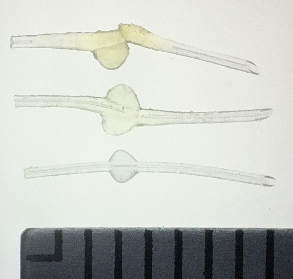
Photographic comparison of MicroShunt illustrating size changes Top: MicroShunt explanted from a patient, exhibiting diffuse swelling with fracture and loss of one fin Middle: MicroShunt explanted from another patient, showing localized swelling around the fin Bottom: Unused MicroShunt (control) Scale: 1 division = 1 mm (Credit: Ryo Tomita)
Laboratory confirmation
Laboratory experiments confirmed the clinical findings. The team immersed unused MicroShunts in petrolatum-based eye ointment to reproduce the swelling seen in clinical cases. Microscopic measurements showed significant changes. After 24 hours in the ointment, the MicroShunt’s outer diameter increased to 1.44 times its original size, and the fin-like portion widened to 1.29 times its initial value.
Chemical analysis identified the cause of this change. After 24 hours of immersion, oil components made up approximately 45% of the MicroShunt’s total weight, rising to 73% after three months. These results confirmed the primary cause of swelling to be the absorption of oil-based ointment constituents into the material.
Clinical implications
The research team emphasises that clinicians should avoid using petrolatum-based ointments on patients with MicroShunt implants, particularly when the device is exposed outside the conjunctiva. Alternative post-operative treatments should be considered, while further research is needed to assess whether swelling impacts MicroShunt performance even when rupture does not occur.
“Our study found that commonly used medical materials can cause unexpected complications if their chemical properties and usage environments are not fully understood,” Noro stated. “From both medical and engineering perspectives, we emphasise the importance of understanding the chemical properties of medical materials and appropriately managing their usage environments.”
Paper information:
Ryo Tomita, Taiga Inooka, Takato Kajita, Hideyuki Shimizu, Ayana Suzumura, Jun Takeuchi, Tsuyoshi Matsuno, Hidekazu Inami, Koji M. Nishiguchi, Atsushi Noro, and Kenya Yuki. (2026) Petrolatum-based ointment application induces swelling of the PRESERFLO MicroShunt. Graefe’s Archive for Clinical and Experimental Ophthalmology DOI: 10.1007/s00417-025-07075-2
A study conducted by University of Louvain (UCLouvain), published in Nature Communications, shows that part of the brain of babies born blind is permanently altered, while another part remains surprisingly intact. Babies’ brains are much more adaptable than previously thought: even if they cannot see at the very beginning of life, they can later learn to recognise the world around them.
Some babies are born with early blindness due to dense bilateral congenital cataracts, requiring surgery to restore their sight. This period of several months without vision can leave a lasting mark on how the brain processes visual details, but surprisingly little on the recognition of faces, objects, or words.
Using brain imaging, the researchers compared adults who had undergone surgery for congenital cataracts as babies with people born with normal vision. The results are striking: in people born with cataracts, the area of the brain that analyses small visual details (contours, contrasts, etc.) retains a lasting alteration from this early blindness. On the other hand, the more advanced regions of the visual brain, responsible for recognising faces, objects, and words, function almost normally. These “biological” results have been validated by computer models involving artificial neural networks. This distinction between altered and preserved areas of the brain paves the way for new treatments. In the future, clinicians may be able to offer visual therapies that are better tailored to each patient.
“Babies’ brains are much more adaptable than we thought,” explains Olivier Collignon, Professor at University of Louvain (UCLouvain). “Even if vision is lacking at the very beginning of life, the brain can adapt and learn to recognise the world around it even on the basis of degraded information.”
These findings also challenge the idea of a single “critical period” for visual development. Some areas of the brain are more vulnerable to early vision loss, while others retain a surprising capacity for recovery. “The brain is both fragile and resilient,” adds Olivier Collignon. “Early experiences matter, but they don’t determine everything.”
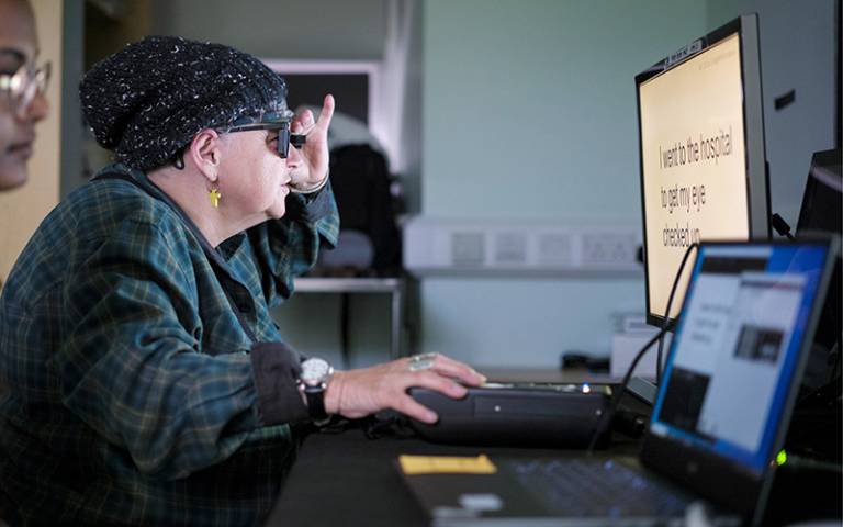
Study participant Sheila Irvine training with the device. Credit: Moorfields Eye Hospital
After being treated with an electronic eye implant paired with augmented-reality glasses, people with sight loss have recovered reading vision, reports a trial involving a UCL and Moorfields clinical researcher.
The results of the European clinical trial, published in The New England Journal of Medicine, showed 84% of participants were able to read letters, numbers and words using prosthetic vision through an eye that had previously lost its sight due to the untreatable progressive eye condition, geographic atrophy with dry age-related macular degeneration (AMD).
Those treated with the device could also read, on average, five lines of a vision chart; some participants could not even see the chart before their surgery.
The trial, with 38 patients in 17 hospital sites across five countries, was testing a pioneering device called PRIMA, with Moorfields Eye Hospital being the sole UK site. All patients had lost complete sight in their eye before receiving the implant.
Dry AMD is a slow deterioration of the cells of the macula over many years, as the light-sensitive retinal cells die off. For most people with dry AMD, they can experience a slight loss of central vision. Through a process known as geographic atrophy (GA), it can progress to full sight loss in the eye, as the cells die and the central macula melts away. There is currently no treatment for GA, which affects 5 million people globally. All participants in this trial had lost the central sight of the eye being tested, leaving only limited peripheral vision.
This revolutionary new implant is the first ever device to enable people to read letters, numbers and words through an eye that had lost its sight.
Mr Mahi Muqit, associate professor in the UCL Institute of Ophthalmology and senior vitreoretinal consultant at Moorfields Eye Hospital, who led the UK arm of the trial, said: “In the history of artificial vision, this represents a new era. Blind patients are actually able to have meaningful central vision restoration, which has never been done before.
“Getting back the ability to read is a major improvement in their quality of life, lifts their mood and helps to restore their confidence and independence. The PRIMA chip operation can safely be performed by any trained vitreoretinal surgeon in under two hours – that is key for allowing all blind patients to have access to this new medical therapy for GA in dry AMD.”
The procedure involves a vitrectomy, where the eye’s vitreous jelly is removed from between the lens and the retina, and the surgeon inserts the ultra-thin microchip, which is shaped like a SIM card and just 2mm x 2mm. This is inserted under the centre of a patient’s retina, by creating a trapdoor into which the chip is posted. The patient uses augmented-reality glasses, containing a video camera that is connected to a small computer, with a zoom feature, attached to their waistband.
Around a month or so after the operation, once the eye has settled, the new chip is activated. The video camera in the glasses projects the visual scene as an infra-red beam directly across the chip to activate the device. Artificial intelligence (AI) algorithms through the pocket computer process this information, which is then converted into an electrical signal. This signal passes through the retinal and optical nerve cells into the brain, where it is interpreted as vision. The patient uses their glasses to focus and scan across the main object in the projected image from the video camera, using the zoom feature to enlarge the text. Each patient goes through an intensive rehabilitation programme over several months to learn to interpret these signals and start reading again.
No significant decline in existing peripheral vison was observed in trial participants.
These findings pave the way for seeking approval to market this new device.
Sheila Irvine, one of Moorfields’ patients on the trial who was diagnosed with age-related macular degeneration, said: “I wanted to take part in research to help future generations, and my optician suggested I get in touch with Moorfields. Before receiving the implant, it was like having two black discs in my eyes, with the outside distorted.
“I was an avid bookworm, and I wanted that back. I was nervous, excited, all those things. There was no pain during the operation, but you’re still aware of what’s happening. It’s a new way of looking through your eyes, and it was dead exciting when I began seeing a letter. It’s not simple, learning to read again, but the more hours I put in, the more I pick up.
“The team at Moorfields has given me challenges, like ‘Look at your prescription’, which is always tiny. I like stretching myself, trying to look at the little writing on tins, doing crosswords.
“It’s made a big difference. Reading takes you into another world, I’m definitely more optimistic now.”
The global trial was led by Dr Frank Holz of the University of Bonn, with participants from the UK, France, Italy and the Netherlands.
The PRIMA System device used in this operation is being developed by Science Corporation (science.xyz), which develops brain-computer interfaces and neural engineering.
Mr Muqit added: “My feeling is that the door is open for medical devices in this area, because there is no treatment currently licensed for dry AMD – it doesn’t exist.
“I think it’s something that, in future, could be used to treat multiple eye conditions.”
More about the device:
The device is a novel wireless subretinal photovoltaic implant paired with specialised glasses that project near-infrared light to the implant, which acts like a miniature solar panel.
It is 30 micrometres/microns (0.03mm) thick, about half the thickness of a human hair.
A zoom feature gives patients the ability to magnify letters. It is implanted in the subretinal layer, under the retinal cells that have died. Until the glasses and waistband computer are turned on, the implant has no visual stimulus or signal to pass through to the brain.
In addition to practising their reading and attending regular training, patients on the trial were encouraged to explore ways of using the device. Sheila chose to learn to do puzzles and crosswords while one of the French patients used them to help navigate the Paris Metro – both tasks being more complex than reading alone.