Navigating patient confidentiality, social media and professional boundaries
Photo by National Cancer Institute on Unsplash
Thursday 13th August 2026
18:00 – 19:45
Earn 2 ethics CPD points
During this webinar, the HPCSA Booklet 5: Confidentiality – Protecting and Providing Information and HPCSA Booklet 16: Ethical Guidelines on social media will be explored from a South African legal and ethical practice perspective. The webinar will offer insights into the complexities of digital communication, including WhatsApp and social media use, consent, online reviews and cybersecurity, while focusing on protecting patient confidentiality and public trust across all forms of communication.
The audience will have an opportunity to listen and engage with clinical, legal and medicolegal subject matter experts. During the webinar, a range of learning opportunities will be offered including short lectures, interactive case studies, audience polling and Q&A.
This webinar will focus on healthcare practitioners engaging in digital communication with patients and colleagues. Administrative staff working in these practices are welcome to join the discussion.
Joining us as panellists will be Emma Sadleir, South Africa’s leading expert on social media law, and Dr Isabel do Vale, a practising medical practitioner and President-elect of APRASSA. Attendance will qualify for 2 Ethics CPD points and EthiQal Recognition Programme points.
Taking antiretroviral therapy as recommended has expanded the lifespan of people with HIV. (Photo: Unsplash)
By Elna Schütz for Spotlight
South Africa’s first set of clinical guidelines focused on older people living with HIV has been released. They offer practical steps in a resource-strained health system to take care of an ageing patient population.
The guidelines are particularly important in South Africa since the country has an ageing population of people living with HIV. Many of these people would only have started treatment relatively long after they contracted the virus, largely because of the government’s reluctance to make antiretroviral treatment available in the early 2000s. The sooner people start treatment after infection, the better their long-term prognosis tends to be.
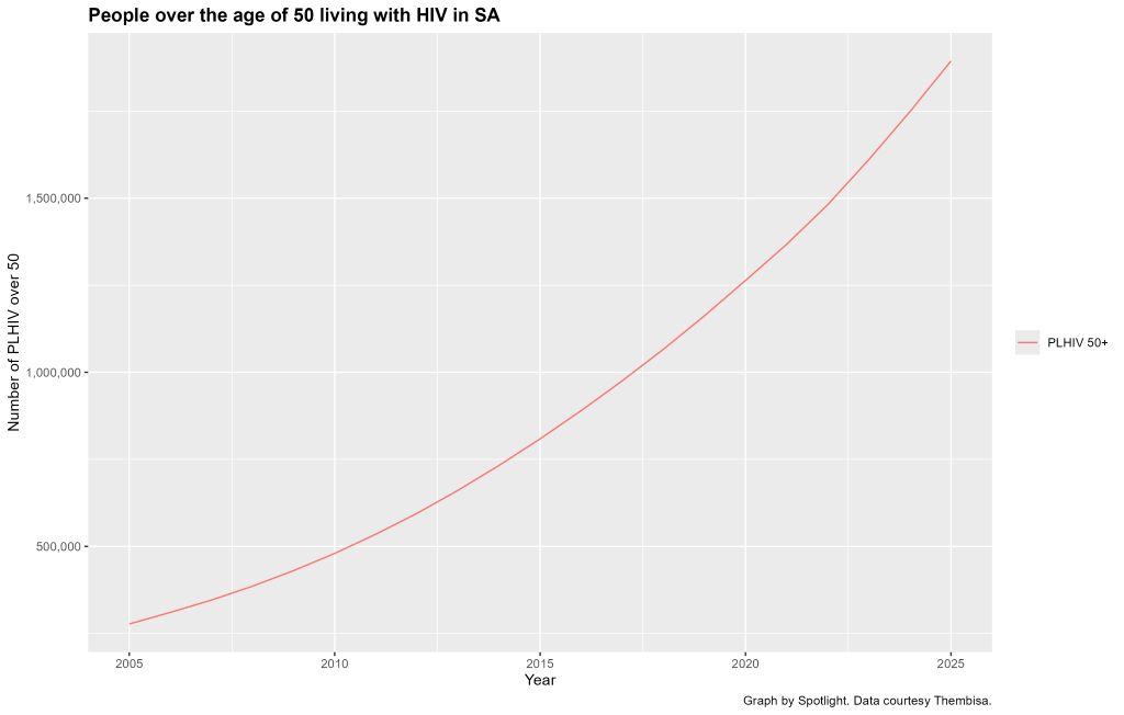
In 2025, there were around 1.9 million people over the age of 50 living with HIV in South Africa, according to Thembisa, the leading mathematical model of HIV in the country. This is 24% of the estimated 7.9 million HIV positive people in the country. The 1.9 million figure is more than double the 800 000 people over 50 who were living with HIV in 2015. This number is projected to rise to over 3.6 million by 2035.
Most people over the age of 50 who are living with HIV contracted the virus before they turned 50. The increase depicted in this graph is thus mainly a function of people who are already living with HIV ageing into the over 50 age group. Some people over 50 do become newly infected with HIV, but those numbers are comparatively small.
The changing make-up of the population of people living with HIV, coupled with the fact that antiretroviral therapy has been crucial for clearing and suppressing HIV in the body was a core driver for developing the new guidelines, Dr Camilla Wattrus, the Clinical Director at the Southern African HIV Clinicians Society, tells Spotlight. She is one of the guidelines’ authors.
“Antiretroviral therapy has expanded the lifespan of people with HIV, but we must now also consider how to preserve the ‘health span’ in this group,” says Wattrus.
She explains that this means increasing the years that are spent in good health with a good quality of life.
Another co-author of the guidelines, Nomathemba Chandiwana, Chief Scientific Officer at the Desmond Tutu Health Foundation, points out that after antiretroviral treatment was introduced in South Africa, the life expectancy of people living with HIV increased dramatically. “We didn’t think people would live as long as they have now, so that’s been a big success,” she says. “But now we have new problems.”
Chandiwana says that older people living with HIV have around 16 fewer years in good health than people without HIV. The 16-year figure (technically 15.3) seems to originate in a study published in 2020 in the JAMA medical journal that compared the health and lifespans of insured people with and without HIV in the United States. For people with HIV who started antiretroviral treatment when they were still healthy (CD4 countes above 500), the difference in healthy years was 9.5 years.
Another broad concern is that clinicians may be focused on HIV-related issues like viral suppression for these patients and not be sufficiently aware of other ageing-related developments. People with HIV get the same ageing related diseases as other people, but there is evidence that they tend to get them earlier.
We know from Thembisa model outputs that on average, people living with HIV today are slightly more likely to die of non-HIV-related causes than AIDS. According to the model, there were 53 000 HIV-related deaths in the year from mid-2024 to mid-2025. This is a thousand fewer than the 54 000 people with HIV who died of non-HIV-related causes over the same period.
What is in the new guidelines
The new guidance states that it is designed to:
Raise healthcare workers’ awareness of the needs and concerns of the population of people living with HIV who are 50 years and older.
Inform healthcare workers about an ageing-related approach to older people with HIV.
Highlight good practices to help healthcare workers provide optimal care for this population.
Provide resources about ageing with HIV for healthcare workers, their patients and their patients’ carers.
Guide clinical settings in implementing geriatric care into HIV clinical practice.
The clinical advice in the guidelines follow the World Health Organisation’s (WHO) principles for Integrated Care for Older People (ICOPE), which emphasises prevention prior to frailty, person-centred assessment, and the involvement of healthcare workers other than doctors.
The guidelines cover a thorough list of challenges faced by older people with HIV that need to be monitored and addressed. For instance, physiologically, there is a risk of comorbid conditions like hypertension and cancer, and an increased risk of complications from polypharmacy, when more than five medicines are used concurrently. Social and behavioural challenges include that older people are perceived to be less likely to get infected with HIV and therefore have lower rates of HIV testing and use of HIV prevention tools.
This population is also at risk of being disregarded or not fully cared for in the healthcare system. The guidelines give examples such as restricted mobility access to health facilities and healthcare workers being unaware of the HIV-related risks in older people. “The health system needs to be equipped to manage their needs in a holistic and integrated way, and that is what this guideline aims to support,” says Wattrus.
The guidelines include a comprehensive schedule of what need to be assessed and screened and at what regularity. There is a particular focus on geriatric syndromes like frailty, cognitive impairment, and managing comorbid non-communicable diseases.
“The idea is that every visit with an older patient involves more than just routine HIV care and that it becomes a conversation about how that person is functioning and living,” says Wattrus.
The guidelines also emphasise how care can be offered by a variety of healthcare providers, depending on the resources available. “Recommendations enable task-shifting, which is a practical necessity in a country where specialists such as geriatricians are scarce, and the bulk of HIV routine care is delivered by healthcare workers at primary care level,” says Wattrus.
Even though the guidelines focus on overall health in older people living with HIV, managing HIV is, of course, a part of this. It cautions that “CD4 recovery may be slower and blunter compared to younger individuals,” but viral suppression is still the primary treatment goal.
The crucial factor here is to choose the correct antiretroviral treatment regimen for the patient. For instance, popular tenofovir disoproxil fumarate (TDF) combinations should be avoided in people at risk of or with osteoporosis, bone fractures, or renal impairment. Regimens with tenofovir-alafenamide or abacavir may be better, though the latter is contraindicated if there is high cardiovascular risk.
The new local guidelines hit largely the same notes as a major commission on HIV and ageing that was published by the journal Lancet HIV to coincide with the AIDS 2026 conference taking place in Rio de Janeiro, Brazil.
“Supporting healthy ageing requires more than sustained viral suppression; it requires care that is informed by what matters most to the individual, with attention to maintaining physical and mental function, minimising healthcare complexity, and addressing multimorbidity, polypharmacy, stigma, and social determinants of health,” the commission found.
Simple systems, big change
Apart from giving healthcare workers a framework for giving better care to older people living with HIV, the guidelines advise how this larger change in the health system can happen for this growing older population. “What is great is that most of the recommendations are not complicated or expensive,” says Wattrus.
She explains that the sensitisation and training of healthcare workers, especially in primary care, is a crucial first step. If they know how to, they can easily incorporate brief screenings, such as those for frailty, into normal appointments. For example, as Chandiwana points out, several geriatric tests need only a chair for the patient to sit down on and get up from. She says it is easier to do these things for people with HIV during their existing appointments, compared to people without HIV who may not be visiting health facilities for regular screenings.
Another relatively easy adaptation is to simply make healthcare services easier to access. “This can be done by having appointments aligned across conditions, fewer unnecessary referrals and genuine attention to broader aspects of their health such as poverty, isolation and limited mobility,” she says.
Chandiwana also suggests that one could consider rolling out geriatric care health cards to track screening, as is often done with children. She would also like to see more community buy-in, in a similar way as there was during the earlier part of the HIV treatment roll-out. For instance, she suggests community health clubs and increased health literacy efforts around ageing.
Avoiding problematic polypharmacy, says Wattrus, is another low-cost, high-yield strategy that does not require specialist input. “Routinely reviewing medication lists, identifying unnecessary drugs, and checking for interactions is straightforward and can make a significant difference,” she says.
More specialists would of course help. Chandiwana says there are fewer than 50 geriatric specialists in the country. She says there is also a much wider need for geriatric-specific training across the healthcare system, including for primary care nurses and community healthcare workers.
Lastly, Chandiwana says the guidelines offer a much-needed look into the unique challenges and needs of older people with HIV as an opportunity for the government to act to prevent a future problem. “So that investment in having scalable, simple systems for people who are ageing, both with HIV and without, I think, would be fantastic, but that needs money,” she says.
Mayo Clinic field trial shows hospitals can reduce emergency department overcrowding using a simple patient-routing checklist based on information already collected at triage.
Photo by Camilo Jimenez on Unsplash
Emergency departments face a common challenge: how to reduce long wait times without adding more beds, hiring more staff or expanding facilities. New research suggests one answer may be hiding in plain sight – making better decisions about where patients receive care.
A new study published in the INFORMS journal Management Science, entitled, “Vertical Patient Streaming in Emergency Departments,” found that a simple, data-driven triage protocol reduced emergency department length of stay by 11 minutes without compromising patient safety or requiring additional resources.
Researchers from Harvard University, Oxford University and Mayo Clinic developed an evidence-based protocol to identify patients who could safely receive care in a seated treatment area – known as a vertical processing pathway – instead of occupying a traditional emergency department bed.
Although many emergency departments already have these areas, decisions about who should be treated there are often made on an ad hoc basis. The researchers sought to replace that variability with a standardised, easy-to-use protocol.
The team first analysed nearly 50 000 emergency department visits at Mayo Clinic Arizona to develop a machine learning model that predicts, using only information collected during triage, whether a patient will ultimately require an emergency department bed. They then combined those predictions with mathematical models of patient flow to determine the most efficient routing strategy before translating the results into a straightforward decision tree that clinicians could implement without new software or changes to hospital IT systems.
To evaluate the approach in practice, the researchers conducted a 13-week prospective field trial involving 11 015 patients at Mayo Clinic Arizona’s new emergency department.
The results demonstrated measurable improvements in efficiency:
11-minute (4.2%) reduction in total emergency department length of stay.
Eight-minute (4.5%) reduction in time from arrival to clinical disposition.
No increase in 72-hour return visits, indicating patient care quality was maintained.
“Our goal wasn’t to add technology to the emergency department,” said Arshya Feizi, lead author of the study and a researcher at Harvard University. “It was to give clinicians a practical, evidence-based way to decide which patients can safely receive care without occupying one of the department’s limited beds.”
The protocol relies only on information hospitals already collect during triage, including a patient’s Emergency Severity Index score, presenting complaint and whether the department is operating over capacity. Because it requires no additional staff, equipment or software integration, the researchers say it could be implemented quickly in many emergency departments.
“Our findings show that improving patient flow doesn’t always require expanding capacity,” said Soroush Saghafian, co-author of the study and professor at Harvard University. “Sometimes the greatest opportunity comes from using existing resources more intelligently.”
The researchers estimate that a medium-sized emergency department treating approximately 40 000 patients annually could recover nearly 6800 bed-hours each year – enough capacity to care for roughly 2000 additional patients while potentially generating approximately $3 million in additional reimbursement, all without expanding facilities or increasing staffing.
Emergency department overcrowding has challenged hospitals for decades, contributing to treatment delays, patient dissatisfaction, clinician burnout and higher healthcare costs. The researchers believe their findings demonstrate that operational improvements grounded in analytics and implemented through simple clinical protocols can produce meaningful gains without sacrificing quality of care.
Cerebrospinal fluid (CSF) protects the central nervous system (CNS). Credit: Scientific Animations Wiki CC-BY 4.0
Deep inside your brain, a clear liquid is constantly on the move. Cerebrospinal fluid (CSF) cushions the brain, delivers nutrients, removes waste and keeps pressure stable. Think of it as an internal tide, circulating through cavities in the brain and around the spinal cord to keep this delicate organ in balance. When that flow is disrupted, the consequences can be serious.
For decades, doctors have relied mainly on static brain scans to guide treatment. But structure tells only part of the story. Researchers at the University of Pretoria (UP) are now focusing on something more dynamic: how fluid actually moves.
Two of the most common neurosurgical conditions worldwide – brain tumours and hydrocephalus (a dangerous build-up of fluid in the brain) – are closely tied to disturbed CSF circulation. Tumours can block or distort the pathways through which fluid moves. Hydrocephalus represents a more obvious breakdown, where fluid accumulates and pressure rises. In both cases, symptoms such as headaches, problems with vision and neurological decline are not simply caused by the presence of disease, but by changes in pressure and pulsating flow inside the skull.
Professor Llewellyn Padayachy, Head of the Department of Neurosurgery at BTC@UP explains: “The optic nerve, which connects the eye to the brain, is surrounded by the same protective layers as the brain itself. CSF flows along this nerve, meaning changes in brain pressure can subtly affect structures at the back of the eye. By using advanced, non-invasive eye imaging, researchers can detect signs of altered fluid flow and pressure without inserting monitors into the brain.”
This matters enormously for children with hydrocephalus and patients with brain tumours who require long-term monitoring. It offers a safer, repeatable way to track disease progression and treatment response. In low- and middle-income countries, where hydrocephalus is common but access to advanced imaging and neurosurgical infrastructure may be limited, such non-invasive tools could reduce reliance on costly technology while still delivering meaningful clinical insight.
The research also helps refine innovation. Modern shunts and endoscopic procedures increasingly aim to restore more natural fluid circulation rather than simply drain excess fluid. Objective eye-based markers provide measurable ways to evaluate whether these technologies truly improve flow.
While the link between the eye and brain pressure has long been recognised, what is new is the integration of advanced imaging, physiological modelling and continuous monitoring. This approach treats CSF flow as a living system, and shifts care from reacting to late damage towards detecting subtle change earlier.
Why this research matters
This work reframes brain disease through a simple but powerful idea: health depends on flow. By learning to read the movements of brain fluid, even through the eye, researchers are paving the way for safer monitoring, smarter surgery and more equitable neurological care worldwide.
Fast fact
The most common surgical treatment for hydrocephalus is the surgical placement of a shunt, which has one of the highest failure rates of any medical device on the market.
Adding oral antihistamines to existing eczema treatments is unlikely to lead to clinically important reductions in eczema and itch severity, and may not reduce sleep disturbance or flare-ups, finds a review of the latest trial evidence published by The BMJ today.
Some antihistamines may also increase side effects such as drowsiness and the risk of patients stopping treatment.
The researchers say this review addresses longstanding uncertainty surrounding the role of antihistamines in treating eczema, and the results do not support their use in routine eczema management.
Atopic dermatitis, commonly known as eczema, is a chronic condition caused by an overactive immune system that leads to dry, inflamed, and intensely itchy skin. It also often impairs quality of life, mental health, and social relationships.
Oral antihistamines are one of the most commonly used drugs for managing eczema, with an estimated one in five UK patients and nearly half of patients in the US using them. Yet despite their widespread use, evidence-based assessments of their benefits and harms in treating eczema remain inconclusive.
To address this uncertainty, researchers reviewed the results of 47 randomised controlled trials involving 6,230 children and adults (average age 20 years; 52% female) with mainly moderate to severe eczema.
The trials compared the effects of adding oral H1 antihistamines, H2 blockers, mast cell stabilizers, or their combinations to placebo (with or without background moisturisers or steroid creams) on eczema severity, itch severity, sleep disturbance, flare-ups and quality of life, as well as harms such as sedation and drowsiness.
The trials were of varying quality, but the researchers were able to assess their risk of bias and certainty of evidence using established tools.
The results show that compared with placebo, H1 antihistamines likely result in a small but clinically unimportant reduction in eczema severity and itch severity and may not reduce sleep disturbance or flare-ups.
First generation (sedating) antihistamines also probably increase cognitive impairment such as sedation and drowsiness, and may increase the risk of stopping treatment due to side effects.
The researchers acknowledge several limitations related to the nature of the included trials, but say these were addressed using standardised and systematic approaches.
As such, they conclude: “This systematic review and network meta-analysis addresses the longstanding uncertainty surrounding the role of antihistamines in treating atopic dermatitis, showing they likely do not meaningfully improve patient important outcomes while probably increasing harms.”
They add: “Our findings do not support their use in routine atopic dermatitis management. We have provided a foundation for an evidence based change in practice, supporting the development of updated atopic dermatitis guidelines that prioritise efficacy, safety, and patient centred care.”
Cardiovascular risk factors are linked to higher risk of heart disease, but it has been unknown how they are related to abrupt closure of the coronary arteries leading to heart attack and sudden cardiac death. A new study by Mass General Brigham Heart and Vascular Institute investigators finds that people with higher modifiable risk factors burden such as high blood pressure, high cholesterol, diabetes, smoking and obesity have more widespread buildup of plaques in major coronary arteries. Furthermore, these modifiable factors were associated with more unstable, high-risk plaques that are prone to rupture and cause heart attacks. Results are published in JACC: Advances.
“The higher the number of vulnerable, or rupture-prone plaques, the greater the chance that one of them will trigger an adverse event,” said senior author Ik-Kyung Jang, MD, PhD, at the Mass General Brigham Heart and Vascular Institute. “Our findings highlight the importance of early, intensive and sustained interventions to control the modifiable risk factors and prevent future events.”
To better understand how modifiable risk factors and non-modifiable risk factors (including age, sex and family history of heart disease) influence dangerous plaque buildup, the research team examined plaque burden and plaque characteristics across all three major coronary arteries. They analysed 534 plaques in 131 patients who underwent optical coherence tomography (OCT), an intravascular imaging modality that can visualise plaques at high resolution.
The researchers reported that patients with more risk factors had more plaques across all three coronary arteries. In addition, greater number of modifiable risk factors was associated with fewer stable plaques and more prevalent vulnerable plaque features such as lipid plaques with thin fibrous caps that are susceptible to disruption. In contrast, non-modifiable risk factors were associated with more stable plaque phenotypes.
The authors note that prospective research involving larger cohorts, including patients who have previously been treated for blocked coronary arteries, is needed to generalise their findings.
As South Africans face rising inflation, pressure from the burden of chronic disease and financial strain, Momentum Health is advocating for a stronger national focus on the prevention dividend: the long-term value created when South Africans use preventative healthcare, wellness programmes, screenings, health assessments and digital tools to manage health risks before they become serious and costly.
This comes as households, employers and the healthcare system face increasing pressure from affordability constraints, chronic disease and mental health challenges. Research referenced by Stellenbosch Business School has estimated that health-related productivity losses cost South Africa around R161 billion a year, driven by absenteeism, presenteeism and untreated mental health challenges.
Momentum Health believes the conversation needs to move beyond healthcare as an expense that pays only when people are ill. Instead, medical aid should be seen as an investment in helping people stay healthier for longer.
“Rather than paying claims after someone becomes ill, modern healthcare needs to focus on helping people understand their risks earlier, take action sooner and build healthier habits that can improve quality of life over time,” says Damian McHugh, Chief Marketing Officer at Momentum Health. “Prevention is one of the most practical ways to support both better health outcomes and greater financial resilience for South Africans.”
Prevention is becoming more urgent
South Africa’s health and economic realities make prevention increasingly important. PwC’s South Africa Economic Outlook notes that South Africa’s real GDP per capita declined by a cumulative 4.2% over the past decade, a signal often associated with pressure on living standards and economic stability.
At the same time, the country continues to carry a significant disease burden. Government and World Health Organisation reporting show that South Africa has made progress in controlling tuberculosis, with TB cases declining significantly since 2015 and treatment coverage improving to 79% in 2023. However, more than half of TB-affected households still face catastrophic costs, showing how illness can deepen financial vulnerability when people are diagnosed late or struggle to access care.
Mental health is also placing pressure on individuals, employers and families. SADAG’s Working Life Survey found that 52% of surveyed employees had been diagnosed with a mental health condition, with depression, stress, anxiety and burnout among the commonly reported conditions.
These figures show why prevention must be personal, practical and accessible,” adds McHugh. “Every member’s health risks are different. The more people know about their own health, the easier it becomes to make informed choices, use the right benefits and take action before problems escalate.”
Healthy habits can create long-term value
Regular health checks, screenings, physical activity, digital healthcare tools and ongoing health management can help members detect risks earlier and make healthier decisions more consistently.
Preventative care can also support employers and the wider economy. When people are healthier, they are more likely to be present, productive and able to participate fully at work and in their communities. This matters in a country where affordability pressures mean many people are looking for healthcare solutions that deliver tangible value, not only financial protection after illness occurs.
Behavioural incentives can play an important role in this shift. When healthy choices are recognised and rewarded, members are more likely to build routines that support their physical, mental and financial wellbeing. This is the principle behind Momentum’s broader health and wellness approach- making it easier for members to take control of their health before problems arise.
Prevention is personal
The next phase of healthcare will be increasingly personalised. Data, health assessments and digital tools can help members better understand their individual health risks and choose the most relevant actions, whether that means completing a screening, increasing physical activity, accessing care digitally or managing a chronic condition more proactively.
“Prevention works best when it is simple, personalised and part of everyday life,” says McHugh. “Our role is to help members connect the dots between their daily habits, their health outcomes and their financial wellbeing. That is how healthcare becomes more than a monthly contribution. It becomes an investment in a healthier future.”
If an HIV prevention pill that provides a month of protection at a time performs well in two ongoing clinical trials, it could become the next big thing in HIV prevention after the lenacapavir injection. A new licensing agreement is paving the way for South Africa’s Aspen Pharmacare to produce the pill should the study findings be positive and the drug be registered.
In June, South Africa’s health department started rolling out the six-monthly lenacapavir HIV prevention injection to around 10% of public sector clinics. While the rollout of this jab still has a long way to go, the next generation of HIV prevention products is already on the horizon.
Two of those new products stand out. One is a new formulation of lenacapavir that looks as if it can provide 12 months of protection at a time. While results so far are promising, the pivotal data on this once-yearly HIV prevention jab is only expected in a year or two.
The other product that has many people in the HIV world excited is a monthly HIV prevention pill that contains a highly potent antiretroviral medicine called alimatravir (it was previously called MK-8527). It starts working within around an hour after someone takes it and appears to provide a month of protection at a time. One benefit of the pill, compared to the lenacapavir injection, is that it would be easier to distribute at scale, given that there is no need for a nurse to administer an injection.
As Spotlight reported in some depth last year, alimatravir looked very promising in a phase 2 study, although for now the jury is still out on the drug’s safety and efficacy. It is currently being evaluated in two pivotal phase 3 clinical trials called EXPRESSIVE-10 and EXPRESSIVE-11. Both these studies started last year and are expected to be completed by around mid-to-late 2027. Medicines are typically only registered for use after positive results in such phase 3 studies.
“The monthly pill offers an alternative for people who would like a long acting, less frequently dosed PrEP but not needle friendly … so it really is about giving more options especially on the pill side,” Professor Linda-Gail Bekker, primary investigator in South Africa on the EXPRESSIVE-10 study, told Spotlight. “You could imagine that just having to remember to take a small easy to swallow pill on the day you pay your bills monthly could be very easy for people. We understand that the packaging is also going to be very user friendly- looking more like a gum packet than a bottle of antiretroviral pills which may also reduce stigma.” (PrEP refers to pre-exposure prophylaxis like HIV prevention pills or injections.)
Licence to make generics
The prospects for future access to alimatravir got a major boost last week when the pharmaceutical company Merck (known as MSD outside of the United States and Canada) announced that it granted licences to seven different companies to produce generic versions of the monthly pill. One of the seven companies is South Africa’s Aspen Pharmacare. The others are Uganda’s Quality Chemical Industries Limited, Kenya’s Universal Corporation Ltd, and Aurobindo, Cipla, Emcure and Viatris in India.
Early responses to the licences have mostly been positive.
Having a generic company with a licence in South Africa is excellent news, said Bekker.
“It is particularly exciting to see manufacturers in Kenya, South Africa and Uganda included in these licenses,” Mitchell Warren, Executive Director of AVAC (a global HIV advocacy group), told Spotlight by e-mail. “These are the first generic PrEP licenses in East and Southern Africa, meaning manufacturing can happen where trials are happening, where need is greatest and where we have the largest PrEP markets.”
“Through our agreement with MSD (Merck), we have the opportunity to support the future supply of an innovative HIV prevention option while strengthening local pharmaceutical manufacturing and healthcare resilience across the continent,” Stephen Saad, Aspen Group Chief Executive, said in a media statement. Under the agreement, the company says it will receive a technical package from Merck, together with licensing rights covering 129 countries, including all African countries.
Speaking to Spotlight, Stavros Nicolaou, Aspen’s Head of Strategic Trade, described alimatravir as “ground-breaking and a potential game-changer”. He commended Merk for starting the licensing process so early. He framed the licence as an important step forward for both South Africa’s HIV response and for local production of antiretrovirals, although he also raised concerns about the procurement of locally manufactured antiretrovirals – the percentage of South Africa’s antiretroviral tenders awarded to local manufacturers has been trending downward.
According to earlier reporting by Business Day, Nicolaou has declined to give any indication as to a potential price for the pill, but he did tell the publication that they could potentially supply it for both South Africa’s public and private sectors.
There are indications that a relatively low price is on the cards. Research being presented at AIDS 2026 this week found that alimatravir could be mass produced and sold at a profit for as little as $15 (around R250 to R300) per person per year. This is less than half the $40 per person per year that South Africa is expected to pay for generic lenacapavir injections in a year or two from now.
“Merck expects to provide initial supply and continue supplying product as needed while licensed generic manufacturers complete development, obtain the necessary regulatory approvals and prepare to provide supply in the licensed territories. The goal is to help avoid delays in access by providing an initial supply pathway until generic manufacturing capacity is established and brought online,” the company said in a media statement.
Earlier licensing
The timing of the licensing announcement is somewhat unusual – such announcements are typically only made after phase 3 trials have been concluded and it is confirmed that the drug is safe and effective.
“Granting licensing agreements to generic manufacturers while clinical trials are still enrolling, before it is known if the product is effective, should significantly reduce the time to market for the product,” Warren said in an earlier AVAC media statement. “The timeline announced today gives us ample opportunity to work with ministries of health, donors, communities, and Merck to plan for broad access to the monthly PrEP pill.”
Warren told Spotlight that the small amount of active drug in alimatravir and the fact that it is an oral dose should make the technology transfer from Merck to generics quite quick. “The hope would be that genetic alimatravir reaches the market within months of the approval of the originator, compared to more than a year for lenacapavir,” he said.
Nicolaou was also upbeat about how quickly things are unfolding. He said that Merck’s decision to execute licences while the phase 3 clinical trials are ongoing allows for an earlier registration pathway (if phase 3 findings are positive, alimatravir will have to be filed for registration with regulators like the South African Health Products Regulatory Authority). He also pointed out that it is a small tablet and that it should be easier to manufacture than HIV prevention injections.
Nicolaou told Spotlight that the plan is for Aspen to do formulation of alimatravir in South Africa, but that they are not currently planning to produce the active pharmaceutical ingredient – this will likely be sourced from Chinese or Indian suppliers.
Some activist criticism
But while the timing has generally been welcomed, there has also been some criticism over the licenses.
A statement from activist group HealthGap points out that Latin American countries like Brazil, Argentina, and Colombia are not included in the list of 129 countries covered by the license, even though some of the phase 3 trial sites for alimatravir are in these countries. The HealthGap statement calls for compulsory licenses to be issued.
In an earlier statement, Merck said that, in recognition of the significant unmet need in Latin America, “Merck is in active discussions with organizations, including Fiocruz (a key player in medicines production and procurement in Brazil), with a goal to enable rapid availability and broad supply of alimatravir in the region”.
Disclosure: The Gates Foundation has provided financial support for clinical trials of alimatravir. Spotlight receives funding from the Gates Foundation, but is editorially independent – an independence the editors guard jealously. Spotlight is a member of the South African Press Council.
An international research team led by Prof. Markus Ege of LMU University Hospital has, for the first time, identified which bacteria in barn air are responsible for the so-called “farm effect,” which can protect against allergies, asthma, and hay fever. Photo by Christopher Stites on Unsplash
An international research team has identified for the first time which bacteria in barn air are responsible for the ‘farm effect’ that can protect against allergies, asthma, and hay fever.
Children who grow up in a farm environment are less prone to allergies, asthma, and hay fever than their classmates. This so-called farm effect has been identified in several observational studies worldwide. It is most likely attributable to the fact that various bacteria present in barn air prevent excessive inflammatory responses of the immune system that are characteristic of such diseases. But which bacteria exactly?
Now, an international team led by Professor Markus Ege from the Dr. von Hauner Children’s Hospital at LMU University Hospital and the Institute of Asthma and Allergy Prevention at Helmholtz Munich has answered this question. The researchers have shown for the first time which specific bacteria in barn air trigger the farm effect, which substances within those bacteria mediate the protection, and which receptors in the body they bind to. Their findings have been published in The New England Journal of Medicine – Evidence.
For years we have been hearing about the hygiene hypothesis, which was first proposed in 1989. This came after three decades of dramatic increases in allergies, asthma, and hay fever among children in Western industrialised countries. These are all conditions in which the immune system mounts an exaggerated inflammatory response and mistakenly attacks the body’s own tissues. According to the hygiene hypothesis, this happens because of under-stimulation in early childhood. The immune systems of children who encounter too few environmental microbes and common cold viruses are more likely to malfunction. “Girls and boys who grow up on farms and are exposed to a wider variety of microbes have the problem far less often,” says Ege. As their immune systems constantly contend with bacterial ‘sparring partners’ from barn air, they are trained to avoid excessive inflammatory responses.
However, the hygiene hypothesis has not been definitively proven, as it is largely based on observational studies. “This kind of research can only show more or less convincing correlations,” says epidemiologist Ege. “But with our new study, we can make a much stronger case, because we can identify the individual links in the proposed causal chain: the bacteria, the relevant microbial metabolic products, and the human receptors.”
The Approach
The researchers analysed data from more than 1000 children participating in European studies in rural areas. For all the children, girls and boys, two types of samples had been collected: nasal swabs and mattress dust. In addition, dust samples were taken from cowsheds for 47 farm children. The team examined which bacteria and fungi were present in these samples, how the different microorganisms were related, and whether specific microbial groups protected the children against asthma.
To this end, the epidemiologists and bioinformaticians employed state-of-the-art genetic and metabolic analyses and developed computer models. Through a process of elimination, they eventually identified the key microorganisms. They also investigated whether the children’s own genes influenced this protective effect. To validate their findings, the researchers additionally used data from France and Finland.
The Results
We identified a small number of bacteria, which we can now pinpoint down to the species level,” explains Giulia Pagani, first author of the study and bioinformatician at the Institute of Asthma and Allergy Prevention at Helmholtz Munich, “such as Romboutsia timonensis and Glutamicibacter arilaitensis. These gram-positive bacteria together mediate two-thirds of the entire farm effect for asthma protection and half of the effect for hay fever and atopic eczema.” The bacteria originate in the cow’s digestive tract, where they produce or metabolize substances like kynurenine, xanthine, alpha-linolenic acid, and stearidonic acid. These compounds are easily inhaled and recognized by two receptors on human airway cells – AhR and PPARγ. “These receptors,” Ege continues, “have multiple functions, including in the immune system, where they appear to prevent excessive inflammatory responses.” Their role in the farm effect was previously unknown. For the first time, therefore, the complete biological chain is visible: cow → barn air → bacteria → metabolic products → human receptors → protection against asthma.
The new findings can now be used by laboratory researchers to decipher the molecular and cellular mechanisms of the farm effect. This opens up the prospect of developing a drug that mimics the farm effect – without the need for children to spend time in barns.
Original publication
G. Pagani et al.: Gram-Positive Bacteria and the Inverse Association between Farm Exposure and Childhood Asthma, NEJM Evidence
Researchers from the Wits Vaccines and Infectious Diseases Analytics (Wits VIDA) Research Unit at the University of the Witwatersrand (Wits) have played a leading role in a landmark international phase 1 / 2 clinical trial demonstrating the safety and immunogenicity of an investigational maternal vaccine against Group B Streptococcus (GBS) – a major cause of life-threatening infections in newborn babies worldwide. The findings have been published in Nature Medicine.
The study evaluated Pfizer’s investigational hexavalent Group B Streptococcus conjugate vaccine candidate (GBS6) in both non-pregnant women and pregnant women, together with their infants. The trial found that the vaccine was generally well tolerated, generated robust immune responses against all six clinically important GBS serotypes, and resulted in efficient transfer of GBS-specific antibodies from vaccinated mothers to their babies.
Wits VIDA was central to the implementation and successful conduct of the clinical trial. South Africa was the primary recruiting country, contributing the overwhelming majority of study participants across both the early and later stages of the trial, reflecting the country’s internationally recognised expertise in maternal and infant vaccine research.
Group B Streptococcus is one of the leading causes of neonatal sepsis, meningitis and pneumonia, accounting for approximately 300 000 serious infections, including 100 000 deaths, in young infants globally each year. The greatest burden of GBS disease in infants occurs in Africa and other low- and middle-income countries, with South Africa reporting among the highest incidence globally (approximately 2-3 cases for every 1000 births). Also, maternal GBS infection could predispose to preterm labour and stillbirths. Current prevention strategies based on intrapartum antibiotics have important limitations, particularly in resource-constrained settings, and do not protect against late-onset disease. Consequently, the WHO has designated a vaccine against GBS, to be given to women during pregnancy aimed at protecting their young infant, as a priority vaccine for development.
Professor Shabir Madhi, Director of Wits VIDA and one of the study’s senior investigators, said: “This publication represents another important milestone in the decades-long effort to develop an effective maternal vaccine against Group B Streptococcus. Maternal immunisation offers the opportunity to protect newborns during the most vulnerable first months of life, when they are at greatest risk of severe infection. These encouraging findings provide further evidence supporting continued development of this vaccine.”
The trial demonstrated comparable safety outcomes between vaccine and placebo recipients. Among pregnant participants, rates of adverse events, serious adverse events and delivery outcomes were similar between the two groups. Infants born to vaccinated mothers likewise had similar safety outcomes to those born to mothers receiving placebo. Importantly, vaccinated mothers developed high concentrations of antibodies that crossed the placenta efficiently, resulting in robust antibody levels in their infants at birth.
The publication builds on Wits VIDA’s longstanding leadership in vaccine research, which has contributed to the development and evaluation of vaccines against respiratory syncytial virus (RSV), rotavirus, pneumococcal disease, influenza virus, COVID-19 and now Group B Streptococcus. The unit continues to play a pivotal role in generating evidence that informs global maternal and child health policy.
The study also highlights the importance of South Africa’s clinical research infrastructure and its contribution to advancing vaccines designed to address diseases that disproportionately affect African populations.
While additional studies are required before the vaccine can be licensed, the results provide strong support for continued clinical development of maternal GBS vaccination as a strategy to reduce newborn deaths and severe infections worldwide.
Wits is currently enrolling in a phase 3 trial for GBS6 (NCT: NCT07160244).
About Wits VIDA
The Vaccines and Infectious Diseases Analytics (VIDA) Research Unit at the University of the Witwatersrand is one of the world’s leading centres for vaccine and infectious disease research. VIDA conducts internationally recognised clinical trials, epidemiological studies and translational research focused on reducing the burden of infectious diseases, particularly among mothers, infants and children in Africa. Its research has contributed to the development and implementation of several life-saving vaccines globally.