Month: February 2022

Hospitals in Ukraine Face Oxygen Shortage, MSF Suspends Operations

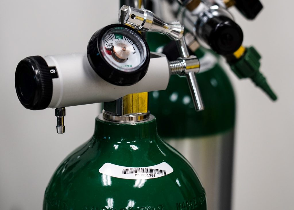
Supplies of medical oxygen in Ukraine are dangerously low due to disruption caused by the Russian invasion, the World Health Organization has warned.

Due to the crisis, the WHO estimates that the country needs an additional 20–25% increase in oxygen supplies over and above its normal needs. As it currently stands, the transport of oxygen cylinders across the country is being disrupted, especially into the capital Kyiv. As of 27 February, many hospitals across the country, including in Kyiv, had less than 24 hours’ supply remaining.

Furthermore, oxygen production facilities are experiencing shortages of zeolite, which is needed for the safe production of oxygen in the pressure swing absorption process

Prior to the conflict, the WHO had worked with Ukraine to improve its oxygen supply infrastructure, especially during the COVID pandemic. “Of the over 600 health facilities nationwide assessed by WHO during the pandemic, close to half were directly supported with supplies, technical know-how and infrastructure investments, enabling health authorities to save tens of thousands of lives,” the WHO said. This progress is threatening to be undone.

“Compounding the risk to patients, critical hospital services are also being jeopardised by electricity and power shortages, and ambulances transporting patients are in danger of getting caught in the crossfire,” the WHO said in its press release.

To offset this, the WHO is working through regional networks to bring in oxygen, as well as providing trauma treatment supplies. These would be brought in through a safe logistics corridor in Poland.

Médecins Sans Frontières (MSF) has announced that it is suspending activities in Ukraine. “These included care for people living with HIV in Severodonetsk; care for patients with tuberculosis in Zhytomyr; and improving access to healthcare access in Donetsk, in eastern Ukraine, where we have been providing much-needed healthcare, including for mental health, to conflict-affected communities,” the organisation said in an announcement.

However, it is working to ensure some continuity of its operations, and are working to provide trauma training to certain hospitals and have provided some trauma supplies.

The Ukrainian capital of Kyiv has also put out a call for donations of medicines, such as the antiviral amixin, the antibiotic nifuroxazide and the haemostatic agent aminocaproic acid.

Source: World Health Organization

To Properly Use AI to Analyse Breast Cancers, Look to Past Mistakes

Source: National Cancer Institute

Doctors writing in an editorial in JAMA Health Forum caution that while using AI to analyse breast cancer tumours has the potential to improve healthcare efficiency and outcomes, similar technological leaps have previously led to higher rates of false-positive tests and over-treatment.

The editorial wasco-written by Joann G. Elmore, MD, MPH, professor of medicine at the David Geffen School of Medicine at UCLA, and Christoph I. Lee, MD, MS, MBA, a professor of radiology at the University of Washington School of Medicine.

“Without a more robust approach to the evaluation and implementation of AI, given the unabated adoption of emergent technology in clinical practice, we are failing to learn from our past mistakes in mammography,” the authors wrote.

One of those “past mistakes in mammography,” the authors said, was adjunct computer-aided detection (CAD) tools, which grew rapidly in popularity in the field of breast cancer screening starting more than two decades ago. CAD was approved by the FDA in 1998, and by 2016 more than 92% of U.S. imaging facilities were using the technology to interpret mammograms and hunt for tumours. However, CAD did not improve mammography accuracy., according to the evidence. “CAD tools are associated with increased false positive rates, leading to overdiagnosis of ductal carcinoma in situ and unnecessary diagnostic testing,” the authors wrote. The US Medicare system stopped paying for CAD in 2018, but by then the tools had run up more than $400 million a year in wasted health costs.

“The premature adoption of CAD is a premonitory symptom of the wholehearted embrace of emergent technologies prior to fully understanding their impact on patient outcomes,” Drs Elmore and Lee wrote. “As AI algorithms are increasingly receiving FDA clearance and becoming commercially available with ROC curves similar to what we observed prior to CAD clearance and adoption, how can we prevent history from repeating itself?”

The doctors suggest a number of safeguards to avoid “repeating past mistakes” such as tying reimbursement to proven efficacy.

Source: UCLA Health

Nintedanib Slows Autoimmune-related Lung Disease

Anatomical model of lungs
Photo by Robina Weermeijer on Unsplash

Findings from a new clinical trial published in Arthritis & Rheumatology reveal that nintedanib,  an intracellular inhibitor of tyrosine kinases, may help patients with fibrosing from autoimmune disease-related interstitial lung diseases (ILDs).

ILDs are a common manifestation of systemic autoimmune diseases such as rheumatoid arthritis. Connective tissue diseases and vasculitides affect all areas of the lungs (bronchioles, parenchyma, alveoles) which is why ILD is a common feature of rheumatology diseases.

The trial enrolled 170 subjects with a fibrosing ILD other than idiopathic pulmonary fibrosis, with diffuse fibrosing lung disease of > 10% extent on high-resolution CT imagery, forced vital capacity (FVC) ≥ 45% predicted and diffusing capacity of the lungs for carbon monoxide ≥ 30% –< 80% predicted. FVC is a predictor of mortality in patients with autoimmune disease-associated ILDs.

Patients were randomised to receive nintedanib or placebo. Investigators assessed patients’ forced vital capacity (FVC). The trial found that the rate of decline in FVC over one year was -75.9 mL/year with nintedanib versus -178.6 mL/year with placebo.

“Until now, therapies that can significantly reduce the rate of decline in lung function in connective tissue disease–related ILDs characterised by progressive fibrosis have been lacking. We now have a therapeutic approach that offers a strategy for reducing the morbidity associated with these diseases,” said lead author Eric L. Matteson, MD, MPH, of the Mayo Clinic College of Medicine and Science.

Source: Wiley

A Synthetic Alternative to Pig-derived Heparin

Photo by Corinna Widmer from Pexels

Scientists have developed a process to synthesise the vital blood thinner heparin, which is normally harvested from pig intestines. This synthetic heparin would help the quality control issues and shortages associated with pig-derived heparin.

The most expensive part of a pig is not a cut of bacon or a chop, but the part of the intestine used to make heparin. About 2000 pigs required to produce a kilogram of heparin, which provides medication to up to 6000 patients. In total, it is estimated that about one billion primarily Chinese food pigs each year also supply intestines for the extraction and processing of heparin.

However, this can cause problems for patients. When making medicines derived from animals, the chemical structure is rarely uniform. There are relatively common but harmless side effects, and in very rare cases severe and life-threatening immune reactions. Additionally, there are ethical and religious concerns for many patients. Bovine- and sheep-derived heparin are also produced but have the same concerns of being of animal origin. In fact, prior to 2000, heparin was derived from cows until the outbreak of mad cow disease.

Therefore, it has long been an ambition among researchers to make heparin in a laboratory to get cleaner heparin without side effects. Now researchers from the Copenhagen Center for Glycomics at the University of Copenhagen are ready with a study that shows that it is possible to make heparin without the use of animals.  which was published in Science Advances.

“By making heparin without the animal, you get a much cleaner and more uniform chemical structure. We show that we can do it in the laboratory, i.e. in a so-called ‘cell factory’, in the same way as many other types of medicine are made. It is a step in the direction of the development that has also happened with insulin, which was previously extracted from the pancreas in pigs before we learned to produce it in the laboratory,” explained Associate Professor Rebecca Miller, who led the study. 

There is already a synthetic alternative to heparin, but it is difficult to dose and can lead to overdose. Because of this, GPs often prescribe pig-derived heparin to their patients.

Heparin is today extracted from pig intestines’ mucosa. Due to the sheer number of patients who need the medicine, the scale of the production is vast, making quality control a recurring problem for manufacturers.

In 2008, a number of stocks of heparin from Chinese pigs were recalled when it was found that the medicine was contaminated. The case ended up costing the lives of more than 100 Americans.

“Of course you want to avoid that, in addition to moving the source from animals to laboratory cells. With our new technology, we have made a design for how to make heparin in a cell that is clean and uniform and it suggests that it has the same medicinal effect as market heparin. In this way, you potentially get a product that leads to neither common nor life-threatening side effects,” said Richard Karlsson, PhD, who has also contributed to the main author of the study.

Right now, the world is facing a shortage of heparin because swine flu has thinned the pig population in China, the largest heparin producer.
Next steps would be to scale up production to provide much larger quantities of the new synthetic heparin. 

Source: University of Copenhagen

Study Reveals the Intricacy of C. Diff’s Armour

The spectacular structure of the protective armour of superbug C. difficile has been revealed for the first time showing the close-knit yet flexible outer layer – like chain mail. This assembly prevents molecules getting in and provides a new target for future treatments, according to the scientists at Newcastle, Sheffield and Glasgow Universities who have uncovered it. Credit: Newcastle University, UK

The spectacular structure of the protective armour of C. difficile has been revealed for the first time showing the close-knit yet flexible outer layer – like a mediaeval knight’s chain mail.

This tight arrangement keeps molecules from getting in and provides a new target for future treatments, according to the scientists who have uncovered it.

Published in Nature Communications, the team of scientists outlined the structure of the main protein, SlpA, that forms the links of the chain mail and how they link up to form a pattern and create this flexible armour.
One of the many ways that Clostridioides difficile has to protect itself from antibiotics is a special layer that covers the cell of the whole bacteria – the surface layer or S-layer. This flexible armour protects against the entry of drugs or molecules released by our immune system to fight bacteria.

Using a combination of X-ray and electron crystallography, the team determined the structure of the proteins and their arrangement.

Corresponding author and lead researcher Dr Paula Salgado said: “I started working on this structure more than 10 years ago, it’s been a long, hard journey but we got some really exciting results! Surprisingly, we found that the protein forming the outer layer, SlpA, packs very tightly, with very narrow openings that allow very few molecules to enter the cells. S-layer from other bacteria studied so far tend to have wider gaps, allowing bigger molecules to penetrate. This may explain the success of C.diff at defending itself against the antibiotics and immune system molecules sent to attack it.

“Excitingly, it also opens the possibility of developing drugs that target the interactions that make up the chain mail. If we break these, we can create holes that allow drugs and immune system molecules to enter the cell and kill it.”

Antimicrobial resistance (AMR), a growing problem, was declared by WHO as one of the top 10 global public health threats facing humanity.
One of the many bacteria that have evolved resistance to antibiotics, C. diff infects the human gut and is resistant to all but three current drugs. Antibiotics only compound the problem, as the good bacteria in the gut are killed alongside those causing an infection and, as C. diff is resistant, it can grow and cause diseases ranging from diarrhoea to death due to massive lesions in the gut. Since the only way to treat C.diff is to take antibiotics, it creates a vicious cycle of recurrent infections.

This knowledge could lead to the development of C. diff specific drugs that break the protective layer, creating holes to allow drug molecules to penetrate and kill the cell.

Dr Rob Fagan, who helped carry out the electron crystallography work, said: “We’re now looking at how our findings could be used to find new ways to treat C. diff infections such as using bacteriophages to attach to and kill C. diff cells – a promising potential alternative to traditional antibiotic drugs.”

Source: EurekAlert!

Antiepileptics in Comatose Cardiac Arrest Survivors are Ineffective, Study Shows

Image by Falkurian Design on Unsplash

A large scale study of comatose intensive care (ICU) patients admitted after cardiac arrest and resuscitation has shown that antiepileptics to treat epilepsy-like brain activity has no effect, and may even prolong ICU stay.

Following a cardiac arrest and resuscitation, patients may need an ICU stay, and are in a coma. By that stage, the cardiac arrest may have damaged the brain to such an extent that half of the patients will not recover from coma. The other half will also have permanent damage, for example of memory functions. It is extremely difficult to predict if a patient will awaken and what their prognosis is, so clinicians make use of EEG (electroencephalography).

In 10–20% of the patients admitted to the ICU after cardiac arrest and resuscitation, there are signs of brain activity that appear similar to epilepsy: unlike an attack this activity is continuous. For a long time, it was unclear if anti-epileptic medication could help better recovery. As a result, some patients received this medication and some did not.

Now, a large-scale study done between 2014 and 2021 on 172 patients has proven that this medication is ineffective: it does not help recovery, even necessitating a longer ICU stay. The researchers, led by Professor Jeannette Hofmeijer of the University of Twente and Rijnstate Hospital in Arnhem, published their findings in the New England Journal of Medicine.

The conclusion from this study is that anti-epileptic medication does not lead to an improved recovery. The findings show that patients may need to stay longer at the ICU: for the patient an undesired situation, and it puts extra pressure on the health care system. 

Aside from patients who show continuous epileptic signals, a small group of patients show signs of a typical epileptic seizure: a short and heavy attack. In these situations, anti-epileptics could help, but this still needs further research.

“Although the outcome of the trial may be disappointing in terms of chances of recovery, it also takes away uncertainties from the family. The signals point at serious brain damage that would lead to a much longer stay at the ICU,” said Prof Hofmeijer.

Source: University of Twente

High CAC and Lipoprotein(a) Scores Greatly Worsen CVD Risk

Healthy red blood cells. Source: NIH

Having both a high lipoprotein(a) and high coronary artery calcium score (CAC) results in a 22% risk of heart attack or stroke over the following 10 years, nearly double the risk of having either condition alone. These are the findings are from a study published in the Journal of the American College of Cardiology (JACC).

Two decades ago, it was recognised that lipoprotein(a) (Lp(a)) concentrations were elevated in patients with cardiovascular disease (CVD). However Lp(a) was not yet proven to be important due to a lack of both Lp(a)-lowering therapy and evidence that reducing Lp(a) levels improves CVD risk. Recent research has added to the evidence 

“We are hopeful that by making the connection between Lp(a) and CAC as dual risk drivers, we can raise awareness in the medical community and improve earlier heart attack prevention for these patients,” said cardiologist Parag Joshi, MD, Associate Professor of Internal Medicine at UT Southwestern. “Our data may also expedite the development of treatments designed specifically for this high-risk population.”

About one sixth of people in the U.S. have high Lp(a), driven largely by genetics. Coronary artery calcium (CAC) is a marker of plaque deposits around the heart. 

Cardiology researchers confirmed the Lp(a) and CAC connection by comparing data from two landmark cardiovascular trials, the Dallas Heart Study, an ongoing comprehensive study of 6000 diverse and heart-healthy patients conducted from 2000 to present, and the Multi-Ethnic Study of Atherosclerosis (MESA) 6000-participant study investigating early-stage atherosclerosis.

The researchers found that participants with combined high Lp(a) and high CAC had a 22% 10-year risk of heart attack or stroke, compared with a 10-15% 10-year risk in patients who had either risk factor alone.

The team identified three distinct risk-related trends:

  • High Lp(a), high CAC: These individuals face the highest 10-year risk of heart attack or stroke.
  • High Lp(a), zero CAC: 10-year heart attack and stroke risk is low when there is no CAC, even if Lp(a) is high.
  • Low Lp(a), high CAC: 10-year heart attack or stroke risk is higher than average but lower than with high LP(a) and high CAC combined.

“Establishing the connection between Lp(a) and CAC means we can move to the important next phase of research, which will be defining and personalizing early screening protocols to identify patients at high risk of heart attack,” said Dr Joshi. “With further research, this could mean selectively scanning patients with high Lp(a) for their CAC score, and studying therapies specifically designed to reduce Lp(a) among patients with high CAC.”

Source: UT Southwestern Medical Center

SA COVID Study: ‘No Longer at Code Red’, Prof Madhi Says

Image from Pixabay

Commenting on a recently published South African study showing a high COVID antibody sero-prevalence and decoupling of hospitalisation and death rates, first author Professor Shabir Madhi said that “we [are] no longer at “code red’.”

The study, published in the New England Journal of Medicine, was conducted in Gauteng from October 22 to December 9, 2021, showed a high sero-positivity rate even as the Omicron wave started. Under-12s (56%) had the lowest rate of sero-positivity, while it was 80% in over-50s and 85% in inner city residents. Unsurprising, vaccinated individuals had much higher rates (93%) than unvaccinated ones (68%). Epidemiologic data showed that the incidence of COVID infection increased and subsequently declined more rapidly during the fourth wave than it had during the three previous waves.

The researchers imputed 10.4 million infections, compared to the <1 million COVID cases recorded before Omicron. The researchers also evaluated COVID epidemiologic trends in the province, including cases, hospitalisations, recorded deaths, and excess deaths from the start of the pandemic through January 12, 2022.

At time of Omicron wave onset, 59159 Covid attributable deaths using excess mortality data (rate 396/100,000) in Gauteng. Infection fatality risk for Gauteng 0.57% pre-omicron (substantially higher than 0.019% imputed for seasonal flu pre-Covid calculated using similar methods).

In Gauteng at the start of the Omicron wave. vaccine coverage 36% for at least 1 dose in Gauteng, but 61% in over-50s (responsible for >80% deaths pre-Omicron). The sero-survey showed that, 70% of vaccinated were also infected pre-omicron, indiciating a substantial prevalence of hybrid immunity

Prof Madhi further noted analysis of the incidence trends shows a “massive decoupling” of COVID cases to hospitalisation and death rates over the course of Omicron dominance, which was seen in all age groups.
Omicron was responsible for only 3% of COVID deaths compared to 50% for those in Delta-dominant waves. In the 50-59 age group, Omicron was responsible for only 2% of deaths compared to 53% of Delta-dominant deaths.

They also found that children under 12 were not seriously affected during the Omicron wave, with the Omicron wave making up 26% of hospitalisations and 17% of deaths versus 39% and 47%, respectively with the Delta wave.

The researchers concluded that the SA experience indicates that we are now moving into the convalescent phase of the COVID pandemic. Prof Madhi noted in his tweets that this is likely to be similar in other countries that have had a low or modest vaccine uptake, but which have also seen high rates of natural infection – which, in low- and middle-income countries, has likely been accompanied by significant under-reporting of COVID fatalities. 

Given low rates of vaccine rollout and donations, Africa should focus on vaccinating its vulnerable elderly population, Prof Madhi recommended.

He tweeted that SA had expressed optimism that the pandemic had reached a turning point “which many in high income countries dismissed as ’empirical’ and not applicable to their settings despite high vaccine coverage,” subsequently materialised around the world wherever COVID was “not [a] zero-sum game.”

Hypersensitivity Link Between MRI and X-Ray Contrast Agents

Photo by Mart Production on Pexels

People with a history of hypersensitivity to iodine-based contrast agents for X-ray based scans, are also susceptible to similar reactions from commonly used MRI contrast agents, according to a large, eight-year cohort study. The study, published in the journal Radiology, also found that premedication or switching to a different MRI contrast agent may reduce risk in patients who have had previous contrast agent reactions.

For a long time, gadolinium-based contrast agents (GBCA) have been used to enhance visualisation of organs, tissues and blood vessels on MRI and provide a more accurate depiction of disease. Though GBCA are relatively safe, recent studies have reported several adverse reactions related to their use, including allergic-like hypersensitivity reactions, such as rash and flushing.

These reactions are increasing in incidence with the widespread use of GBCA, prompting an urgent need for research into risk factors, according to the study’s senior author Hye-Ryun Kang, MD, PhD.

Analysing more than 330 000 cases of GBCA exposure in 154 539 patients over an eight-year period, the researchers found 1304 cases of allergic-like hypersensitivity reactions, for a rate of 0.4%. In patients who had a previous GBCE reaction, the average recurrence rate was 15%.

Acute allergic-like hypersensitivity reactions, or those that occur within one hour of contrast administration, accounted for 1178 cases, while a far smaller number of 126 cases were delayed allergic-like hypersensitivity reactions, or those that occur beyond the first hour and mostly within one week after exposure.

The risk of allergic-like hypersensitivity reactions to GBCAs was higher in those with a history of similar reactions to iodinated contrast media. Normally, having a history of iodinated contrast media hypersensitivity was not thought to be a risk factor for hypersensitivity to GBCAs and vice versa, because of their structural and compositional differences.

“The results of our study challenge this idea,” Dr Kang said.

An underlying predisposition to drug allergies in susceptible patients could be the cause, Dr Kang said, as opposed to any cross-reactivity associated with structural similarities between iodinated contrast media and GBCA. In fact, the risk of hypersensitivity reactions to iodinated contrast media was also higher in those who previously experienced a similar reaction to GBCA.

“Thus, physicians should be aware that patients with a history of hypersensitivity to one of iodinated contrast media or GBCA are at greater risk of developing hypersensitivity reactions to the other,” she said.

Analysis of the data showed that premedication, typically with steroids and antihistamines, and changing the GBCA showed preventive effects in patients with a history of acute allergic-like hypersensitivity reactions. Patients who received premedication and before MRI or were switched to a different GBCA showed the lowest rate of recurrence. Only premedication significantly reduced the incidence of reactions in patients with a history of delayed reactions.

“As the most important preventive measure is avoidance of the culprit agent, a precise record of previously used GBCA should be kept for all patients,” Dr. Kang said. “Physicians should discuss appropriate premedication strategies with their patients prior to MRI procedures.”

Dr Kang nevertheless stressed that contrast-enhanced MRI examinations are invaluable in the diagnosis and follow-up of various diseases, and the overall risk remains low.

“As most of these reactions are mild, we believe the benefits of MRI outweigh the potential risks associated with GBCA use,” she said.

Dr Kang recommended that in all patients receiving an MRI with GBCA exposure, a detailed history of previous hypersensitivity allergic reactions be conducted, and when necessary, appropriate prevention measures should be implemented, such as using premedication and switching to different GBCA types.

Future work would be to perform studies with larger populations to identify possible risk factors and effective preventive strategies for delayed hypersensitivity reactions to GBCA.

Source: Radiological Society of North America

Severe COVID Raises Risk of Pregnancy Complications

Source: Pixabay

A University of Oxford study of over 4000 pregnant women indicates that severe COVID in pregnancy increases the risk of pre-labour caesarean birth, a very or extreme preterm birth, stillborn birth, and the need for admission to a neonatal unit.  

The study, published in Acta Obstetricia et Gynecologica Scandinavica, included 4436 pregnant women hospitalised in the UK with symptomatic COVID from March 1, 2020 to October 31, 2021, of whom 13.9% of had severe COVID. As well as having increased risks of adverse pregnancy-related outcomes, women with severe infection were more likely to be aged 30 years or over, be overweight or obese, be of mixed ethnicity, or have gestational diabetes compared with those with mild or moderate infection.  

“This new analysis shows that certain pregnant women admitted to a hospital with COVID face an elevated risk of severe disease. However, it shows once again the strongly protective effect of vaccination against severe disease and adverse outcomes for both mother and baby,” said senior author Marian Knight, FMedSci, of the University of Oxford. “This study emphasises the importance of ensuring that interventions to promote vaccine uptake are particularly focused towards those at highest risk.”

Source: Wiley