New Liver Perfusion Technology Marks a Breakthrough for Transplant Care in South Africa at Wits Donald Gordon Medical Centre
Johannesburg, 12 June 2026: For a patient waiting for a liver transplant in South Africa, the hardest part is not the surgery. It is the wait and the knowledge that an organ may never come. In a country facing severe organ shortages, every decision to accept or decline a donor liver carries immense weight and every viable organ that goes unused represents a lost opportunity to save a life.
At the centre of changing this reality is the Wits Donald Gordon Medical Centre (WDGMC), home to one of the leading liver transplant programmes in Africa and a unit internationally recognised for its contribution to specialised transplant care, research and surgical training. Having performed over 1 000 liver transplants, the programme represents decades of expertise, innovation and collaboration.
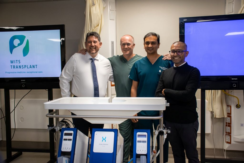
Now, WDGMC, in partnership with Surgeons for Little Lives and with support from key corporate sponsor Weelee, has introduced a state-of-the-art liver perfusion machine, becoming the first transplant centre on the African continent to implement this technology for liver transplantation.
This technology keeps donor livers viable outside the body while clinicians assess, monitor and actively improve the condition of the organ before transplantation. By allowing transplant teams to better maintain organ viability, the machine has the potential to increase organ utilisation, reduce complications and improve transplant outcomes for patients who may otherwise not survive the wait.
“As a transplant programme, our responsibility extends far beyond the operating theatre,” says Professor Jerome Loveland, Head of Solid Organ Transplantation at WDGMC. “This technology will help us better assess donor organs and increase the number of livers that can safely be transplanted, whilst simultaneously improving results. In a country where every donor organ matters, this will have a significant impact on organ utility and patient outcomes.”
South Africa’s transplant programmes continue to achieve strong outcomes despite operating within a severely resource-constrained environment and against the backdrop of ongoing organ shortages. As a result, transplant teams are often required to make difficult decisions under significant pressure.
“This technology changes the level of information we have available before transplantation. Traditionally, organs are preserved on ice and assessment is limited. Machine perfusion allows us to monitor how the liver is functioning outside the body. Beyond the valuable information it provides, the machine has the ability to resuscitate the liver by delivering oxygen to the liver cells, creating the best metabolic environment outside the body. This helps us make more informed clinical decisions and potentially increases the number of organs that can safely be transplanted,” says Dr Sharan Rambarran, Transplant Surgeon at WDGMC.
The introduction of the machine is also expected to contribute to reduced post-operative complications, shorter hospital stays and improved recovery outcomes.
“Too many patients in South Africa deteriorate while waiting for a transplant because there are simply not enough donor organs available,” says Dr Bilal Bobat, Transplant Hepatologist at WDGMC. “Anything that helps us safely expand organ utilisation has the potential to directly impact survival and quality of life for patients and families facing end-stage liver disease.”
“Weelee is always looking for opportunities to contribute to causes that create real and lasting impact,” says Errol Levin, CEO of Weelee. “Supporting advancements in liver perfusion technology aligns perfectly with our commitment to innovation that improves lives. This ground-breaking initiative has the potential to save countless lives and we are proud to be associated with a project of such significance.”
WDGMC plays a unique role within South Africa’s healthcare system. As a private academic hospital affiliated with the University of the Witwatersrand, the Centre combines highly specialised clinical care with academic medicine and collaboration across both the private and public healthcare sectors.
While the technology represents an important advancement in liver transplantation, clinicians stress that increasing organ donation awareness remains critical to improving access to life saving transplants in South Africa.
For the transplant teams, this marks not only a clinical advancement but the beginning of a broader effort to continue strengthening transplant medicine in South Africa.