In the study, investigators analysed 2003–2018 data from 1044 US women participating in the National Health and Examination Survey: 174 with and 870 without cervical cancer. With these data, the team looked for an association between cervical cancer and the Composite Dietary Antioxidant Index (CDAI) – a validated composite score that reflects an individual’s intake level of dietary antioxidants, based on dietary recall interviews.
After adjusting for other factors that might affect cancer risk, the researchers found that each 1-unit increase in the CDAI was associated with a 11.4% lower prevalence of cervical cancer. Participants in the highest CDAI tertile had a 55.8% lower prevalence of cervical cancer than those in the lowest tertile. Additional analyses indicated a linear inverse association between the CDAI and the prevalence of cervical cancer.
“These findings suggest that dietary antioxidant intake may play a potential role in the prevention of cervical cancer. However, further prospective studies are needed to confirm this association and clarify the underlying mechanisms,” the authors wrote.
Growing evidence links higher consumption of ultra-processed foods (UPFs) to a range of health problems. Research has identified several potential biological pathways through which UPFs may contribute to cancer. These include metabolic disruption, inflammation, oxidative stress, and changes to the gut microbiome. However, data are sparse about whether men who consume high amounts of UPFs have greater risks of prostate cancer.
Now, new research from Florida Atlantic University’s Charles E. Schmidt College of Medicine provides evidence that men with higher consumption of UPFs have increased risks of prostate cancer. Published in The American Journal of Medicine, the study analyses a large and nationally representative survey from 17 024 US adults males from 2003 through 2023. Researchers used two 24-hour dietary recalls to calculate the percentage of daily calories participants consumed from UPFs. Using the validated and widely used NOVA classification system, they divided participants into four groups, ranging from less than about 20% of calories from UPFs to more than 44%.
Men in the three higher-consumption groups of UPFs had a 29% greater risk of prostate cancer, while those in the two highest-consumption groups had a 30% greater risk, compared to those in the lowest consumption group. After adjustments for age, race and ethnicity, smoking, and poverty status, the increased risk remained significant, ranging from 24% to 31%. Men with the highest UPF intake alone had a possible 34% higher risk, although the finding did not reach statistical significance, potentially reflecting the smaller number of prostate cancer cases in that group.
“Among adult men in this nationally representative US population, those who consumed higher amounts of UPFs had significantly higher risks of the subsequent development of prostate cancer,” said Charles H. Hennekens, MD, FACPM, FACC, senior author and the First Sir Richard Doll Professor of Medicine and Preventive Medicine in the Department of Medicine and the Department of Population Health at FAU’s College of Medicine. “These findings add to emerging evidence that UPFs have major adverse health effects and underscore the need for large-scale observational studies and randomised trials to adequately test the hypothesis.”
Previous studies have shown that people who consume larger amounts of UPFs have higher risks of overweight and obesity, metabolic disease, markers of inflammation, cardiovascular disease, principally heart attacks and strokes, as well as premature death.
This original research adds to the armamentaria of a major battle in which the priorities of the food industry do not align with the needs of the U.S. public. The authors opine that just as the dangers of tobacco began to emerge during the middle of the prior century, decades passed before the preponderance of evidence and the efforts of forward-thinking health officials prompted policy changes to discourage cigarette use. The authors believe that this is likely to be a similar path for UPFs.
“Reducing consumption of UPFs is a complex public health challenge, particularly given how prevalent and accessible these products are,” said co-author Timothy De Ver Dye, PhD, professor and chair of FAU’s Department of Population Health. “Healthcare providers should recognise that many patients face barriers to accessing and affording healthier food options. Addressing these challenges will require efforts that extend beyond individual choices to support greater access to nutritious, minimally processed foods.”
According to the International Agency for Research on Cancer, nearly 1.5 million men worldwide were diagnosed with prostate cancer in 2022, and nearly 400 000 died from the disease.
“All healthcare professionals should encourage patients to adopt healthy lifestyle practices and evidence-based therapies, while also helping them reduce their consumption of ultra-processed foods,” Hennekens said. “Patients and their healthcare providers should consider this finding as part of broader efforts to promote healthier dietary choices.”
Study co-authors are FAU medical students Hunter Scott and John Dunn as well as Yanna Willett, a doctoral student at the University of Oxford in the United Kingdom.
Real-world study reveals the precise level associated with patient prognosis.
Credit: Darryl Leja National Human Genome Research Institute National Institutes Of Health
After treatment for metastatic prostate cancer, a drop in prostate-specific antigen (PSA) levels in the blood is an indicator that the treatment is working. A recent real-world study indicates that reaching a PSA level below 0.2 ng/mL is an indicator of improved patient survival. The study is published by Wiley online in CANCER, a peer-reviewed journal of the American Cancer Society.
Multiple phase 3 clinical trials have shown that a PSA level below 0.2 ng/mL is the best cut-off for determining a patient’s prognosis. Real-world data are lacking, however, and physicians often use other metrics, such as percentage of PSA decline, to assess treatment response. To determine whether there is a PSA response target associated with the most favourable outcome in community-dwelling adults with metastatic prostate cancer, investigators analysed Veterans Health Administration data on 4890 patients who received testosterone deprivation therapy with or without other treatments in 2018–2023 for metastatic hormone-sensitive prostate cancer.
Over a median follow-up of approximately 2 years, 896 patients died. Patients whose PSA levels dropped below 0.2 ng/mL within 9 months of starting treatment were 54% less likely to die than patients whose levels did not drop this low, whether their PSA levels also decreased by at least 90% or not. Patients who had at least a 90% PSA decline but did not achieve a PSA of less than 0.2 ng/ml had no improvement in survival. The findings suggest that patients who do not experience a PSA drop below 0.2 ng/mL could be candidates for more aggressive treatments. Also, patients who received testosterone deprivation therapy plus another drug to further block testosterone were more likely to reach a PSA below 0.2 ng/mL.
“These data show that we need to target a PSA below 0.2 ng/ml as our metric of success to optimize outcomes for our patients with metastatic prostate cancer,” said corresponding author Stephen J. Freedland, MD, professor of Urology at Cedars-Sinai and Staff Physician at the Durham VA Medical Center.
ADAR1 inhibitor researched in outer space holds promise for AML and myelofibrosis now – and possibly cancers like glioblastoma multiforme in the future
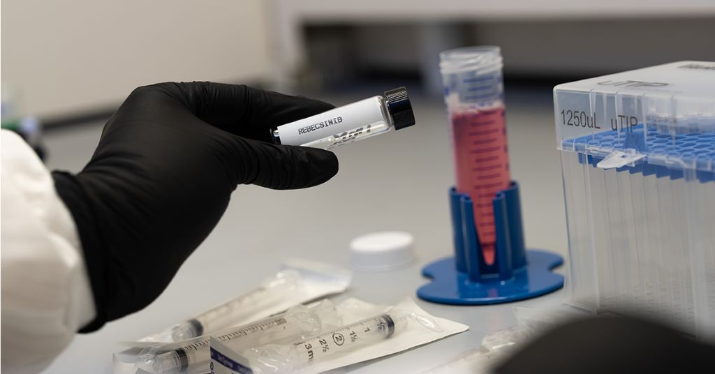
A member of the lab of Catriona Jamieson, MD, PhD, prepares a vial of investigational new drug rebecsinib ahead of the launch of Axiom 4 in July 2025. The drug – which inhibits the gene ADAR1, implicated in the growth of more than 20 cancers – is now available via clinical trial at UC San Diego for patients 18 years of age and older who have secondary acute myeloid leukaemia (AML) that has either recurred or not responded to treatment, or higher-risk myelofibrosis.
A clinical trial of rebecsinib – a first-in-class investigational drug that inhibits the ADAR1 gene involved in the proliferation of more than 20 cancers – is underway at UC San Diego.
The first patient was treated July 6, according to principal investigator James Mangan, MD, PhD, professor of medicine at UC San Diego School of Medicine and a haematologist and oncologist at UC San Diego Health. He called the drug “promising.”
“This trial has great science behind it,” Mangan said. “It uses a totally novel mechanism and really is for patients who have a desperate, unmet need.”
The Phase 1 clinical trial, sponsored by Aspera Biomedicines, is open to adults 18 years of age and older who have secondary acute myeloid leukaemia (AML) that has either recurred or not responded to treatment. It’s also open to patients with higher-risk myelofibrosis. Both are rare blood cancers for which few treatment options exist initially – and no good options if they return.
For AML and myelofibrosis patients, the rebecsinib clinical trial means hope, Mangan said: “If this works, it’s a good option for those who don’t otherwise have targeted agents available to them.”
UC San Diego Sanford Stem Cell Institute Director Catriona Jamieson, MD, PhD, a haematologist and researcher who discovered the drug, said she is “thrilled to take it from bench – and a bench on the International Space Station (ISS), no less – to the bedside of patients who need it most.”
“Rebecsinib shows all the promise in the world not only to halt the progression of multiple cancers, but to shrink them, as well as prevent their spread to multiple sites in the body,” she added.
One of the First Drugs Tested in Space
The U.S. Food and Drug Administration green-lit rebecsinib for clinical trial in March of last year, making it the first and only ADAR1 inhibitor with an investigational new drug application.
It’s one of the very first drugs studied in the cosmos. Jamieson, who is also a professor of medicine at UC San Diego School of Medicine and chief of its Division of Regenerative Medicine, has sent multiple research payloads to the ISS, testing the drug on various types of highly lethal cancers with ADAR1 involvement like ovarian cancer, metastatic breast cancer, AML and glioblastoma multiforme – experiments made possible by millions in grants from NASA’s In-Space Production Applications program.
In fact, in the summer of 2024, Jamieson received the prestigious ISS National Laboratory Compelling Results Award in Biology and Medicine for her discovery that the drug blocks the activation of ADAR1 in cancer – in space.
“Seeing Dr Jamieson’s cancer stem cell research launch on SpaceX CRS-34 – mere weeks before the first patient received rebecsinib in clinical trial – was nothing short of extraordinary,” said donor Rebecca Moores, whose funding of Jamieson’s lab made possible the drug’s development. “Hope is literally on the horizon for patients with blood cancer – and, hopefully, soon, those with other types of cancer as well.”
Scientists are still learning about the distinctive properties of space that threaten human health, including microgravity and galactic cosmic radiation. Such conditions create a uniquely stressful environment that mimics an accelerated version of aging and disease progression on Earth. Depending on the experiment, one month in microgravity can give researchers a preview of a few years, if not more than a decade, of maturation on Earth. This allows them to quickly see how a patient’s cells might age or how a medical condition like cancer might manifest in extended time. It also gives them a quick preview of how a drug might work long-term on a patient’s cells, whether a tumour or a miniature organ created from stem cells.
The landmark NASA Twins Study of 2015-2016 found that space can affect the immune system, gut bacteria, body weight, serum metabolites, immune system, gene expression and cognition of astronauts, among other health factors. Jameison’s research found that space also activates ADAR1, which, in turn, produces ADAR1p150, a protein that promotes tumor growth by hiding cancer from the immune system.
“Space gives Dr Jamieson a tremendous chance to see a lot of changes in stem cell DNA in a short period of time,” Mangan said.
Rebecsinib, he added, could be “a therapeutic mechanism to restore stem cell function after space travel” for astronauts. “If that’s true, it could also be very applicable to an analogous situation that occurs not in a two-week space journey, but over the course of 60 years of life as a human being, over which we accumulate similar stresses to, and mutations in, stem cells.”
‘Every Patient Needs Hope’
Rebecsinib’s June clinical trial launch is only the beginning. The trial may eventually expand to other ADAR1-involved cancers, including lymphoma, glioblastoma multiforme and metastatic breast cancer.
Among those hopeful for the impact of rebecsinib is patient advocate Andrew Schorr, 75, who has lived with myelofibrosis and chronic lymphocytic leukemia (CLL), another blood cancer, for decades.
His myelofibrosis is relatively stable at the moment, he said. If that were to change, however, rebecsinib “might be another option – and I would be grateful.”
Schorr is no stranger to clinical trials. He has participated in two over the years – one for CLL and another for deep vein thrombosis – and has covered many over his career as a medical journalist.
“Every patient needs hope for what could be their next treatment, because these drugs peter out,” he said. “Cancer finds a way around them. They’re not as effective over time, as your disease progresses. You’re always left wondering what the next option is. The fact that there could be a next option gives me a lot of hope.”
A single blood test could in future provide a more comprehensive picture of cancer than current methods. In a review article published in the journal Genome Medicine, researchers at Karolinska Institutet describe how several different biological signals can be analysed simultaneously from the same blood sample to detect and monitor cancer diseases.
Researchers and clinicians currently use so-called liquid biopsies, in which material from tumours can be detected in the blood. The method is less invasive than traditional tissue samples and can be used to monitor disease progression over time. In the current review article, researchers summarise developments in a growing field of research in which several different molecular signals are combined in the same analysis. These signals may come, among other things, from free DNA and RNA in the blood, as well as from changes in the structure of the genome and chemical markers.
The researchers call the approach ‘multifeature sequencing-based liquid biopsy’ (MSLB). The idea is not only to look for a single change linked to cancer, but to combine several types of information to provide a broader picture of the tumour’s characteristics.
“By analysing several biological signals simultaneously from the same blood sample, we can potentially gain a more complete picture of the biology of cancer than by studying each signal separately,” says Mariano A. Molina Beitia, researcher at the Department of Laboratory Medicine, Karolinska Institutet.
The article describes several research studies in which combinations of different signals have been used to detect cancer or monitor the disease over time. For example, analyses of DNA methylation, fragment size and chromosomal changes in blood have shown promising results for the early detection of several types of cancer. The researchers also describe how advanced bioinformatics methods and machine learning are used to interpret the large amounts of data generated.
Challenges remain
At the same time, the researchers emphasise that the technology still faces several challenges. Many studies have been carried out in limited patient groups, and the results need to be confirmed in larger prospective studies. In addition, the methods are technically complex, and there are still no common standards for how the analyses should be performed and quality-assured across different healthcare centres.
“For the technology to be widely used in healthcare, standardised workflows, independent validation and studies demonstrating the benefits of the analyses for patients are needed,” says Daniel Hagey at the Department of Laboratory Medicine and senior researcher in the study.
The researchers believe that the first clinical applications will most likely be in monitoring cancer patients, assessing treatment effects and situations where repeated tissue samples are difficult to obtain. In the longer term, the method could contribute to a more integrated and dynamic picture of cancer development based on a simple blood sample.
Koo Govender, Non-Executive Board Member and CEO of Publicis Groupe
The spirit of Christmas was alive and well at PinkDrive’s annual Christmas in July fundraiser dinner, recently held at the Indaba Hotel in Johannesburg. Twinkling fairy lights, festive décor, generous giving and heartfelt stories created an atmosphere of hope, reminding guests that the greatest gift anyone can give is the chance to save a life.
Supported by long-standing corporate partners, the themed ‘Night of Giving Back’ once again united business, cancer survivors and the community behind PinkDrive’s mission to make early cancer detection accessible to more South Africans.
Each year, this non-profit organisation (NPO) makes a measurable difference to thousands of lives. Relying on donations, corporate sponsorships and community support, it’s dedicated to the early detection of gender-related cancers in underserved communities. PinkDrives mobile units travel across the country, offering screenings such as mammograms, clinical breast examinations, pap smears, and PSA testing directly to people who might otherwise have limited access to healthcare.
The evening struck a thoughtful balance between festive celebration and meaningful reflection, honouring cancer survivors while remembering those who lost their lives to the disease. One of the most moving moments came through a testimonial video featuring women whose lives were saved thanks to PinkDrive’s mobile screening units. Their stories vividly illustrated the impact of accessible healthcare and reinforced the organisation’s commitment to taking cancer screening to communities where it is needed most.
The message was echoed by guest speaker Nicole Fuller, elite athlete and cancer survivor, who shared her personal journey following an early diagnosis that enabled her to avoid chemotherapy and radiation treatment. Inspired by her own experience, Fuller has since become the first South African to receive specialist training in cancer recovery and rehabilitation. Encouraging guests to prioritise regular screenings, she highlighted how early detection, combined with appropriate rehabilitation and physical activity, can significantly improve both recovery and long-term wellbeing.
An address by Koo Govender, Non-Executive Board Member and CEO of Publicis Groupe, spoke to the strength of PinkDrive’s leadership and the collective expertise steering its mission. Sound governance and committed partnerships, she noted, are what allow the NPO to keep expanding its reach and deepening its impact.
Closing the formal programme, PinkDrive founder and CEO Noelene Kotschan took guests back to where it all began: a single mobile screening unit, and a mission to reach those that healthcare so often overlooks. From that modest start, she traced PinkDrive’s growth into a nationally recognised force in cancer awareness and early detection. She closed with heartfelt thanks to all those whose continued support keeps that mission alive.
The fundraiser’s philanthropic spirit continued throughout the evening with raffle draws and a lively auction at the end. Guests bid enthusiastically for luxury prizes including diamond earrings and dream holiday experiences, helping to raise significant funds for PinkDrive’s awareness, education and mobile screening programmes.
To add to the Christmas spirit, spot prizes and hampers, generously sponsored by PinkDrive’s corporate partners, sent guests home with some festive spoils too. And as part of its ongoing support, Lee-Chem Laboratories, through its Mandy’s brand, ensured every guest received a generously filled Mandy’s goodie bag, adding another thoughtful touch to an evening centred on generosity, hope and community.
For Lee-Chem, that generosity runs deeper than a gesture on the night.
“You can’t put a price on the moment someone gets the all-clear because they were screened in time. That’s what keeps us partnering with PinkDrive year after year, and why we’re proud to stand with so many others who believe in that same mission,” says Bhavna Sanker, Marketing Manager at Lee-Chem Laboratories.
Because while Christmas may only appear on the calendar once a year, the spirit of giving and the opportunity to change someone’s future are always in season.
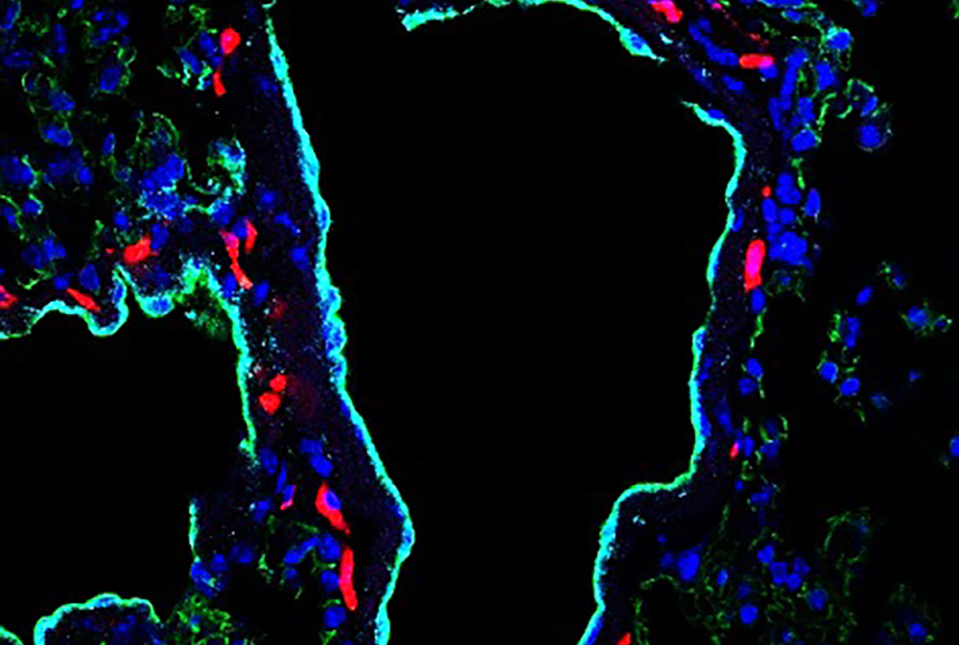
Confocal image of a lung from a tumour-bearing mouse before cancer cells arrive. Red fluorescent dextran highlights leakage from blood vessels (cyan), while veins are labelled in green, illustrating how tumours prepare distant organs for future metastasis by increasing vascular permeability. Credit: Dr Shani Dror
A protein secreted by common cancer types makes blood vessels leakier to allow metastatic tumours to form in the lungs, according to a study led by investigators at Weill Cornell Medicine and Memorial Sloan Kettering Cancer Center. The discovery could lead to new ways of monitoring and reducing lung metastasis risk in cancer patients.
The researchers, whose findings appear Aug. 5 in Nature Cancer, sought to explain how some tumours enhance their ability to metastasise to the lungs by secreting tiny capsules and particles that carry proteins, DNA and other molecules. These extracellular vesicles and particles (EVPs) are secreted by virtually all cells as a way of signalling to other cells and helping to shape their environments, and tumours have adapted them for their own purposes. In the study, the researchers identified a protein called integrin alpha-5 as the cargo in tumour-derived EVPs that promotes lung blood vessel leakiness and lung metastasis in several types of cancer.
The recognition that EVPs secreted by tumour cells represent an important layer of cancer biology has come only in the past two decades. As Lyden’s and other labs have shown, tumours use the molecular cargo of EVPs to create a local “pro-tumour” environment and prepare distant organs for metastasis. Tumour EVPs and the molecules they carry are now viewed as promising targets for cancer therapies and for cancer detection and monitoring technologies called liquid biopsies that rely on analysis of blood samples.
In earlier work, Lyden lab researchers found that some tumours’ EVPs have a potent ability to facilitate lung metastasis by inducing a leakiness in blood vessels that allows circulating tumour cells to enter the lungs. In the new study, they looked for the EVP cargo responsible for this effect.
They confirmed in animal models that for some cancers – melanoma, osteosarcoma, and colorectal, in particular – tumour implantation elsewhere in the body can trigger large increases in lung blood vessel leakiness, doing so via tumour-secreted EVPs. They found that these EVPs have the same effect when injected on their own into healthy, tumour-free mice.
“This effect on lung vessels is surprisingly rapid – we could measure it just an hour or two after injecting the EVPs,” said study first author Dr Shani Dror, a research associate in the Lyden lab.
The researchers expected to find that these EVPs cause vessel leakiness by acting directly on the endothelial cells lining blood vessels. They found instead that the EVPs work indirectly via immune cells called macrophages that reside close to lung blood vessels and exert their effect only when they carry integrin alpha-5. Even a breast tumour model that is not prone to metastasise did so readily when EVPs containing integrin alpha-5 were added. The scientists showed that integrin alpha-5 works on lung macrophages by inducing them to secrete the pro-inflammatory protein IL-6, which then causes nearby endothelial cells to loosen up vessel linings.
To confirm that the results were relevant beyond animal models of cancer, the researchers analysed a large dataset of gene expression in human tumours, finding that high expression of integrin alpha-5 in several tumour types, including colorectal tumours, is associated with poorer overall survival. They also found tumour samples from patients with intermediate to advanced colorectal cancer released EVPs containing integrin alpha-5 and were indeed capable of inducing vessel leakiness.
The results reveal a key metastasis-promoting mechanism in some cancers and have potential clinical implications.
“One possibility is that we could try to detect these integrin alpha-5-containing EVPs during cancer surgery, to assess the risk of metastasis,” said study co-corresponding author Dr Jacqueline Bromberg, a breast oncologist at MSK and an associate professor of medicine at Weill Cornell Medicine. “If we had a good drug for blocking integrin alpha-5, we could give this at the time of surgery to potentially reduce the risk of metastasis.”
A new study from The Wistar Institute has uncovered an unexpected link between fructose and the spread of an aggressive form of ovarian cancer. Published in Nature Aging, the study found that cancer cells not killed by chemotherapy send signals to neighbouring tumour cells, driving metastasis. The researchers identified fructose as a key messenger in this process, revealing a previously unrecognised way that treatment-surviving cancer cells may promote metastasis.
“Some cancer cells that survive chemotherapy aren’t dividing anymore, but they’re still biologically active,” said Aidan Cole, PhD, a postdoctoral fellow in the lab of Katherine Aird, PhD, at The Wistar Institute and first author on the study. “Instead, they continue to release molecules that send signals to nearby cells. Our study is among the first to show that a nutrient – in this case, fructose – can act as one of those signals.”
Ovarian cancer is treated almost universally with platinum-based chemotherapy, and many patients respond well at first. However, the disease recurs in most patients and almost always spreads through the abdominal cavity. That spread, called metastasis, accounts for roughly 90% of deaths from the disease.
Prior research has suggested that cancer cells not killed by chemotherapy play a role in recurrence in part through the ability of these cells to release a complex mix of signalling molecules. Cole and his colleagues began their research by designing a unique experiment: they collected the molecules released by chemotherapy-surviving cells and found these factors alone could significantly increase the spread of cancer cells.
“As far as we know, this is the first time anyone has shown, in a preclinical model rather than just a dish, that it’s the molecules these cells release – not the cells themselves – that drive the cancer’s spread,” said Cole.
That finding sent the team looking for what was being released that caused the cancer cells to spread. Fructose, it turned out, was being made by the surviving cells and sent as the signal for increased spread. Even more interesting, they found that without chemotherapy treatment, consuming the high levels of fructose found in sugary drinks can also signal cancer to spread. The fructose finding is especially compelling because it raises the possibility that simple dietary changes could help shape how cancer progresses. This is particularly important given the prevalence of fructose consumption in the United States, with high fructose corn syrup accounting for ~8-20% of the daily caloric intake in some individuals. Unlike many cancer risk factors outside of patient control, fructose consumption can be modified by dietary choices. While the effectiveness of limiting fructose intake hasn’t yet been tested directly in patients, the study raises the possibility that nutrition could influence cancer progression in previously unrecognised ways.
The researchers next aimed to understand why fructose increases cancer cell spread. Through a variety of large-scale analytical techniques, including the use of a CRISPR screen, they discovered that fructose suppresses cholesterol within the neighbouring cells. Cholesterol is important for cells to stick to each other like a biological glue, so decreased cholesterol allows cells to more easily escape and spread.
The finding that fructose lowers cholesterol production has important clinical implications. Statins, which are taken by 39 million people in the United States, lower cholesterol production. The team found that statins alone decreased the glue between cells to promote escape. The research team is now examining whether these medications could be interfering with the effects of chemotherapy – though they stress this isn’t a reason for patients to stop taking them.
“We haven’t tested this effect in patients yet, but it raises questions about combining cholesterol-lowering drugs with chemotherapy, especially since ovarian cancer is most common in postmenopausal women who are often already on statins,” said Katherine Aird, PhD, professor and co-leader of the Molecular and Cellular Oncogenesis Program in the Ellen and Ronald Caplan Cancer Center at The Wistar Institute, and senior author of the study.
The researchers are also looking into how this mechanism might extend beyond ovarian cancer.
“We think other cancers that spread within the torso – pancreatic, colon, liver – could behave similarly. We can’t call it universal yet, but we think the effects are not just limited to ovarian cancer,” said Aird.
She and Cole have already started designing follow-up experiments to test the reproducibility of their findings in a variety of other cancer types.
An international research team led by the Medical University of Vienna reports on a new treatment strategy that has made a kidney transplant possible for a patient with no realistic chance of receiving a suitable donor organ. As the case study demonstrates for the first time, the use of a new drug from the field of cancer medicine can achieve a substantial and sustained reduction in antibodies against potential transplants, a level not previously attained. This significantly improves the prospects of a successful organ transplant even in cases with a particularly unfavourable initial immunological profile. The results have recently been published in the New England Journal of Medicine and could open up new perspectives in transplant medicine.
The new drug Teclistamab is currently used to treat blood cancer (myeloma). It specifically eliminates those cells in the blood and bone marrow that produce antibodies against foreign structures. Due to this unique mechanism of action, the substance has now also come to the attention of transplant medicine: the research team led by Georg Böhmig and Martina Schatzl (Clinical Department of Nephrology and Dialysis, Department of Medicine III, MedUni Vienna) applied it for the first time as part of a case study in a dialysis-dependent patient with a highly unfavourable initial immunological profile.
The 37-year-old had developed particularly pronounced HLA sensitisation following two previous kidney transplants. In this process, the immune system produces antibodies against tissue markers of potential donor organs, known as HLA (Human Leukocyte Antigens). These antibodies significantly limit the availability of suitable organs. In the specific case of the study, the calculated probability (cPRA value) of ever finding a compatible donor kidney for the patient was actually zero. Consequently, the study participant’s name had been on the waiting list for more than twelve years, whilst his condition progressively deteriorated.
Successful transplant after 31 weeks of therapy
Treatment with teclistamab over a period of 31 weeks turned the tide: the drug achieved such a substantial and sustained reduction in antibodies against tissue antigens as is not possible with the methods currently available for HLA sensitisation. “During the course of therapy, HLA markers from donor kidneys, against which there had previously been strong antibody reactions, were gradually classified as acceptable,” reports lead author Martina Schatzl. Eventually, a suitable organ was found and successfully transplanted. “The patient is doing very well today; his kidney function is excellent, and he no longer needs dialysis,” adds study leader Georg Böhmig.
Further studies on benefits and risks needed
20 to 30 per cent of patients on the waiting list for donor kidneys are affected by significant HLA sensitisation, some of whom have no chance of receiving a suitable organ. Existing procedures for so-called desensitisation aim to reduce the number of antibodies prior to transplantation, but are only effective to a limited extent and for a short period. The treatment approach described in the case study, by contrast, directly intervenes in antibody production and sustainably reduces the immune response over a longer period. “This could herald a paradigm shift in transplant medicine and open up new prospects for a group of patients who have been particularly disadvantaged until now,” says Böhmig.
Detailed immunological results from the current case study also suggest that the new treatment strategy might also be applicable in xenotransplantation – that is, the transplantation of organs from genetically modified pigs – as well as in blood-group-incompatible transplants. “Looking ahead, an extension to other forms of organ transplantation, such as heart transplantation, as well as use in the post-transplant setting to treat antibody-mediated rejection reactions, also seems conceivable,” says Böhmig. However, before the new treatment strategy can be used in clinical practice, its benefits and risks must be systematically investigated. A study of this kind is already being planned at MedUni Vienna.
By Dr Fatima Hoosain, specialist surgeon and Principal of Apffelstaedt, Hoosain & Associates, with a clinical focus on breast and endocrine surgery.
Photo by National Cancer Institute on Unsplash
When people think about breast cancer screening, the conversation often begins and ends with one message: screen more women.
As clinicians, we know it is not quite that simple.
There is no question that screening saves lives. Regular mammography reduces breast cancer mortality and gives us the opportunity to diagnose disease when it is smaller, more treatable and associated with significantly better outcomes. Few interventions in medicine demonstrate such a clear benefit.
The challenge is that good breast care is not defined simply by how many mammograms we perform. It is defined by the quality of the decisions that surround them.
This was the focus of my presentation at the recent Board of Healthcare Funders (BHF) Conference, where we explored how clinicians can balance the burden of breast cancer with evidence-based screening decisions while remaining mindful of both underdiagnosis and overdiagnosis.
Those competing risks are encountered by every clinician involved in breast care.
We all worry about the patient whose cancer is diagnosed later than it should have been. Earlier diagnosis frequently means less extensive surgery, more treatment options and, ultimately, better outcomes. The survival difference between early-stage and advanced disease is substantial, making timely diagnosis one of the most important contributors to long-term prognosis.
At the same time, screening is not without consequences.
Not every abnormality detected on imaging will become life-threatening, yet every suspicious finding understandably creates anxiety. Additional imaging, biopsies and sometimes treatment may follow. Our responsibility is therefore not simply to detect abnormalities, but to interpret them appropriately within the context of each patient’s overall clinical picture.
This is why breast screening should never be approached as a uniform process. Risk matters.
A woman with an inherited genetic mutation or a strong family history should not necessarily follow the same screening pathway as someone at average risk. Likewise, imaging should answer a clinical question. Mammography remains the cornerstone of breast screening, but dense breast tissue, patient age and individual risk factors may require supplementary investigations such as ultrasound or MRI. More imaging is not automatically better medicine. Appropriate imaging is.
These decisions have become even more complex within the South African healthcare environment. International guidelines provide an excellent evidence base, but they do not remove the practical realities we face every day. Access to imaging differs between regions. Advanced investigations may not always be readily available. Medical scheme funding, co-payments and affordability inevitably influence what is possible for many patients. These factors cannot be ignored when discussing best practice because they form part of the reality in which clinical decisions are made.
Fortunately, the treatment landscape continues to evolve.
Advances in oncoplastic surgery, targeted therapies, immunotherapy and modern radiation techniques have transformed outcomes for many patients diagnosed with breast cancer. These developments are encouraging, but they should not distract us from one fundamental principle: the earlier we diagnose clinically significant disease, the greater the opportunity to offer patients treatments that are both effective and less invasive.
Diagnosis, however, is only the beginning of the journey.
Long-term follow-up remains an essential part of breast cancer care. Ongoing surveillance, adherence to endocrine therapy where appropriate, management of treatment side effects and supporting patients through the psychological and financial impact of a cancer diagnosis all influence outcomes. Good breast care extends well beyond the operating theatre or oncology unit.
As our healthcare system continues to face increasing clinical and financial pressures, I believe we need to move beyond simplistic conversations about screening uptake alone.
The more important discussion is whether we are making consistently good clinical decisions. Are we identifying the patients who stand to benefit most? Are we investigating appropriately? Are we avoiding unnecessary intervention when the evidence suggests it is unlikely to improve outcomes?
Those are not easy questions, but they are the ones that matter.
Ultimately, breast cancer screening is not about doing more. It is about doing what is right for the patient sitting in front of us. That remains the most important clinical judgement we make.