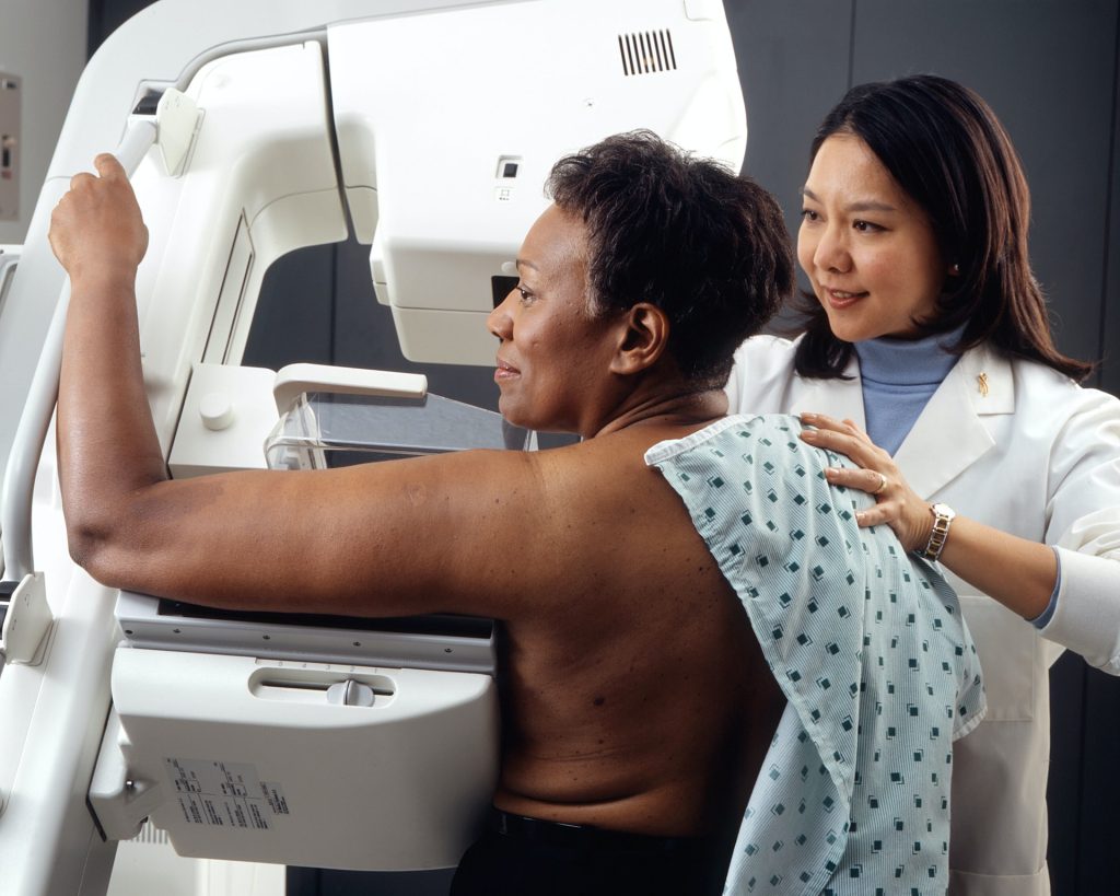
Mayo Clinic researchers and collaborators have shown that an artificial intelligence (AI) tool can analyse routine pathology slides to help clinicians classify meningiomas, the most common primary brain tumour in adults, and better understand a patient’s risk of tumour recurrence.
The study, published in The Lancet Digital Health, demonstrates that deep learning models can support the extraction of molecular and prognostic information from standard haematoxylin and eosin, or H&E, slides – the same type of tissue images already used in routine clinical care. These insights are typically obtained through DNA methylation profiling, an advanced genetic test which provides valuable diagnostic and prognostic information but can be costly, time-consuming and is unavailable in many hospitals.
“This is one of the many studies where we can harness the strength of digital pathology by capturing the last two decades of genomic and molecular knowledge into AI algorithms,” says Gelareh Zadeh, MD, PhD, chair of the Department of Neurologic Surgery at Mayo Clinic in Rochester and Chief Medical Officer for Mayo Clinic Platform.
Making advanced tumor insights more accessible
Meningiomas can vary widely in behaviour. Some grow slowly and may never return after treatment, while others are more aggressive and more likely to recur. Understanding that risk is critical for patients and care teams deciding whether additional treatment, such as radiation therapy, may be needed after surgery.
Molecular testing can help identify which tumours are more likely to recur and which may respond differently to treatment. But these tests require specialized technology and expertise, limiting access for many patients.
Using tissue samples, pathology images and clinical data from 672 patients, researchers developed and tested AI models designed to help identify patterns linked to a tumour’s biology. Drawing on multiple de-identified datasets, including data resources from Mayo Clinic Platform, the models supported classification of meningioma subtypes and recurrence risk prediction using standard pathology slides that are already part of routine patient care.
The findings suggest that, with further validation, AI-based tools could one day help clinicians obtain more detailed tumour information to inform patient care, without requiring every patient to undergo advanced genetic testing.
Helping guide treatment decisions
For patients with meningiomas, recurrence risk can influence follow-up care, imaging frequency and whether radiation therapy should be considered. The study found that AI-based predictions remained useful even after accounting for traditional clinical factors such as tumour grade, the extent to which surgery was able to remove the tumour and patient age.
Researchers also found that the AI models could identify patterns of tumour heterogeneity – differences within the same tumour – that may help explain why some tumours behave more aggressively or respond differently to treatment.
The researchers note that additional prospective studies are needed before the AI models can be used routinely in clinical care. Still, they say the findings lay the groundwork for more accessible, personalised care for patients with meningiomas – and potentially for similar AI approaches in other cancers.
As with any clinical decision-support tool, the researchers emphasise that these models would require rigorous evaluation, validation and ongoing physician oversight before being considered for routine care. “The aim is to make these algorithms readily and simply accessible for use globally, improving patient care across many healthcare settings,” says Dr Zadeh.
For a complete list of authors, disclosures and funding, review the publication.
Urine samples. Credit: Cancer Research UK CC-BY4.0
Cambridge scientists hunting tell-tale killer ‘zombie’ cells that signal early lung cancer have developed a world-first urine test that could transform diagnosis and survival for thousands of patients.
[The test] could one day be used easily in GP surgeries and hospitals to help detect recurrence in this hard-to-treat cancer much earlier.
Ljiljana Fruk
As published this week in Nature Aging, the team has shown that this simple and affordable test could detect the earliest signs of lung cancer months, or even years, before symptoms appear, as well as monitor whether treatment is working and identify potential relapse.
It works by identifying the presence of senescent cells in the lungs – so called ’zombie cells’ – that stop dividing but linger and release abnormal inflammatory signals that damage surrounding tissue and help create an environment that lowers the body’s ability to fight the cancer.
The study, funded by Cancer Research UK, marks a major leap towards more precise therapy and a test for early cancer and treatment efficiency that could be rolled out across the NHS one day.
Lung cancer is the UK’s most common cause of cancer death taking the lives of around 32,800 people every year. Thanks to huge strides in prevention, detection and treatment, in the UK, lung cancer has seen a 22% reduction in death rates in the last decade. And around two in three people (65%) with lung cancer in England survive their disease for five years or more when diagnosed at the earliest stage. But when diagnosed at the latest stage, this falls to 5 in 100 (5%).
This new test could save and improve thousands more lives in the future.
The researchers created an injectable sensor that interacts with proteins released by senescent cells. When these proteins are present, the sensor triggers the release of a detectable compound that appears in urine – signalling the earliest biological signs of therapy resistance and lung cancer development.
The researchers say that early identification is critical to saving more lives, as the disease often relapses silently with few or no symptoms until it has already spread. By detecting signs of lung cancer development and therapy resistance early, their simple urine test can spot lung cancer and treatment resistance early, helping doctors to tailor and adapt the treatment to the patient and start that treatment earlier when it works best.
The team confirmed their results using real patient samples and large genetic datasets.
Professor Ljiljana Fruk, from the Department of Chemical Engineering and Biotechnology at Cambridge, said: “The sensor has not yet been tested in humans, next is the clinical trials and it is likely it will take few years to bring it to patients, but it is a first big step and it could one day used easily in GP surgeries and hospitals to help detect recurrence in this hard-to-treat cancer much earlier.”
Nearly half (46%) of lung cancers in England are diagnosed at the latest stage.
Professor Daniel Munoz-Espin from the Early Cancer Institute and co-lead for the Cancer Research UK Cambridge Centre Thoracic Cancer Programme, said: “Our previous studies showed that senescent cells in response to chemotherapy can cause treatment resistance and an aggressive lung cancer relapse. We also found that senescent immune system cells promote lung cancer development by causing immunosuppression.
“Our urine nano sensor may allow primary care detection of therapy resistance and lung cancer early development in future clinical settings.”
Professor Robert Rintoul of the Department of Oncology, and co-lead for the Cancer Research UK Cambridge Centre Thoracic Cancer Programme said: “Novel approaches for lung cancer detection and response to treatment are urgently needed to improve patient outcomes. This work forms the basis for testing within clinical trials with a view to future use in the clinic.”
Cancer Research UK’s spokesperson for the East of England, Patrick Keely, said: “With new technologies opening doors to new discoveries, we’re living in a golden age of research, which is powerfully underlined by this innovative new urine test to detect early lung cancer.”
Adapted from a press release from Cancer Research UK
With colorectal cancer a growing concern among younger people, the American Cancer Society has endorsed two new types of stool tests to encourage people to get screened while also recommending a limited role for new blood tests many patients find appealing.
The recommendations are an update to the ACS’s screening guidelines – an update led by Andrew Wolf, MD, a cancer-prevention expert at UVA Health. He and a blue-ribbon panel of cancer experts conducted a systemic review of the available colorectal cancer tests to determine which are most effective. In addition to recommending a next-generation DNA stool test and a new type of RNA stool test, the group is advising doctors to recommend blood tests only to patients who decline all other options.
The recommendations come with a dose of pragmatism: “The most effective screening test,” the panel concludes, “is the one that the patient completes.”
“The new guidance adds a stool RNA test and an updated stool DNA test to the menu of preferred options for colorectal cancer screening, which currently include colonoscopy and stool tests that detect tiny amounts of blood, among other options,” said Wolf, a professor emeritus at the University of Virginia School of Medicine. “Although the idea of a blood test for colorectal cancer sounds very attractive, they aren’t yet as good as the other tests at detecting precancerous growths and early-stage cancer, so we don’t believe they are as effective as a screening test. That said, we’re very hopeful that broadening the array of options will get more folks screened and reduce the burden of suffering from colorectal cancer.”
About Colorectal Cancer
Colorectal cancer is the second-leading cause of cancer deaths in the United States, killing 55 000 people in 2026. Improvements in detection, screening and treatment have contributed to declining colorectal cancer death rates over the last several decades, but that decline has been accompanied since 2013 by an alarming increase in the cancer among people under the age of 50. Among that age group, colorectal cancer is now the leading cause of cancer death for men and the second-leading cause for women.
In response, the American Cancer Society in 2018 lowered the recommended age for initial colorectal cancer screening from 50 to 45 for people at average risk. It also affirmed the importance of screening tools such as stool-based tests as well as visual exams such as colonoscopies. Since then, however, new, multi-target stool tests and blood-based screening tests have become available. The new blood tests proved popular in a patient survey, with 53% of respondents saying they would prefer blood testing every three years to taking a stool test every year or receiving a colonoscopy every 10.
For the latest guideline update, Wolf and his colleagues examined the effectiveness of the new tests to provide doctors with guidance on if, how and when they should be used. The experts conclude that the DNA and RNA tests had high sensitivity for detecting colorectal cancer and moderate sensitivity for detecting advanced precancerous lesions that are about to turn into cancer. The blood tests, on the other hand, showed lower sensitivity for both advanced precancerous lesions and stage 1 cancers.
“While colorectal screening blood tests may not be as effective as other options, they are certainly better than not screening,” Wolf said. “So if a patient declines a stool test or a visual exam like a colonoscopy, a blood test would be the way to go, as long as the patient understands it is not as effective, and, if it is positive, they will still need to have a colonoscopy.”
Based on their results, the experts endorse the stool tests for patients at average risk but urge doctors to reserve the blood tests for patients who refuse other screening options. And they recommend that anyone who tests positive on any stool or blood test should receive a colonoscopy promptly.
It’s important, they note, that doctors explain to patients the strengths and weaknesses of the available tests so that patients can make informed decisions.
“Currently, almost a third of adults are not up to date with colorectal cancer screening, and among those ages 45 to 49, it’s twice that number,” Wolf said. “We hope these new options will help to close this gap. The most important message is that colorectal cancer is a disease you don’t have to die from, and there’s a screening test out there that’s right for you.”
Better preventing, detecting and treating cancer is the core mission of UVA Comprehensive Cancer Center, one of only 57 cancer centers in the nation to earn the prestigious “comprehensive” designation from the National Cancer Institute. That designation is awarded only to elite cancer centers with the most outstanding cancer care and research programs in the country.
Credit: Darryl Leja National Human Genome Research Institute National Institutes Of Health
Prostate cancer screening compares favourably to screening for breast cancer in identifying significant cancers, reducing mortality and avoiding unnecessary harms, according to new research. The findings are presented on Sunday 15 March 2026 at the European Association of Urology Congress (EAU26) in London. The research is also accepted for publication in European Urology.
The researchers maintain that the similarities between the two forms of screening mean it is no longer rational to reject prostate cancer screening on one hand while endorsing screening for breast cancer on the other. Nevertheless, they recommend some caution given their research compares a trial with a population-based screening programme and across two different cancers.
Although breast and prostate cancer are the most commonly diagnosed cancers in Europe amongst men and women respectively, screening for the diseases is vastly different. Organised breast cancer screening programmes have been established across Europe for more than three decades. Prostate cancer screening has lagged behind, primarily due to concerns around the effectiveness of the PSA blood test and the risks of overdiagnosis and overtreatment. Nevertheless, many men undergo variable, ‘opportunistic’ screening for the disease, mostly based on self-referral.
Several prostate cancer screening trials in Europe have now reported long-term outcomes, showing a reduced risk of death from prostate cancer [1]. This risk reduction is similar to that seen in breast screening programmes.
The new analysis compares the two types of cancer screening in terms of the effectiveness of the diagnostic tests and levels of overdiagnosis. The researchers, from the German Cancer Research Centre in Heidelberg, Germany, drew on data from the PROBASE prostate cancer screening trial in Germany and the country’s breast cancer screening programme.
They used data from 39,392 men who underwent an initial PSA blood test as part of the PROBASE trial at age 45 or 50. They compared this with data from just over 2.8 million women, aged 50–69, who had a mammography as part of Germany’s organised breast cancer screening programme. They found:
PSA blood testing followed by an MRI scan leads to a higher number of false positives than mammography (37-42% vs 10%).
A similar proportion of men and women were referred for biopsy (0.8-2.4% for men and 1.1% for women) as men in the PROBASE trial were triaged before referral using various factors to determine the likelihood of significant cancer (known as risk stratification)
Biopsies were far more likely to identify significant cancer in prostate screening than in breast screening (50-68% vs 10%), indicating that fewer men were referred for biopsy unnecessarily.
The percentages of invasive cancers identified were similar across both prostate and breast cancer screening (60-74% vs 73%).
Prostate cancer screening was more likely to identify non-aggressive cancers than breast cancer screening (26-31% vs. 22%). However, in prostate cancer the option of active surveillance is well-established, and the researchers maintain this would limit the risk of overtreatment. Active surveillance involves monitoring lower grade cancers and only starting treatment (radiotherapy or surgery) if they progress.
Dr Sigrid Carlsson, who leads Clinical Epidemiology of Early Cancer Detection at the German Cancer Research Centre (DKFZ) in Heidelberg, is lead author of the research. She said: “Until we have a population-based screening programme for prostate cancer, we can’t make an exact like-for-like comparison with breast cancer. But we can make some informed assumptions based on the data from our trial, which shows that if prostate cancer screening were extended to the wider population, then the outcomes are likely to be very similar to breast cancer. Although our study used German data, the findings are applicable to other countries. The final question we now need to answer is: what will this cost compared to what we are already paying for opportunistic screening? And that work is already underway.”
Tobias Nordström is a clinical urologist and Associate Professor at the Karolinska Institute, Sweden and a member of the EAU Scientific Congress Office. He said: “There is much that prostate cancer screening can learn from breast cancer screening and that is why this analysis is an important addition to our knowledge base. As these kinds of comparisons are very challenging, the results do need to be taken with a level of caution. That said, the clear overall similarities between the outcomes for breast and prostate cancer screening show that we are moving in the right direction, ensuring prostate cancer screening offers more benefits than harm.”
Small cell lung cancer cells (green and blue) that metastasised to the brain in a laboratory mouse recruit brain cells called astrocytes (red) for their protection. Credit: Fangfei Qu
Artificial intelligence tools are increasingly being developed to predict cancer biology directly from microscope images, promising faster diagnoses and cheaper testing. But new research from the University of Warwick, published in Nature Biomedical Engineering, suggests that many of these systems may be using visual shortcuts rather than true biology – raising concerns that some AI pathology tools are currently too unreliable for real-world patient care.
“It’s a bit like judging a restaurant’s quality by the queue of people waiting to get in: it’s a useful shortcut, but it’s not a direct measure of what’s happening in the kitchen,” says Dr Fayyaz Minhas, Associate Professor and principal investigator of the Predictive Systems in Biomedicine (PRISM) Lab in the Department of Computer Science, University of Warwick, and lead author of the study.
“Many AI pathology models are doing the same thing, relying on correlations between biomarkers or on obvious tissue features, rather than isolating biomarker-specific signals. And when conditions change, these shortcuts often fall apart.”
To reach this conclusion, the researchers analysed more than 8000 patient samples across four major cancer types – breast, colorectal, lung and endometrial – and compared the performance of leading machine learning approaches. While the models often achieved high headline accuracy, the team found this frequently came from statistical “shortcuts.”
For example, instead of detecting mutations in the cancer-associated BRAF gene, a model might learn that BRAF mutations often occur alongside another clinical feature such as microsatellite instability (MSI). The system then learns to use this combination of cues to predict BRAF status rather than learning the causal BRAF signal itself – meaning accurate cancer predictions work only when these biomarkers co-occur and become unreliable when they do not.
Kim Branson, SVP Global Head of Artificial Intelligence and Machine Learning, GSK and co-author says, “We’ve found that predicting a BRAF mutation by looking at correlated features like MSI is often like predicting rain by looking at umbrellas – it works, but it doesn’t mean you understand meteorology.
“Crucially, if a model cannot demonstrate information gain above a simple pathologist-assigned grade, we haven’t advanced the field; we’ve just automated a shortcut. The roadmap for the next generation of pathology AI isn’t necessarily bigger models; it’s stricter evaluation protocols that force algorithms to stop cheating and learn the hard biology.”
When performance of AI models was assessed within stratified patient subgroups, such as only high-grade breast cancers or only MSI-positive tumours, accuracy fell substantially, revealing that the models were dependent on shortcut signals that disappear once confounding factors are controlled.
For certain prediction tasks, the performance advantage of deep learning over human-derived clinical information was modest. AI systems achieved accuracy scores of just over 80% when predicting biomarkers, compared with around 75% using tumour grade alone – a measure already assessed by pathologists.
Machine learning methods can still prove valuable for research, drug development candidate screening and for clinical triaging, screening, or supplementary decision support. However, the researchers argue that future AI tools must move beyond correlation-based learning and adopt approaches that explicitly model biological relationships and causal structure.
They also call for stronger evaluation standards, including subgroup testing and comparison against simple clinical baselines, before looking at deployment in routine care.
Dr Minhas concludes, “This research is not a condemnation of AI in pathology. It is a wake-up call. Current models may perform well in controlled settings but rely on statistical shortcuts rather than genuine biological understanding. Until more robust evaluation standards are in place, these tools should not be seen as replacements for molecular testing, and it is essential that clinicians and researchers understand their limitations and use them with appropriate caution.”
People living with HIV are at an increased risk of developing anal cancer, particularly if they have compromised immune systems. Photo by Lorenzo Turroni on Unsplash
By Elna Schütz
South Africa has the world’s largest population of people living with HIV, which both heightens the risk of anal cancers and their severity. However, neither the collection of data nor the efforts for prevention and screening are in line with the likely impact. Experts say significant change is needed.
“Almost everyone has an anus,” Dr Daniel Surridge, a colorectal surgeon at Joburg Colorectal, says with a smile. He is one of a group of specialists trying to draw attention to arguably one of the most neglected areas in cancer.
“We’re quite a weird niche group who talk about bums all day, but most people are really in denial that they have an anus,” jokes Dr Tim Forgan, another colorectal surgeon, working in the private and public sector in Cape Town.
“It’s such an essential part of your daily life and you need your anus,” adds Dr Mark Faesen, specialist gynaecologist with the Clinical HIV Research Unit (CHRU), who runs an anal cancer screening clinic at Helen Joseph Hospital in Johannesburg, as far as we know, the only one in the country.
The stigma surrounding this particular body part, unfortunately, does no one any favours when it comes to cancer awareness and treatment.
A tricky hidden cancer
Anal cancers occur in the last few centimetres towards the external opening of the rectum. They can be associated with rectal, colon, or genital issues.
Professor Michael Herbst, health specialist consultant for the Cancer Association of South Africa, explains that the vast majority of these cancers are anal squamous cell carcinomas, meaning they develop in the skin cells of the anal canal.
Most anal cancers are caused by Human Papillomavirus (HPV), a virus that also causes most cases of cervical cancer.
“Patients and doctors often misdiagnose those early symptoms as haemorrhoids,” Herbst says, explaining that the disease is asymptomatic at first. Later, it may present with itching, discharge, bleeding or a palpable lump.
Ideally, a diagnosis is made of a pre-malignant lesion, which is a fairly flat, slightly dark growth. This can be found through a rectal exam or smear. A biopsy under anaesthesia may be needed to confirm the diagnosis.
Premalignant lesions can be treated topically if caught early. Otherwise, the skin may have to be surgically removed, which is often a difficult and risky surgery in this part of the body.
Once a lesion has progressed to cancer, treatment involves high doses of chemotherapy and radiation, which Surridge says is intense and only treats about half of patients effectively. “The rest go to a surgery where you have to remove the anus along with the rectum and put in a permanent colostomy bag,” he says.
In comparison to the rectal and colon cancers that Surridge sees in his work, he describes anal cancers as less predictable and more aggressive, with painful consequences. “It’s going to hurt like hell,” he says. “It stinks like you’re rotting from the inside, so no one wants to come near you.”
Anal cancers are also particularly resistant to chemotherapy, Surridge says, and run the risk of spreading through the lymph system, leading to a dismal outcome, possibly leading to death.
People living with HIV are at an increased risk of developing anal cancer, especially if they have compromised immune systems.
Faesen says that internationally, in the general population, the incidence of anal cancer is around 2 per 100 000 people per year. “If you’re HIV positive long enough, so over the age of 45, the risk is 20 to 40 per 100 000 per year,” he says. For men who have sex with men, the incidence can be as high as 60 or 130 per 100 000.
Those with HPV and patients with immune systems not working as well as they should, such as those who have received an organ transplant, are at risk. Furthermore, groups who engage in high-risk sexual activities, like men who have anal sex with multiple male partners, should be aware of the risk. However, sexual orientation and anal sex do not directly lead to an increase in anal cancer risk.
Rare but not that rare
Anal cancer may be considered a rare cancer, but the few local experts on it see it as a concerning cancer because of South Africa’s high number of people who are at increased risk.
“Anal cancer is strangely common in South Africa. It’s not extremely common, but it is reasonably common,” says Forgan.
The National Cancer Registry’s latest numbers, from 2023, has the cancer reported in around 300 women and 220 men, making up less than 0,7% of reported cancers. A recent analysis of the registry’s numbers found that the cancer’s incidence has significantly increased between 1994 and 2021. The paper found that younger black women and older white women were most likely to get the cancer. A study at the University of the Witwatersrand in 2023 found that three-quarters of their anal cancer cohort were female and 80% were HIV positive.
“We don’t actually know the true incidence in South Africa,” says Dr James Pattinson, Head of Colorectal Surgery at Chris Hani Baragwanath Academic Hospital, explaining that the disease is likely under-reported. Anecdotally, he says the cancer seems common in Gauteng. He says his unit alone sees around 100 new cases of anal cancer a year, making up around 30% of new reported colorectal cancers.
Surridge says it is getting more common, and “it is certainly raging through Gauteng”.
The challenges
The doctors agree that the reported numbers are likely lower than the real prevalence and that many cases could be avoided or caught early with intervention. A key factor is the lack of education and patient hesitancy to get tested. “The natural stigma and embarrassment associated with anal conditions cause patients to wait until the condition is severe before seeking medical help,” Pattinson says.
“The lack of awareness doesn’t stop at the door of the Department of Health,” Faesen says. He laments that few healthcare workers are well-informed about this cancer. This leads to misdiagnoses and problems being missed. This is aggravated by financial and resource constraints. But, he says, this is not a “blame game”, since the greater awareness of anal cancer is fairly new.
In that study, of over 4 000 people, progression to anal cancer was more than 50% lower in people who received treatment for precancerous lesions than in people who did not. The study provided a compelling rationale for increased screening, since it is only through finding precancerous lesions in the first place that they can be treated and progression to cancer be prevented.
Reaching the level of common-place awareness for anal screening that there is around cervical pap smears is still a while away. “It took 50 to 60 years to get there, but we’ve just started,” Faesen says. “We are at the absolute beginning of anal cancer awareness.” He does however note that the incidence of anal cancer in some South African populations is already much higher than that of cervical cancer when routine screening for that was started.
What to do
The lack of screening for anal cancer is one clear issue that needs to be addressed. “Hopefully, we can demonstrate with more and more screening that there is a need for it,” Faesen says. He hopes that this will catch the problem before it progresses to a serious disease in more patients.
However, Pattinson notes that screening in other countries has been historically focused on high-risk populations such as men who have sex with men. “This is obviously not feasible in South Africa, as high-risk individuals are the millions of people living with HIV.”
Screening could potentially be focused on certain sites, like HIV-specific clinics or doctors who particularly work with HPV and cervical screening. Expanding screenings for high-risk groups to include anal would not be incredibly expensive but would add an extra burden on staff, Forgan says. “And it’s a very easy thing to screen for. You just have a look.”
There is also a preventative solution, the HPV vaccine. A two-strain form of this vaccine is already offered to girls aged 9 to 12 years old by the Department of Health. This does not cover other strains and is mostly focused on cervical cancer.
Surridge says that focusing on vaccinating only girls means boys aren’t protected, and creates a possible lag in protection against anal cancer. He says the vaccine, ideally one with more strains, if possible, should be given to as many people as possible.
“If you’re in a higher risk group, like those (who are) immuno-suppressed, with HIV, or solid organ transplant recipients, you should be vaccinated,” Forgan says. “Then you wouldn’t need a screening programme, per se, because you had prevented it from happening.”
Beyond this, increasing education around the disease and eventually instituting local guidelines would be crucial.
The National Department of Health did not respond to questions from Spotlight about their plans relating to anal cancer.
Prof Llewellyn Padayachy is pioneering work in non-invasive techniques to assess and measure raised pressure inside the skull.
Paediatric neurosurgeon Professor Llewellyn Padayachy, Head of the Department of Neurosurgery at the University of Pretoria’s (UP) Steve Biko Academic Hospital, is redefining how brain-related diseases are diagnosed and treated, especially in low-resource settings. He’s at the forefront of pioneering work in non-invasive techniques to assess and measure raised pressure inside the skull, known as intracranial pressure (ICP).
As part of his PhD 15 years ago, Prof Padayachy set out to find safer methods for earlier diagnosis of brain tumours in children, a patient group that often presented far too late, with tumours already dangerously large. This trend of delayed diagnosis shifted his research focus to detecting raised ICP, pressure within the skull – a critical marker when diagnosing life-threatening neurological conditions. Traditionally, assessing this pressure involves invasive procedures and highly specialised equipment, resources that are often unavailable in rural or primary care settings.
“Ultimately, this non-invasive system offers a ‘thermometer for the brain’ – a simple yet powerful diagnostic tool that enables earlier treatment, better outcomes and more equitable healthcare access,” Prof Padayachy explains. “This research provides a lifesaving bridge between innovation and accessibility, especially on a continent where neurosurgery is severely under-resourced.”
At the heart of this innovation is the concept of the eye as a window to the brain. Initially using ultrasound imaging to measure the optic nerve sheath – along with technologies like optical coherence tomography (which uses light waves to take cross-sectional images of eye tissue), intraocular tonometry (to measure pressure inside the eye) and retinal scanning – his team has refined methods for non-invasively assessing ICP, without radiation or surgical intervention. This offers a faster, safer and more portable method for diagnosing neurological diseases.
Prof Padayachy’s initial work has since expanded to include adult patients, and now plays a crucial role in identifying a range of central nervous system disorders, including brain tumours, hydrocephalus, infections and intracranial bleeding, conditions where early detection is essential for effective treatment. This non-invasive approach has major benefits for both patients and health systems.
Early detection of conditions like brain tumours and hydrocephalus allows for intervention when symptoms are still mild and treatment is most effective. Detecting tumours earlier is the best modifier of outcome.
This eye-based technique is designed for point-of-care diagnosis. It is a simple, rapid method that can be employed in GP practices, rural clinics or by assistant nurse, with minimal training. By analysing high volumes of data using machine-learning algorithms, a “traffic light” system has been developed to streamline diagnosis: green for normal, orange for uncertain and red for urgent intervention.
The reduced risk and cost of this approach eliminates the dangers of invasive testing and reliance on expensive imaging tools like magnetic resonance imaging (MRI) and computed tomography (CT) scans, which are often unavailable in rural areas.
It can support broader disease management by aiding in the diagnosis of not just tumours but various central nervous system disorders, including bleeds, infection, strokes and traumatic brain injuries. This technology is also being tested in countries like Norway and Germany, and is applicable to astronauts who experience raised intracranial pressure in microgravity.
A solution for Africa, with global impact
According to the World Health Organization (WHO), more than two billion people around the world lack access to safe surgical care, with low- and middle-income countries carrying the greatest burden. Africa faces immense challenges in neurosurgery, such as severe underfunding, a lack of training positions and a high burden of disease.
There is one neurosurgeon per four million people, far below the WHO’s recommendation of one per 200 000. This shortage, compounded by the lack of a central brain tumour registry and limited access to diagnostics, severely impacts patient outcomes. In South Africa alone, limited infrastructure and only a handful of neurosurgical training posts mean that even the brightest medical talent can be lost in the system.
“We have more than 70 applicants for a single registrar training post,” Prof Padayachy says. “This is completely inadequate. This research demonstrates how innovation born out of necessity can help us overcome these hurdles.”
This non-invasive technique isn’t just capable of transforming care in Africa; its application in diagnosing visual impairment due to raised intracranial pressure in astronauts, where a conventional tool like lumbar puncture is difficult to use, highlights its versatility. Ultrasound, which is portable and radiation-free, is the only imaging modality suitable for space. The same “thermometer for the brain” now being tested in orbit began in the clinics of South Africa.
“With the right support, we can create a self-sustaining model for research in Africa, by Africans,” Prof Padayachy says. “We certainly have the talent, and we can develop the tools to lead the world in non-invasive brain diagnostics.”
Study shows it would lead to increases in stages I–III diagnoses and a large decrease in stage IV diagnoses.
Photo by National Cancer Institute on Unsplash
Routine screening is limited to only a few cancer types. New research indicates that routine liquid biopsy testing (multi-cancer early detection testing) could substantially reduce late-stage cancer diagnoses, allowing patients to receive treatment at earlier cancer stages, which are more likely to respond to interventions. The findings are published by Wiley online in CANCER, a peer-reviewed journal of the American Cancer Society.
Currently, routine screening is only recommended for four types of cancer, leaving approximately 70% of new cancer cases to be detected only after symptoms appear, often at an advanced stage when survival rates are lower. Multi-cancer early detection tests offer a revolutionary approach by screening for multiple cancer types simultaneously from a single blood draw.
To evaluate the impact of one such test, Cancerguard, investigators used epidemiological data from the Surveillance, Epidemiology, and End Results database and developed a simulation model of 14 cancer types, which account for nearly 80% of cancer incidence and mortality. The researchers simulated 10-year disease progression for 5 million US adults aged 50–84 years and assessed the effects of incorporating an annual blood-based multi-cancer early detection test into standard care.
The model estimated that over 10 years, supplemental multi-cancer early detection testing would lead to a 10% increase in stage I diagnoses, a 20% increase in stage II diagnoses, a 30% increase in stage III diagnoses, and a 45% decrease in stage IV diagnoses, relative to standard care. The largest absolute reductions in stage IV diagnoses were in lung, colorectal, and pancreatic cancers. The largest relative reductions were in cervical, liver, and colorectal cancers.
“Our analysis shows that multi-cancer blood tests could be a game changer for cancer control,” said Jagpreet Chhatwal, PhD, the study’s lead author and Director of the Institute for Technology Assessment at Massachusetts General Hospital and Harvard Medical School. “By detecting cancers earlier – before they spread – these tests could potentially improve survival and reduce the personal and economic burden of cancer.”
Women who miss their first mammogram run a higher risk of being diagnosed with advanced breast cancer and dying from the disease. This is shown in a new study from Karolinska Institutet published in The BMJ.
Since the early 1990s, women in Sweden have been offered regular mammograms, which has contributed to a decrease in breast cancer mortality. Despite this, a significant proportion choose not to attend their first examination. The researchers behind the new study wanted to investigate the long-term consequences of this.
The study is based on data from the Swedish mammography screening program and national health registries, and covers almost 433 000 women in Stockholm between 1991 and 2020, with follow-up for up to 25 years.
The results show that 32% of all women who were invited to their first screening declined. These women were also less likely to participate in future examinations, which often led to a later diagnosis and poorer prognosis.
“Skipping the first mammogram is a strong indicator of who is at risk of late detection and higher mortality. Our results show that missing the first mammogram is not just a one-time choice, but often marks the beginning of a long-term pattern of not attending check-ups,” says the study’s first author, Ziyan Ma, a doctoral student at the Department of Medical Epidemiology and Biostatistics, Karolinska Institutet.
Were detected at a more advanced stage
When women who skipped their first screening were later diagnosed with breast cancer, the disease was more often detected at a more advanced stage. The risk of developing stage III cancer was approximately 1.5 times higher, and for stage IV, the risk was as much as 3.6 times higher compared to those who participated in the first mammogram. Over a 25-year follow-up period, almost 1 percent of those who did not participate had died of breast cancer, compared with 0.7 percent among the participants – a difference that corresponds to a 40 percent higher risk of dying from the disease.
However, the total proportion of women who developed breast cancer was almost the same in both groups, approximately 7.7%. According to the researchers, this shows that the increased mortality is mainly due to delayed detection rather than more cases of the disease.
“Family history is a well-known, unchangeable risk factor for breast cancer. Our study shows that missing the very first screening examination carries a similar mortality risk – but unlike family history, this is a behaviour that we can change. Since over 30 percent of women skip their first screening, increased participation could save many lives. Since this group can be identified early, decades before deaths occur, healthcare providers have a chance to intervene with reminders or support to encourage participation, says the study’s last author, Kamila Czene, professor at the Department of Medical Epidemiology and Biostatistics, Karolinska Institutet
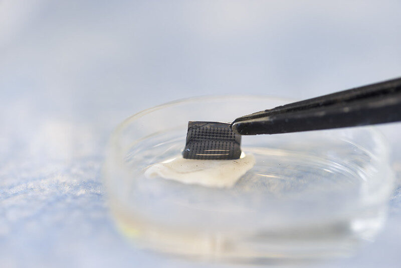
A microneedle patch captures cancer biomarkers in the top-most layer of skin to detect melanoma in animal tissue samples
The newly designed ExoPatch being removed from a sample of mouse skin successfully distinguished melanoma from healthy skin in mice. A gel coating the microneedles picks up cancer indicators from the top-most layer of the skin. Dissolving the gel releases exosomes into a solution, which is then used on a two-lined test strip, similar to an at-home COVID-19 test. Image credit: Jeremy Little, Michigan Engineering.
Melanoma testing could one day be done at home with a skin patch and test strip with two lines, similar to COVID-19 home tests, according to University of Michigan researchers. Developed with funding from the National Institutes of Health, the new silicone patch with star-shaped microneedles, called the ExoPatch, distinguished melanoma from healthy skin in mice.
The patch and test move toward rapid at-home melanoma testing, helping patients catch the most aggressive form of skin cancer early without a biopsy or blood draw.
“The star-shaped needles make puncture easier and less painful, but they are so small that they only go through the top-most layer of the skin, the epidermis, and do not draw blood,” said Sunitha Nagrath, the Dwight F. Benton Professor of Chemical Engineering at U-M and co-corresponding author of the study published in Biosensors and Bioelectronics.
The ExoPatch microneedles, at just 0.6mm long with a width of less than 100 nm (0.0001 mm) at the tip, are coated with a gel that picks up exosomes, tiny packages released by cells, from the interstitial fluid that fills the spaces between cells in the epidermis.
Once thought to be trash ejected from cells for cleanup, exosomes actually contain DNA and RNA fragments that cells use to communicate with each other. Cancer cell exosomes can help tumours spread by preparing tissues to accept tumour cells before arrival, and detecting them can catch cancer earlier than past methods.
The gel that coats the ExoPatch contains a protein called Annexin V that attracts and sticks exosomes to the microneedles’ surface. Once removed from the skin, placing the patch in an acid dissolves the gel, which releases the exosomes into a solution. After dipping a test strip into the solution, two lines form if the sample contains melanoma exosomes, and one line forms for a negative test – the same way an at-home COVID-19 test strip works.
“A fair-skinned person with moles must go to the doctor about every six months to send off a biopsy to see if they’re malignant or benign. With this test, they could instead test at home, get the results right away and follow up with a dermatologist for a positive result,” Nagrath said.
As a first step in the proof-of-concept study, the researchers tested the ExoPatch on a tissue sample of pig skin, which closely resembles human skin in thickness and composition. Using a microscope, they found the microneedles penetrated about 350 to 600nm into the skin. For scale, the epidermis on the human forearm is about 18 300nm thick.
To test whether the ExoPatch could capture melanoma exosomes from skin tissue, the research team tested tissue samples of mouse skin, half from healthy mice and half from mice injected with a fragment of a human melanoma tumour. After a 15-minute application, the ExoPatch was placed under a powerful microscope.
“When looking at microscopy images, I was happy to see how nicely the exosomes adhered to the microneedles and were within the 30 to 150 nanometre size range we expect,” said Scott Smith, U-M doctoral student of chemical engineering and co-lead author of the study.
After confirming the exosomes stuck to the ExoPatch, the researchers dissolved the gel and ran the sample through the test strips. The test successfully distinguished between melanoma and healthy tissues with a 3.5-fold darker line in melanoma samples.
The ExoPatch isolated 11.5 times more exosomal protein from melanoma tissue samples compared to healthy tissue, showing it can specifically target cancerous exosomes.
A pilot study in humans followed by a series of clinical trials will be the next steps to move the technology toward use. Beyond melanoma, the ExoPatch gel coating could be modified to detect exosomes released by other cancers with a solid tumour, including lung, breast, colon, prostate and brain cancer.
“This is the first patch designed to capture disease-specific exosomes from fluid under the skin. The potential applications are huge,” said Nagrath.