Black women experience disproportionately elevated risks of developing and dying from early-onset breast cancer. New research published by Wiley online in CANCER, a peer-reviewed journal of the American Cancer Society, reveals the genes that are most likely to be mutated to contribute to these increased risks.
In the study of 686 young Black women diagnosed in Florida and Tennessee with invasive breast cancer at age 50 or younger in 2005–2018, genetic testing showed that 15.3% of the women carried a gene variant with a suspected link to breast and/or ovarian cancer, with most occurring in the BRCA1 and BRCA2 genes and fewer in PALB2, ATM, and other genes. A family history of breast cancer was common in women with mutations in BRCA1, BRCA2, and PALB2. Triple-negative breast cancers (one of the most aggressive forms) were most often seen in women with BRCA1 mutations. Also, most of the women with BRCA1 mutations were diagnosed at or below age 40, whereas the age at diagnosis was more evenly distributed up to age 50 for women with variants in the other genes.
The study’s findings point to the importance of breast cancer genetic testing for young Black women, a group that is less likely to receive such screening compared with other racial and ethnic groups. Such tests could identify women most likely to benefit from more frequent screening and preventive measures to safeguard their health.
“We must test at-risk women across all populations – testing is essential to personalise treatment strategies and enable life-saving prevention for future cancers, and it may empower at-risk family members to get tested so they too can benefit from this information,” said senior author Tuya Pal, MD, of Vanderbilt University Medical Center. “Equitable access to inherited cancer testing ensures that all women, regardless of race, can benefit from precision medicine and take control of their genetic health.”
Patients with vitamin D deficiency may benefit from supplements before operations
Photo by Tima Miroshnichenko on Pexels
Vitamin D deficiency is associated with more moderate to severe pain following breast cancer surgery and an increased consumption of opioid drugs, finds research published online in the journal Regional Anesthesia & Pain Medicine. Breast cancer patients with low levels of vitamin D (below 30nmol/L) may benefit from taking supplements before undergoing a radical mastectomy, suggest researchers.
There is emerging evidence suggesting that vitamin D helps control how pain is felt and processed by the body. This is likely due to its anti-inflammatory effects and action on the immune system. Vitamin D deficiency is also commonly reported among patients with breast cancer. A team of researchers set out to examine the relationship between vitamin D deficiency and postoperative pain in patients undergoing breast cancer surgery.
Their prospective observational study, carried out at Fayoum University Hospital in Egypt between September 2024 and April 2025, included 184 breast cancer patients who were scheduled to undergo surgical removal of one entire breast.
Half of the patients were classified as vitamin D deficient (below 30 nmol/L) and half were classified as vitamin D sufficient (above 30nmol/L). Both groups had similar characteristics with an average age of 44 in the vitamin D deficient group and 42 in the vitamin D sufficient group.
Patients were managed according to the hospital’s routine protocol both during and after surgery. Clinical staff involved in their care were unaware of the patients’ vitamin D levels.
The opioid fentanyl was administered during the operation to manage acute pain. Following surgery, all patients were given paracetamol through a drip every 8 hours. In addition, patients could control how much tramadol (another opioid analgesic) they were given by directly pressing a button.
Patients reported their pain levels at zero, 6 hours, 12 hours, 18 hours and 24 hours after surgery. Nausea and vomiting, sedation score and days in hospital following surgery were also recorded.
Patients with vitamin D deficiency were three times more likely to report moderate to severe postoperative pain at any time point during the first 24 hours than those with sufficient vitamin D levels, the study found.
The researchers noted, however, that no patient in either group reported severe pain (7 or over on a scale of 0 to 10) so the difference was due entirely to a reduction in moderate pain (4-6 on the pain scale).
Vitamin D deficient patients received, on average, 8 μg more fentanyl during surgery, which the researchers described as a modest difference.
However, the study found those in the vitamin D deficient group used substantially more tramadol (112mg) after surgery than those who had sufficient vitamin D levels. This strong opioid was controlled directly by the patient up to a maximum dose of 50mg per hour.
Opioid drugs can cause a number of side effects including nausea, vomiting, drowsiness and confusion, while also carrying risks of dependency and addiction.
Postoperative nausea was more common in the vitamin D deficient group, and vomiting occurred only in that group, although the difference in vomiting was small and not statistically significant.
The study had some limitations. It was observational and conducted at a single centre, so no firm conclusion can be drawn about cause and effect. The researchers also did not assess inflammatory markers so could not explore the mechanisms underlying the relationship between vitamin D and pain. Data was also not collected on anxiety, depression, cancer stage, treatment or sleep disturbance before the surgery was carried out.
Nevertheless, the researchers conclude, “Vitamin D deficiency is associated with a higher occurrence of moderate to severe postoperative pain and increased opioid consumption in patients undergoing unilateral modified radical mastectomy.”
They suggest, “Preoperative vitamin D supplementation in breast cancer patients with vitamin D levels below 30 nmol/L may have a role in modulating postoperative pain.”
A new study from Karolinska Institutet shows that gene analysis of breast cancer tumours can identify patients who do not benefit from chemotherapy given before surgery. The findings, published in the journal Nature Communications, could in the long term contribute to more personalised treatment.
The study included 179 patients with hormone dependent, HER2 negative breast cancer who took part in the Swedish PREDIX LumB trial. Before surgery, all patients received both treatments, but in different sequences. They were given either chemotherapy followed by hormone blocking therapy together with the drug palbociclib, which slows the division of cancer cells, or the reverse sequence.
When the researchers analysed the results, they found that the treatments led to similar reductions in tumour size overall. Survival was also similar regardless of whether treatment started with chemotherapy or with palbociclib and hormone blocking therapy.
Not all tumours responded
At the same time, the analyses showed that there was a subgroup of tumours with a poorer response to chemotherapy but a better response to palbociclib in combination with hormone‑blocking therapy.
To understand why some tumours did not respond to chemotherapy, the researchers analysed tumour gene expression, how active different genes are in the tumour, in tissue samples taken before treatment started. Based on these analyses, they developed a model called CDKPredX, which can identify tumours that respond poorly to chemotherapy but better to palbociclib combined with hormone blocking therapy.
“Today, we lack reliable ways to determine in advance which patients will actually benefit from chemotherapy before surgery. Our results show that tumour gene expression can provide important information in this respect,” says first author Alexios Matikas, docent at the Department of Oncology‑Pathology, Karolinska Institutet.
The model is based on patterns of gene expression in the tumour, including genes involved in cell division, hormone signalling and the immune system. When the researchers tested the model in other patient groups, they observed similar patterns.
Further studies are needed
“In the longer term, this type of analysis could help patients avoid treatments that do not benefit them, such as chemotherapy, and instead receive treatment that has a better chance of working. At the same time, further studies are needed before the method can be used in clinical practice,” says senior author Theodoros Foukakis, professor at the same department.
The researchers emphasise that the study is exploratory and that the genetic analysis is not yet ready for clinical use. Nevertheless, the results provide new insights into why different tumours respond differently to treatment.
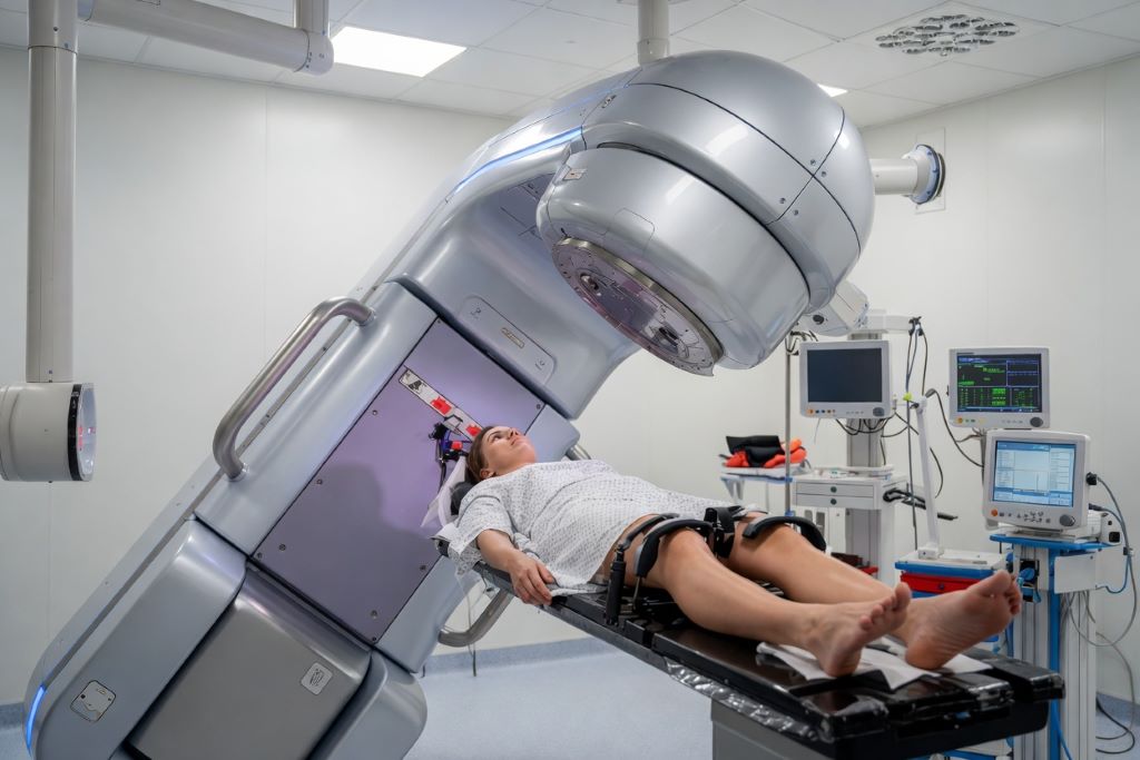
New UK research has found that a one-week course of post-surgery radiotherapy is just as safe and effective as the traditional three-week course for people with early-stage breast cancer.
The FAST-Forward trial, led by Keele’s Professor Murray Brunt and sponsored by The Institute of Cancer Research, London, followed more than 4000 patients for a decade after their treatment.
The 10-year results of this phase III randomised trial, funded by the National Institute for Health and Care Research (NIHR) and published in the Lancet Oncology, show that a shorter, five-day radiotherapy schedule provides the same level of cancer control as the traditional three-week treatment.
These findings build on previous five-year results that have already driven a shift in clinical practice. Since 2020, it is estimated that tens of thousands of people in the UK have already benefited from the shorter course on the NHS. Researchers expect this approach will also reduce the burden on people undergoing treatment for breast cancer worldwide and expand access to life-saving radiotherapy.
Breast cancer is one of the most common cancers globally, and radiotherapy plays a critical role in reducing the risk of recurrence after surgery. A shorter treatment course is not only more convenient for patients – it also reduces hospital visits and eases pressure on radiotherapy services, making treatment more accessible.
The research team compared the traditional schedule of 15 treatments over three weeks with two shorter schedules that used five treatments over one week. The two shorter courses gave slightly different amounts of radiation to allow the research team to work out the best dose.
After 10 years, cancer coming back in the treated breast was very low in all three groups: 3.6% for the standard three-week treatment, 2.9% with the one-week treatment with a slightly higher dose, 2.1% with the one-week treatment with a slightly lower dose.
The lower dose one-week treatment had side effects that were very similar to the standard approach, with no increase in long-term breast or chest wall changes. Because of this, this dose and schedule are now the recommended option.
Professor Murray Brunt, chief investigator of the FAST-Forward study and Professor of Clinical Oncology at Keele University, said: “These 10-year results provide definitive long-term evidence that one-week radiotherapy given at an appropriate dose to the breast is a safe, effective, and more practical option for people with breast cancer.
“By reducing treatment from 15 sessions to just five, we can offer patients the same excellent cancer control with fewer hospital visits, less disruption to their daily life, and reduced pressure on healthcare services. This approach has already transformed practice in the UK and has the potential to improve access to life-saving treatment for people with cancer worldwide.
“Hearing patients talk about how much it helps to only need one week of radiotherapy has been really encouraging for everyone involved.”
Professor Judith Bliss, Professor of Clinical Trials at The Institute of Cancer Research, London, who co-led the trial, said: “The FAST-Forward trial is transforming cancer treatment by reducing standard breast radiotherapy from 3 weeks to just one week, without compromising effectiveness.
“The streamlined schedule has made radiotherapy more accessible, particularly for people who find it difficult to attend hospital and those in lower-income countries.”
“The FAST-Forward trial is part of a long-term programme of research into improving breast cancer radiotherapy at The Institute of Cancer Research (ICR).
“These final 10-year results mark a significant milestone in breast cancer treatment and reinforce the growing shift toward more efficient radiotherapy approaches. The success of FAST-Forward has led to the ongoing NIHR-funded FAST-Forward Boost trial, which is investigating whether more extensive radiotherapy – including an additional ‘boost’ dose for some patients – can also safely be delivered in five days.
Professor Anthony Gordon, Director for NIHR’s Health Technology Assessment (HTA) Programme, added: “The legacy of the FAST-Forward trial is clear to see, with thousands of women benefiting from shorter courses of radiotherapy and fewer hospital visits, helping the health and care sector to achieve more effective and efficient use of resources. The new 10-year results show the benefit of investing in high-quality, long-term research to improve the health and wealth of the nation.
“NIHR’s research aims to tackle the most urgent health and social care challenges, targeting the areas of greatest need and where the most significant impact can be made. The findings from the FAST-Forward trial have already made a considerable difference to the treatment regimens that breast cancer patients undergo, making them more efficient, while at the same time, easing pressure on NHS services.”
Low doses of the investigational medicinal product endoxifen reduce breast density to the same extent as the standard treatment tamoxifen, but without causing such troublesome side effects. These are the findings of a new study from Karolinska Institutet which appears in the Journal of the National Cancer Institute. The results may have implications for future preventive treatment of breast cancer.
Tamoxifen is a well-established drug that has been used for more than 40 years to reduce the risk of recurrence in patients with breast cancer. The drug is also approved for prevention of breast cancer in women at increased risk.
However, the side effects of tamoxifen are a major problem. Many women experience menopausal-like symptoms, such as hot flushes, which means that many do not complete the treatment.
Endoxifen is the most active metabolite formed when tamoxifen is broken down in the body. The new study investigated whether endoxifen in tablet form could provide the same biological impact and a more predictable effect than tamoxifen.
A total of 240 healthy, premenopausal women were randomised to receive a placebo or 1 or 2 mg of endoxifen daily for six months. The researchers then measured mammographic breast density. High mammographic density can contribute to an increased risk of breast cancer but a reduction during treatment can be a good measure of therapeutic outcome.
The results show that 1mg of endoxifen reduced breast density by an average of 19% and 2mg by 26%. Data from a previous study show that 20mg of tamoxifen reduces density by approximately 18.5%. The effect of low-dose endoxifen thus corresponded to that seen with tamoxifen.
Participants who received 2mg of endoxifen reported a greater worsening of hot flushes and night sweats compared with the lower-dose group, whilst the 1 mg group had a safety profile similar to that of the placebo with respect to serious side effects and biomarkers.
”Our results suggest that a lower dose may be sufficient to affect breast density, whilst also appearing to be better tolerated,” says Mattias Hammarström.
The study is a so-called proof-of-concept trial, meaning it is designed to demonstrate that a treatment produces the expected biological effect before larger and longer trials are conducted. However, the study cannot show whether endoxifen reduces the risk of breast cancer or recurrence.
Rapid detection, treatment of infections could avoid complications, additional surgeries after mastectomy
Many of those women opt to have their breasts surgically reconstructed, most commonly with implants, but a relatively high percentage develop infections after implant surgery, requiring intravenous antibiotics and often removal of the implant. This can lead to additional surgeries, delays in cancer care and increased costs, as well as added emotional distress for women already under strain from cancer diagnosis and treatment.
To address this problem, researchers at Washington University School of Medicine in St. Louis have developed a new tool to detect reconstruction-related infections early, before they cause symptoms. This method, reported in the Journal of Clinical Investigation, could allow for preemptive treatment that preserves implants, improves patient outcomes and reduces the psychological and financial burden on patients.
Led by Jeffrey P. Henderson, MD, PhD, a WashU Medicine professor, the study identified biomarkers of infection in fluid drained from reconstruction patients’ breasts days or even weeks before symptoms appeared. This represents a major opportunity for improvement over existing diagnostic methods, which rely heavily on clinical symptoms, such as redness and inflammation, that take time to appear and can overlap with normal reactions to surgery.
The findings are available online and will publish in print Feb. 16 in the Journal of Clinical Investigation.
“The ability to identify with a molecular signature early on that a patient will go on to have an infection opens up the possibility of surveillance as part of standard care,” Henderson said. “This has the potential to enable earlier treatment that would be far more effective – and potentially curative – in patients who would otherwise progress to prolonged courses of treatment and surgery, or even implant removal and reconstructive failure.”
Small molecules, big impact
The study originated when Henderson’s WashU Medicine colleague Margaret A. Olsen, PhD, a retired professor of medicine in the Division of Infectious Diseases who studies hospital infections, noticed high rates of infection among US patients who had reconstruction with implants after mastectomy. The discovery prompted Henderson and Olsen, a co-author on the study, to ask WashU Medicine plastic surgeons who performed breast reconstruction what they would need to improve outcomes in these patients. Their answer was simple: a clear yes/no test for infection.
To develop such a test, Henderson and lead author John A. Wildenthal, an MD/PhD student, leveraged their expertise in metabolomics, the study of metabolites that are created or broken down during cellular processes in the body. Metabolites can indicate the presence of an infection because they include byproducts of both the body’s response to pathogens and the metabolic activity of the pathogens themselves. By analysing changes in metabolite levels, scientists can identify patterns that are characteristic of infections, enabling early diagnosis.
Henderson and colleagues coordinated with WashU Medicine plastic surgeons to obtain fluid samples from 50 patient volunteers during several routine follow-up visits after surgery. The patients included women who later developed infections after post-mastectomy reconstruction and those who did not.
The researchers analysed the samples for differences between the two groups and identified metabolites that were significantly associated with infection and that appeared days to weeks before clinical signs and symptoms of infection. Further, they found that the presence of certain metabolites indicated more serious infections that might require more aggressive treatment.
“Originating from clinical intuition and validated through a clinical study, the evidence in this paper now supports proactive, targeted interventions to predict and address infections before they become clinically significant,” said Justin M. Sacks, MD, a co-author on the paper. “Such interventions can substantially reduce the burden of complications, implant loss and reconstructive failures in these patients.”
For instance, the findings could lead to the development of a point-of-care test that could be provided during a woman’s routine post-operative visits, noted co-author Terence M. Myckatyn, MD, a professor of surgery at WashU Medicine, who performs plastic and reconstructive surgery for breast cancer patients.
“If the test is positive, antibiotics can be started preemptively in these select patients to thwart infection,” Myckatyn said. “And perhaps just as important, we would not give antibiotics to those with a negative test, thereby adhering to a thoughtful approach for antibiotic stewardship.” Such careful use of antibiotics is important for preventing antibiotic resistance, he said.
In the near term, the team is planning additional studies to validate the results. Then a diagnostic tool could be developed and tested in clinical practice. In the future, the broader metabolomic findings about the development of tissue infection in humans could allow physicians to more selectively target a variety of post-surgical infections, for example, by revealing new drug targets.
“While better techniques are always being sought, the reality is that infections still occur despite a meticulous surgical approach,” said Myckatyn. “To be able to identify biomarkers that can portend an infection days before it develops is huge.”
Mass General Brigham’s evaluation of low-field MRI performance lays potential groundwork for this technology to be a lower-cost, accessible option for breast imaging
Photo by National Cancer Institute on Unsplash
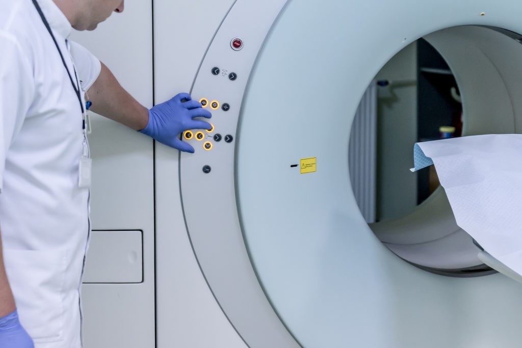
Researchers at Mass General Brigham have demonstrated the technical feasibility of using ultra-low field (ULF) magnetic resonance imaging (MRI) for breast imaging. With further refinement and evaluation, the technology could offer an alternative to existing breast cancer screening methods and may reduce barriers to screening. Results are published in Scientific Reports.
“These results are a very encouraging proof of principle, though larger studies are needed to establish diagnostic performance,” said project principal investigator and co-senior author Matthew Rosen, PhD, an associate professor of Radiology and director of the Low Field MRI laboratory in the Athinoula A. Martinos Center for Biomedical Imaging in the Mass General Brigham Department of Radiology. “They motivate our continued pursuit of safe, comfortable, lower-cost screening approaches that can expand access for patients.”
Current U.S. guidelines recommend screening mammography for women aged 40 to 74 years. Unlike mammography, ULF MRI doesn’t require breast compression, which many patients find uncomfortable. Another benefit of ULF MRI is that it doesn’t use ionizing radiation.
While higher risk patients may receive MRI screening for breast cancer, standard MRI machines are not used in routine breast cancer screening because they are expensive and not widely available. ULF MRI systems cost less than 5% of the price of standard MRI systems and have lower long-term operating costs.
In this study, ULF MRI scans were performed on 14 participants, including 11 women with no history of breast cancer, two women with a prior breast cancer diagnosis, and one woman with a benign mass.
When interpreting the ULF MRI scans, three radiologists could reliably identify essential breast features and distinguish fibroglandular tissue from adipose tissue. The authors note that discrepancies were likely related to the novelty of ULF MRI and may be reduced with additional training and experience.
“This early evidence suggests that ULF MRI can detect essential breast features and some abnormalities without radiation or injected contrast,” said co-first author Neha Koonjoo, PhD, an investigator at the Martinos Center. “These findings point to the potential for ULF MRI as an option that could complement existing screening tools in the future.”
“Even at very low magnetic field, the radiology team was able to make observations about the breast,” said co-principal investigator and co-senior author Kathryn E. Keenan, PhD, from the US National Institute of Standards and Technology. “We attempted this study in hopes that the breast features would be visible, but you don’t always have success. We’re very motivated by this study to continue our work on ultra-low-field MRI for breast screening.”
The researchers note that further study is needed to determine the diagnostic accuracy of ULF MRI for breast cancer screening, including studies in larger cohorts that include patients with benign and malignant lesions. They also emphasize that further refinements in ULF MRI technology are needed to meet clinical resolution standards for breast cancer screening.
“These results will guide the next engineering steps to improve image quality and enable a more comfortable exam and help bring screening to more settings and more patients,” said co-first author Sheng Shen, PhD, of the Martinos Center for Biomedical Imaging.
The potential effect of induced abortion and miscarriage on the risk of breast cancer has remained debated and has been a persistent source of misinformation. Many previous studies have been small and based on self-reported data. Now, a study published in Acta Obstetricia et Gynecologica Scandinavica found that prior abortion or miscarriage was not linked with an increased risk of developing pre- or postmenopausal breast cancer.
In the nationwide Finnish registry-based study, investigators analysed data on 31 687 women with breast cancer diagnosed in 1972–2021 and 158 433 women without breast cancer.
The risk of breast cancer was found to be similar among women with a history of induced abortion and women with no history of abortion, both before and after 50 years of age. Risks were also similar among women with and without a past miscarriage.
In addition, breast cancer risks did not vary significantly by the number of abortions or miscarriages, nor by the time of first abortion or miscarriage.
“Miscarriage or induced abortion as potential risk factors for breast cancer has continued to raise concerns and has led to the spread of misinformation. In this study using high-quality Finnish registry data, we can reliably eliminate these concerns,” said corresponding author Oskari Heikinheimo, MD, PhD, of the University of Helsinki and Helsinki University Hospital. “Induced abortion or miscarriage are not risk factors for breast cancer, even if there are several of them. This information is important and reassuring for millions of women around the world.”
A drug mimicking the hormone progesterone has anti-cancer activity when used together with conventional anti-oestrogen treatment for women with breast cancer, a new Cambridge-led trial has found.
In the two-week window that we looked at, adding a progestin made the anti-oestrogen treatment more effective at slowing tumour growth. What was particularly pleasing to see was that even the lower dose had the desired effectRebecca Burrell
A low dose of megestrol acetate (a synthetic version of progesterone) has already been proven as a treatment to help patients manage hot flushes associated with anti-oestrogen breast cancer therapies, and so could help them continue taking their treatment. The PIONEER trial has now shown that the addition of low dose megestrol to such treatment may also have a direct anti-cancer effect.
Around three-quarters of all breast cancers are ER-positive. This means the tumours are abundant in a molecule known as an oestrogen receptor, ‘feeding’ on the oestrogen circulating in the body. These women are usually offered anti-oestrogens, medication that reduces level of oestrogen and hence deprives the cancer of oestrogen and inhibits its growth. However, reducing oestrogen levels can bring on menopause-like symptoms, including hot flushes, joint and muscle pain, and potential bone loss.
In the PIONEER trial, post-menopausal women with ER-positive cancers were treated with an anti-oestrogen with or without the progesterone mimic, megestrol. After two weeks of treatment, those that received the combination saw a greater decrease in tumour growth rates compared to those treated with an anti-oestrogen only.
Although further work is required in larger patient cohorts and over a longer period of time to confirm the findings, researchers at the University of Cambridge say the trial suggests that megestrol could help improve the lives of thousands of women for whom anti-oestrogen medication causes uncomfortable side-effects and can lead to some women stopping taking the medication.
PIONEER was led by Dr Richard Baird from the Department of Oncology at the University of Cambridge and Honorary Consultant Medical Oncologist at Cambridge University Hospitals NHS Foundation Trust (CUH). He said: “On the whole, anti-oestrogens are very good treatments compared to some chemotherapies. They’re gentler and are well tolerated, so patients often take them for many years. But some patients experience side effects that affect their quality of life. If you’re taking something long term, even seemingly relatively minor side effects can have a big impact.”
Some ER-positive breast cancer patients also have high levels of another molecule, known as progesterone receptor (PR). This group of patients also respond better to the anti-oestrogen hormone therapy.
To explain why, Professor Jason Carroll and colleagues at the Cancer Research UK Cambridge Institute used cell cultures and mouse models to show that the hormone progesterone stops ER-positive cancer cells from dividing by indirectly blocking ER. This results in slower growth of the tumour. When mice treated with anti-oestrogen hormone therapy were also given progesterone, the tumours grew even more slowly.
Professor Carroll, who co-leads the Precision Breast Cancer Institute and is a Fellow of Clare College, Cambridge, said: “These were very promising lab-based results, but we needed to show that this was also the case in patients. There’s been concern that taking hormone replacement therapy – which primarily consists of oestrogen and synthetic versions of progesterone (called progestins) – might encourage tumour growth. Although we no longer think this is the case, there’s still been residual concern around the use of progesterone and progestins in breast cancer.”
To see whether targeting the progesterone receptor in combination with an anti-oestrogen could slow tumour growth in patients, Dr Baird and Professor Carroll designed the PIONEER trial, which tested adding megestrol, a progestin, to the standard anti-oestrogen treatment letrozole.
A total of 198 patients were recruited at ten UK hospitals, including Addenbrooke’s Hospital in Cambridge, and randomised into one of three groups: one group received only letrozole; one group received letrozole alongside 40mg of megestrol daily; and the third group received letrozole plus a much higher daily dose of megestrol, 160mg. In this ‘window of opportunity’ trial, treatment was given for two weeks prior to surgery to remove the tumour. The percentage of actively growing tumour cells was assessed at the start of the trial and then again before surgery.
In findings published today in Nature Cancer, the team showed that adding megestrol boosted the ability of letrozole to block tumour growth, with comparable effects at both the 40mg and 160mg doses.
Joint first author Dr Rebecca Burrell from the Cancer Research UK Cambridge Institute and CUH said: “In the two-week window that we looked at, adding a progestin made the anti-oestrogen treatment more effective at slowing tumour growth. What was particularly pleasing to see was that even the lower dose had the desired effect.
“Although the higher dose of progesterone is licenced as an anti-cancer treatment, over the long term it can have side effects including weight gain and high blood pressure. But just a quarter of the dose was as effective, and this would come with fewer side effects. We know from previous trials that a low dose of progesterone is effective at treating hot flushes for patients on anti-oestrogen therapy. This could reduce the likelihood of patients stopping their medication, and so help improve breast cancer outcomes. Megestrol – the drug we used – is off-patent, making it a cost-effective option.”
Because women in the trial were only given megestrol for a short period of time, follow-up studies will be needed to confirm whether the drug would have the same beneficial effects with reduced side-effects over a longer period of time.
The research was funded by Anticancer Fund, with additional support from Cancer Research UK, Addenbrooke’s Charitable Trust and the National Institute for Health and Care Research Cambridge Biomedical Research Centre.
Personalised and precise cancer treatments underpin the focus of care at the future Cambridge Cancer Research Hospital. The specialist facility planned for the Cambridge Biomedical Campus will bring together world-leading researchers from the University of Cambridge and its Cancer Research UK Cambridge Centre and clinical excellence from Addenbrooke’s Hospital under one roof in a brand-new NHS hospital.
Adding breast magnetic resonance imaging (MRI) to a diagnostic mammogram did not reduce five-year cancer recurrence rates for patients with stage I/II hormone receptor (HR)-negative breast cancer, according to researchers at The University of Texas MD Anderson Cancer Center.
The Phase III Alliance A011104/ACRIN6694 trial found that five-year locoregional recurrence rates were 6.8% in patients who received an MRI as part of a diagnostic work-up and 4.3% in those who did not. These data were presented today at the San Antonio Breast Cancer Symposium (SABCS) by principal investigator Isabelle Bedrosian, MD, professor of Breast Surgical Oncology (Abstract GS2-07).
“We have long assumed that finding more breast cancer on an MRI and removing it with surgery would help lower the chance of a patient’s cancer coming back,” Bedrosian said. “When you look at our findings alongside earlier trials, the message is clear: adding MRI before surgery doesn’t improve results for patients – and may not have to be used as a standard part of the diagnostic process.”
No additional MRI benefit in this group
The trial enrolled 319 patients between 2014 and 2018 with newly diagnosed stage I or II HR-negative breast cancer. These patients were eligible for lumpectomy and did not have germline BRCA1/2 mutations, bilateral breast cancer or a history of prior breast cancer. All patients had undergone diagnostic mammography with or without ultrasound prior to trial enrolment. Patients were randomly assigned to undergo additional imaging by breast MRI (161 patients) or to receive no further imaging (158 patients).
Not only did breast MRI not impact five-year recurrence rates, but there were also not significant differences between groups for five-year distant recurrence-free survival nor overall survival.
A small subset of patients with tumour subtypes (HR- HER2+ and HR-HER2-) and those over the age of 50 at diagnosis also showed no benefit to MRI.
Pre-op MRI not finding anything important
Breast MRI is a common part of the diagnostic evaluation because it can reveal cancer that mammography might not detect. However, the evidence that it improves surgical outcomes for patients has been limited.
“We believe the reason MRI did not reduce recurrence rates may be twofold,” Bedrosian said. “It is possible that MRI didn’t uncover many lesions that mammography hadn’t already found, or perhaps identifying and surgically removing those additional lesions was not important to reducing risk of the cancer coming back. It’s possible that in the group that did not receive MRI, radiation and chemotherapy effectively treated the occult areas of disease”.
Experts are now analysing how often breast MRI identified additional lesions in the trial population to better understand why breast MRI did not impact oncologic outcomes.
Study limitations
Limitations included that most patients involved in the trial had breast cancer that hadn’t spread to their lymph nodes, which may partly explain why recurrence rates were low overall. Despite being open to women of all ages, the study enrolled mostly older women who may have been less likely to benefit from breast MRI.