Study indicates the need for a revised definition of functional high-risk multiple myeloma
Depiction of multiple myeloma. Credit: Scientific Animations
Researchers have found that with new treatments for multiple myeloma, a serious type of blood cancer, clinicians should use different criteria for identifying patients with poor odds of survival. The study is published by Wiley online in CANCER, a peer-reviewed journal of the American Cancer Society.
Functional high-risk (FHR) multiple myeloma, which affects a subset of patients with blood cancer, is commonly defined as a multiple myeloma that progresses within 18 months of starting treatment, and it comes with a survival prognosis of less than 2 years after progression. Recently, however, the use of a 4-part treatment regimen – including anti-CD38 antibodies, proteasome inhibitors, immunomodulatory agents, and dexamethasone – followed by autologous stem cell transplantation, has helped to slow the cancer’s progression.
To update the definition of FHR multiple myeloma in the current era of combination therapy, investigators from the University of Alabama at Birmingham and the CoMMiT consortium analysed information on 310 patients with newly diagnosed multiple myeloma who received this therapy and were followed for a median of 3.5 years.
Survival analyses indicated that cancer progression within 36 months of the onset of combination therapy identified patients whose survival was likely under 2 years from the time of progression. Adding another treatment called T-cell redirecting therapy helped slow progression. This “FHR36” patient population, corresponding to 16.4% of all treated patients, should be prioritised as candidates for early use of T-cell redirecting therapy and for clinical trials of medications with novel mechanisms of action.
“The findings will help physicians choose therapies for this important minority of patients who have disease progression in the first 3 years of diagnosis and identify an important population in greater need for treatment innovations to be addressed in the next generation of clinical trials,” said senior author Luciano J. Costa, MD, PhD, of the University of Alabama at Birmingham.
By Palesa Mokomele, Head of Community Engagement and Communication at DKMS Africa
For years, Black South African patients with blood cancer or blood disorders searching for a stem cell donor were told the odds were not in their favour. Their ancestry, the thinking went, carried too much genetic diversity for a donor search to reliably succeed. New research published in Blood Global Hematology says otherwise. The genetics, it turns out, are not the problem. What stands between many Black patients and a lifesaving transplant is the registry, and who is on it.
A study of nearly 57 000 South Africans mapped the HLA profiles of all four of the country’s population groups to calculate each community’s real odds of finding a match. HLA markers are what doctors use to determine whether a donor’s stem cells are compatible with a patient’s. In a same-community registry of a million donors, a Black South African patient’s probability of finding a full match is 80%. For a White patient, the figure is 81%.
The Registry Gap
The registry does not reflect the country it serves, and that gap is costing lives. South Africa is approximately 81% Black African and 7% White. In the study cohort, Black South Africans made up roughly 37% of donors and White South Africans 45%. Donor drives went where they were easiest to run, and the communities hardest to reach were left out.
The findings revealed large areas of the country where Black African donor representation is almost absent. Entire provinces contribute too few donors for researchers to meaningfully analyse matching patterns. Of the fifteen subpopulations large enough to analyse by language and province, seven were White communities.
The two least genetically diverse groups in the study were Afrikaans-speaking White people in Mpumalanga and isiXhosa-speaking Black Africans in the Western Cape. Because people in these communities are more genetically similar to one another, each new donor has a better chance of matching someone who needs them.
The registry is at least heading in the right direction. Fifty-six percent of new registrations come from people of colour. But it holds just 200 000 donors across all population groups, and the destination is a registry of a million Black African donors alone.
The Human Cost
Consider what that means for a patient like Sbahle, a six-year-old girl diagnosed with aplastic anaemia before she could pronounce the word. She has spent four years, most of her life, waiting for a donor. She is stable, back at school, and still without a match. Not long ago, a family in her position would have been told the odds were against them, and there was little to be done. We know better now.
For a patient still waiting, the donor who could save them may already be out there, alive, healthy, and simply not on the registry.
Not Everyone’s Odds Are the Same
The progress is not uniform. Coloured and Indian/Asian South Africans face a harder search: their greater genetic diversity means common profiles cover far less of the group, around 21% for Coloured patients, and full matches are rarer.
The outlook is not bleak. In a registry of 100 000 donors from their own group, a Coloured patient has roughly a 51% chance of a nine-out-of-ten match and 92% of an eight-out-of-ten. Advances in transplant medicine are also shifting what a near-match means in practice. Recent research has shown that post-transplantation cyclophosphamide, a drug used to prevent graft-versus-host disease, significantly reduces the impact of donor mismatches, to the point where a single mismatch may be clinically negligible. For Coloured and Indian/Asian patients who cannot find a full match, that development changes the calculation considerably.
From Research to Reality
This research also points to where investment should go. The biggest gains will not come from a new algorithm or a better search tool. They will come from signing up donors in the communities the registry has missed. That is slow, expensive, deeply unfashionable work, and it is the thing that actually gets a patient to a transplant.
Recruitment has to reach into Limpopo, KwaZulu-Natal, the Eastern Cape, the Northern Cape and the rural areas with no donor base at all, and it has to be permanent, not a run of once-off drives.
Science has now answered one of the biggest questions in stem cell transplantation: Black South Africans are not inherently harder to match. The challenge is building a registry that reflects the country itself. Every new donor brings that goal, and patients like Sbahle, closer.
UP Vice-Principal: Research, Innovation and Postgraduate Education Prof Sunil Maharaj, OMT chair Rebecca Oppenheimer, Prof Mike Sathekge, head of the Department of Nuclear Medicine at UP and Steve Biko Academic Hospital, as well as President and CEO of NuMeRI, UP Vice-Chancellor and Principal Prof Francis Petersen and OMT CEO Tracey Webster.
Breast cancer is the leading type of cancer among women in South Africa, and globally. Too often it is discovered too late – but a new approach promises a radical change in survivability for patients.
The solution, called theranostics, stems from the field of nuclear medicine. It holds the potential to turn the tide against breast cancer and, like the fight against HIV/Aids, change it from an outright killer to a manageable disease.
“Theranostics, brings diagnosis and treatment together, is a combination of early diagnosis with treatment that is personalised and precise down to mere cells, which allows us to exactly detect and assess tumours, devise specific treatment regimens and assessment of treatment response over time,” says Sathekge.
“The earlier the breast cancer is detected, the more accurately it is assessed and the more precise the treatment, the exponentially better the patient’s prognosis.”
In South Africa, however, this is too often not the outcome. A comprehensive study into the availability of breast cancer services in the public healthcare sector, published recently in the South African Medical Journal, found that 67% of patients had late-stage breast cancer at diagnosis. In other words, their cancer had metastasised and spread to other parts of their bodies, and their prognosis was poor.
The study also noted that South Africa is expected to face a substantial rise in cancer cases over coming decades, driven by population growth, ageing and changing disease patterns. Sathekge’s Harry Oppenheimer Fellowship Award nomination was one of 80 received by OMT, covering a wide range of academic fields. Finalists shortlisted included proposals on a system for measuring the environment, air and our health; frost exposure in tropical Africa; a model for testing modified gravity; conversion of CO2 into useful products (such as fertilisers); and tissue T-cell response profiling of tuberculosis. “There were so many excellent applicants for this year’s award, touching on vital issues impacting the world we live in, and worthy of further research and development,” says OMT chairperson Rebecca Oppenheimer.
“This made our selection panel’s final decision all the more challenging, but I believe we have made an exciting choice that will have far-reaching, positive ramifications for South Africa’s public healthcare system and the people who use it.
“In developing a technology that makes diagnosing and treating cancers more effective, affordable and available, Prof Sathekge and his colleagues hold in their hands the potential for a quantum leap forward in improving South African patients’ health outcomes and human dignity, as well as for beating breast cancer globally. OMT is proud to support his endeavours.”
Sathekge’s solution is, essentially, one whose time has finally come. First conceived around 15 years ago, it capitalises on a protein called trophoblast cell-surface antigen 2, or Trop2. This molecule, found in high levels in breast cancers (and others, including cervical, pancreatic and lung cancers), helps the cancer multiply and makes it stubborn to treat.
Although Trop2 is already recognised as an important target in several cancers, there is still no widely established, clinically scalable way to show exactly where Trop2 is present across a patient’s entire cancer burden.
The answer may lie in nanobodies: tiny, engineered antibody fragments that are designed to bind specifically to Trop2. Their small size allows them to reach tumours rapidly and clear from the bloodstream faster than conventional antibodies, making them particularly attractive for same-day PET imaging.
Through a long-standing collaboration with Prof Frederik Cleeren, assistant professor in the Laboratory for Radiopharmaceutical Research, in the Department of Pharmaceutical and Pharmacological Sciences, and his team at KU Leuven in Belgium and the Joint Research Centre (JRC) in Karlsruhe, who bring expertise in nanobody engineering and radiolabelling, Sathekge’s team is combining these strengths with South Africa’s capabilities in molecular imaging, actinium-225 radiopharmaceutical development and targeted radionuclide therapy.
Together, the collaborators are developing a Trop2-targeted theranostic approach that links diagnosis with treatment. The first step uses a tiny targeting protein, known as a nanobody, designed to bind specifically to Trop2 on cancer cells. This nanobody is labelled with fluorine-18, a short-lived radioactive tracer that allows doctors to visualise Trop2-positive tumours on a PET/CT scan.PET/CT is an advanced imaging method that uses a small amount of radioactive tracer to show biological activity inside the body. In this case, it could help clinicians map Trop2 expression across a patient’s full cancer burden, including disease that may not be accessible for repeated biopsy. It may also allow doctors to monitor changes in the target and treatment response over time.
Where imaging confirms sufficient Trop2 expression, the same targeting strategy can be developed for treatment using actinium-225, a powerful alpha-emitting isotope. Actinium-225 can deliver highly localised radiation over a very short distance, with the aim of concentrating treatment in Trop2-positive cancer cells while limiting radiation exposure to surrounding healthy tissue.
The ambition is to move beyond treating patients based on limited information from a single biopsy, towards a more personalised approach: seeing the target throughout the body, selecting patients more accurately, and laying the foundation for future Trop2-targeted alpha therapy.
Sathekge’s work puts South Africa at the forefront of worldwide research into effective responses to breast cancer, says the University of Pretoria’s Vice-Chancellor and Principal, Prof Francis Petersen.
“South Africa urgently needs better ways to detect, understand and treat aggressive breast cancer. Too many patients still present late, when the disease is more difficult to manage and treatment options are limited.
“Prof Sathekge’s work at NuMeRI brings together advanced imaging, radiopharmaceutical science and targeted treatment in a way that could help doctors make more informed, patient-specific decisions. The research aims to improve how cancer is identified, how treatment is selected and how response is monitored over time.
“It also demonstrates the depth of scientific talent, innovation and academic rigour in South Africa. Through work of this calibre, African researchers are not only responding to local health challenges, but helping to shape the global future of cancer care. We look forward to seeing this research strengthen South African capacity and contribute to better outcomes for patients here and internationally,” says Petersen.
For Sathekge, the most exciting element of his work is how it centres on the patient, giving them dignity and the opportunity to live long and fulfilling lives.
Men and women experience many diseases very differently. Certain diseases present more commonly in one sex than in another. Some conditions like heart attacks may cause different symptoms in men and women. Some treatments work better or not at all for one sex over the other.
Cancer is no exception. There are major differences in male and female immune systems, a system critical for cancer’s growth and for successfully becoming cancer-free. For example, some immunotherapies work better in men than in women and vice versa.
Glioblastoma, the most common and fatal form of brain cancer, is more common and more deadly in men than in women. The reasons behind this difference and how the cancer’s biology differs between men and women remain largely unclear.
Now, a study has identified a cellular mechanism that differs between male and female laboratory models with glioblastoma. The study was published in the journal Nature Cancer and led by Defne Bayik, PhD, assistant professor of molecular and cellular pharmacology at the University of Miami Miller School of Medicine, and Asmita Pathak, PhD, a former postdoctoral fellow in the Bayik Lab.
“We have a growing appreciation that cancer doesn’t act the same way in men and women. There are differences in incidence rates. There are differences in treatment responses. There are differences in outcomes,” Dr Bayik said. “But we don’t really have a good, fundamental understanding of the mechanisms underlying these observational studies.”
Delving Into Immune Differences
To uncover that mechanism for glioblastoma, Dr Bayik and her colleagues focused on a certain class of immune cells in the brain known as myeloid-derived suppressor cells, or MDSCs. As their name suggests, these cells suppress other cells’ immune activity, especially that of T cells. In healthy contexts, their activity is important for regulating the immune system and keeping inflammation under control. But in the context of cancer, these cells are often recruited by tumours to suppress surrounding T cells and other immune cells, protecting cancerous cells from the rest of the immune system and allowing them to grow unchecked.
In previous work, Dr Bayik found sex-specific differences in the immune landscape of glioblastoma, with higher levels of monocytic myeloid-derived suppressor cells associated with disease in male laboratory models. Granulocytic MDSCs play a more prominent role in females. In studies of human glioblastoma tumours, she observed a similar pattern. Men tend to have more monocytic MDSCs within their tumours, Granulocytic MDSCs, or proteins associated with these cells, correlate with worse outcomes for women but not for men.
Women still constitute 40% of glioblastoma patients. By identifying these differences, we can better tailor treatments for both men and women.
Dr Defne Bayik
In the new study, Dr Bayik and her colleagues wanted to understand what drives this difference. How do granulocytic MDSCs act to promote cancer growth in female but not male laboratory models? In Dr Bayik’s previous study, she’d found several drug candidates that are predicted to act on granulocytic MDSCs. A few of these candidate drugs target proteins related to GABA, a brain signaling molecule also known as a neurotransmitter.
By exposing different populations of MDSCs to GABA in the lab, the scientists found that the neurotransmitter specifically affects cellular metabolism only in female granulocytic MDSCs. The process is unaffected in male MDSCs. They also found that this reprogramming of the cells’ metabolism by GABA made the granulocytic MDSCs more immunosuppressive. Finally, they found that blocking the GABA receptor in female laboratory models with glioblastoma improved their outcome. This had no effect on male laboratory models with the cancer.
Potential for treatment personalised by sex
Dr Bayik and her colleagues found that many of their lab findings held up in human samples donated by patients with glioblastoma. Tumour biopsies from women had higher levels of GABA and the GABA receptor in granulocytic MDSCs than did those from men. They also found that GABA reprograms granulocytic MDSC metabolism in women as it does in lab models.
These findings point to the potential for a sex-specific treatment for glioblastoma, Dr. Bayik said. She’s currently working to understand the basis for the difference in cellular metabolism in these immune cells between male and female laboratory models. Further uncovering the mechanism of this sex difference will help her and other scientists find new potential drug targets for the disease. MDSCs are involved in many other types of cance. Drugs that target these cells could have broader applications than just glioblastoma.
“Glioblastoma may be more common in men, but women still constitute 40% of patients,” said Dr Bayik. “By identifying these differences, we can better tailor treatments for both men and women.”
Data from more than 91 000 participants in the UK Biobank who wore activity monitors for seven days revealed an association between prolonged sedentary behaviour and the risk of cancer death
Each additional hour of prolonged, uninterrupted sedentary behaviour in a person’s day is associated with a 9% higher risk of cancer death, according to a study published July 2nd in the open access journal PLOS Medicineby Frederick Ho of the University of Glasgow, UK, and colleagues.
Previous studies have shown that spending more total time on sedentary behaviour, such as sitting, reclining or lying down while awake, is linked to poorer health outcomes. However, most sedentary behaviour guidelines focus on total time spent sedentary, rather than whether that time is accumulated in many short intervals or fewer prolonged intervals.
In the new study, researchers analysed data from 91 292 UK Biobank participants who had worn activity monitors for 7 days and were followed for a median of 12.38 years afterward. Activity was categorised as either prolonged sedentary (bouts of at least 30 minutes with at least 90% of time sedentary), interrupted sedentary behaviour (which lasted less than 30 minutes or was broken up with more than 10% non-sedentary time), or varying degrees of physical activity.
Prolonged sedentary behaviour was associated with a higher risk of cancer mortality (HR 1.09; 95% CI 1.06, 1.11), overall cancer incidence, obesity-related cancers (such as oesophageal, liver, kidney, pancreatic, colorectal, breast, ovarian, and thyroid cancers), and type 2 diabetes-related cancers. Interrupted sedentary behaviour showed the opposite pattern, associated with lower risk across all outcomes. Replacing one hour per day of prolonged sedentary behaviour with light physical activity was associated with a 12% lower risk of cancer death (HR 0.88; 95% CI 0.79, 0.99).
As a single-cohort study of UK Biobank volunteers, who have known health volunteer bias and higher physical activity levels than the general UK population, the findings may not be generalisable and do not prove causality. The researchers also had no data on the context of sedentary behaviour, such as whether it was during work or driving.
“Our findings suggest that the health effects of sedentary behaviour may depend not only on total sedentary time, but also on whether that time is accumulated in prolonged bouts or interrupted by activity,” the authors say. “This pattern is biologically plausible: experimental studies have shown that interrupting prolonged sitting with short bouts of activity can improve metabolic responses compared with uninterrupted sitting.”
The authors add, “Current health guidelines focus heavily on moderate or vigorous exercise, but our findings show that light movement shouldn’t be ignored. Moving forward, clinical trials will help us move beyond blanket advice and develop personalised strategies for breaking up sitting time.”
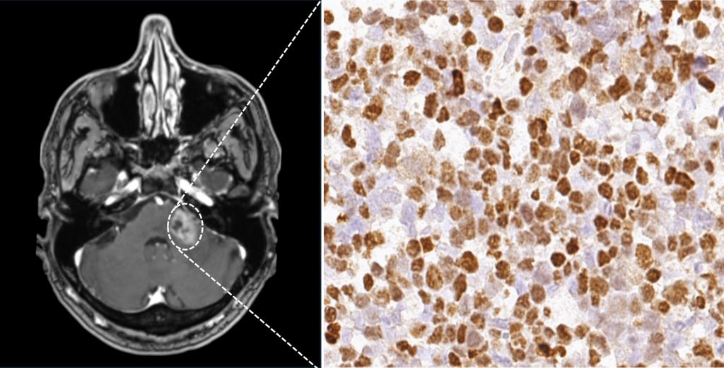
Primary CNS lymphomas in immunocompromised patients are among the rarest and at the same time most aggressive cancers – yet evidence-based recommendations for diagnosis and treatment have been lacking. An international research team led by Heidelberg Faculty of Medicine at Heidelberg University and the German Cancer Research Center has now identified characteristic imaging features of these tumours and developed a prognostic model to better assess disease outcomes. The researchers found that tumours positive for Epstein-Barr virus were associated with particularly poor prognosis. The findings, published in the journal Blood, may help guide future diagnostic and treatment strategies.
Primary CNS lymphoma is a rare cancer that arises from malignant white blood cells. Affected individuals develop tumours in the brain and, more rarely in the spinal cord, the eyes, or within the cerebrospinal fluid. These lymphomas can occur in people with weakened immune systems, for example after organ transplantation, in autoimmune diseases, or in association with HIV infection. This subtype, known as immunodeficiency-associated primary CNS lymphoma (ID-PCNSL) affects approximately 50 people per year in Germany. Researchers at the Heidelberg Faculty of Medicine at Heidelberg University had already shown in earlier work that ID-PCNSL is not simply a variant of classical CNS lymphoma, but a distinct disease entity characterised by specific genetic alterations.
A recent study by the International Primary CNS Lymphoma Collaborative Group – an international network dedicated to the study of CNS lymphomas – has now provided further insights into the diagnosis and treatment of ID-PCNSL. Including data from 308 patients treated at 23 hospitals across seven countries, the study is the largest conducted to date on this rare cancer. The researchers analysed clinical findings, magnetic resonance imaging scans, and tumour tissue samples. Scientists from the Heidelberg Faculty of Medicine at Heidelberg University and the German Cancer Research Center (DKFZ) played a leading role in the study.
Epstein-Barr Virus influences imaging and prognosis
Epstein-Barr virus (EBV) is known to play a central role in this cancer and was detected in 79.2 percent of the tumours examined. “Our analyses show that EBV-positive tumours often follow a more aggressive course and are associated with an unfavourable prognosis,” says first author Dr Leon Kaulen. He conducts research at the Heidelberg Faculty of Medicine at Heidelberg University and at the DKFZ and is a physician at the Department of Neurology at Heidelberg University Hospital (UKHD). EBV-positive tumours also showed characteristic imaging features including different patterns of contrast enhancement compared with EBV-negative tumours.
The researchers developed a prognostic model based on three factors that can help to better predict the course of ID-PCNSL and patient survival more accurately. The three identified factors are the detection of EBV in tumour tissue, age, and the patient’s performance status. Depending on the combination of these three factors, disease courses differed markedly among the patients studied. If only one of the three risk factors was present, the median survival was 135 months. With two risk factors, it decreased to 29 months. If all three factors were present, median survival was reduced to three months. “The prognostic model, with its clear stratification, represents a major advance. It will enable us to assess patients much more precisely in the future and to tailor therapies more effectively to the individual clinical situation,” says senior author Professor Wolfgang Wick, Heidelberg Faculty of Medicine at Heidelberg University, Chair of the Department of Neurology at UKHD, and Head of the Clinical Cooperation Unit Neurooncology at DKFZ and UKHD.
Considering immunodeficiency and cancer together
So far, no standardised therapy has been established for patients with immunodeficiency-associated primary CNS lymphoma. “Our research provides important insights into which approaches may be associated with more favourable outcomes,” says Dr Leon Kaulen. Patients whose immune system could at least partially be reconstituted – for example through adjustment of immunosuppressive medication or effective treatment of HIV infection – and who additionally received combination chemotherapy with rituximab and methotrexate typically (85 percent) responded well to treatment. In a substantial proportion of patients, the disease also remained stable in the long term and became undetectable.“ Our data suggest that the interplay between the tumour and the weakened immune system plays a central role,” says Dr Kaulen. “Both aspects should therefore be addressed together in treatment.”
“With the current study, a robust evidence base for this rare disease is now available for the first time,” summarises Professor Wick. “This represents an important step toward precision medicine even in rare diseases. Relevant research in rare tumour entities can often only be carried out in large international consortia. International scientific collaboration therefore deserves particular attention.”
Immune system aging linked to earlier lung cancer; adipose tissue aging linked to earlier colorectal cancer
Photo by Malvestida on Unsplash
Cancer is often considered a disease of aging. Older adults are at higher risk because they have had more time to accumulate cellular damage that can trigger tumour formation. But as cancer rates in younger adults rise, with each successive generation facing higher risks than the one before it, researchers are asking whether cellular damage is accumulating faster in recent generations, accelerating their body’s biological aging.
A new study led by researchers at Washington University School of Medicine in St. Louis provides evidence that younger generations are indeed aging faster biologically than their older counterparts. The causes remain under investigation around the world, including global efforts led by research members of Siteman Cancer Center, based at Barnes-Jewish Hospital and WashU Medicine, and Cancer Grand Challenges, a global initiative co-founded by the National Cancer Institute and Cancer Research U.K.; but importantly, the new research links this accelerated aging to an increased risk of early-onset cancers in younger generations. In general, early-onset cancers are those diagnosed at age 55 or younger.
The larger the gap between biological age — that is, how old our bodies appear to be — and chronological age — which is how many years we have actually lived — the higher the cancer risk, according to the researchers. They found that people in more recent birth cohorts had larger age gaps than those in older birth cohorts, which may help explain the rise in early-onset cancer in recent generations.
Their study also identified links between faster aging in particular organ systems and increased risks for certain cancers. For instance, an immune system that appears older than its actual age was associated with early-onset lung cancer. Similarly, fat tissue that appears older than its chronological age was associated with early-onset colorectal cancer.
The study, published June 22 in the journal Nature Medicine, suggests that measures of accelerated aging could help identify individuals at higher risk of early-onset cancer and guide new strategies for cancer prevention and early detection.
“Our ultimate goal is to decode how modern environments become biologically embedded to drive cancer risk, transforming prevention from broad recommendations to personalised interventions,” said Yin Cao, ScD, a molecular epidemiologist and an associate professor of surgery and of medicine at WashU Medicine. “This brings us closer to identifying risk earlier and developing prevention strategies that are tailored to an individual’s biology.”
Exploring biological aging
Cao’s team has been at the forefront of identifying individual factors that influence cancer risk across the life course, such as obesity, metabolic dysregulation, alcohol consumption, sedentary behaviour, poor diet quality and caesarean delivery. Although these discoveries have revealed important clues to the origins of cancer at younger ages, the contribution of any single factor is modest.
With that in mind, Cao, also a research member of Siteman, and her colleagues have sought ways to capture the influence of multiple risk factors operating together to spur cancer development. With support from Cancer Grand Challenges, Cao, as co-lead of Team PROSPECT, has been able to go after this problem.
For the current study, Cao’s team analysed data from more than 154,000 young adults in the UK Biobank, a large biomedical dataset containing biological, health and lifestyle data, and from more than 10,000 individuals in the U.S. participating in the National Institutes of Health’s (NIH) All of Us Research Program, an effort to build a comprehensive health dataset on more than 1 million people living in the U.S.
To estimate the level of biological aging — or age gap — the researchers, including first author Ruiyi Tian, a doctoral student in the Cao lab, examined aging at two levels: across the body as a whole, known as systemic aging, and within individual organs, known as organ-specific aging. For systemic aging, the researchers used established measures, including clinical biomarker-based measures such as PhenoAge and the Klemera-Doubal Method, as well as a metabolomic age score, which provides a measure of individual metabolism.
PhenoAge, for example, measures nine blood biochemistry markers such as albumin, made by the liver, and creatinine, a waste product removed by the kidneys. For organ-specific aging, the researchers used blood proteomic data, which measure levels of multiple proteins linked to specific organ systems, to estimate biological aging in individual organs.
The researchers calculated the average age gap for each birth cohort and used standard deviation to describe how much each group differed from the study average. Standard deviation is a measure of how spread out data points are around the average.
The researchers found that individuals in the UK born between 1965 and 1974 had systemic aging that was 23% of one standard deviation higher compared with those born between 1950 and 1954, after accounting for chronological age. In other words, people in the younger birth cohort showed a modest shift toward older biological profiles than people in the older birth cohort when at the same chronological age.
The researchers observed a similar pattern in the U.S cohort. Participants born between 1990 and 1999 had systemic aging that was 92% of one standard deviation higher compared with those born between 1965 and 1969.
This increased systemic aging in the younger group was associated with an 8% increased risk of early-onset solid cancers, especially lung, gastrointestinal and uterine cancers. When participants were divided into three groups based on their level of systemic aging, those with the most advanced systemic aging had a 15% increased risk of early-onset solid cancer compared with those with the least advanced systemic aging. According to the analysis, the increased risk persisted even after controlling for inherited genetic risks of cancer and genetic susceptibility to accelerated aging.
By zooming into organ-specific aging, the researchers found that advanced immune system aging was associated with increased risk of early-onset lung cancer, and advanced adipose (fat) tissue aging was associated with increased risk of early-onset colorectal cancer.
“If we can identify younger people with the highest cancer risk when they are still healthy, we can focus on prevention and early-detection strategies for the individuals who will benefit most from early interventions,” Cao said.
A recent study found that factors such as a person’s birthweight, sex, ethnicity, and father’s age may affect the risk of being diagnosed with colorectal cancer at a young age. The findings are published by Wiley online in CANCER, a peer-reviewed journal of the American Cancer Society.
In the study of 1,221 people born and diagnosed with early-onset colorectal cancer – defined as being diagnosed before age 50 – in California in 1988–2021 and 61 050 matched individuals without cancer, men had a 34% higher risk of early-onset colorectal cancer compared with women. Also, Hispanic ethnicity was linked with a 43% higher risk compared with white ethnicity. Having a foreign-born mother was associated with a 15% lower risk of early-onset colorectal cancer. Among females, every 500g increase in birthweight was associated with a 10% increase in early-onset colorectal cancer risk and having a father aged 35 years or older was associated with a 56% higher risk. Investigators did not observe any links between early-onset colorectal cancer risk and other demographic, birth, and parental characteristics.
Additional research is needed to uncover potential mechanisms behind these associations.
“Evaluating demographic, birth, and parental characteristics is important in understanding what’s causing the rising incidence of early-onset colorectal cancer,” said lead author Sunny Siddique, MPH, PhD, of the Yale School of Public Health. “Our findings warrant future studies aimed to understand the mechanisms through which factors such as male sex, Hispanic ethnicity, birthweight, maternal birthplace, and paternal age may influence risk of early onset colorectal cancer.”
Hollard Daredevil Run Hands over Funds Raised and Launches 2026 Campaign
Hollard has donated R1 200 000 to the Cancer Association of South Africa (CANSA) and the Prostate Cancer Foundation, with every rand raised by the purple speedo-clad heroes who took part in the 2025 Daredevil Run. At the same event, the 2026 campaign was officially launched and South African men will be called upon to lace up, strip down and do it all again!
Seventeen years in and the Hollard Daredevil Run shows no signs of putting its trousers back on. Under this year’s tagline “Lekker Balls; Lekker Life”, the 2026 cheque handover and campaign launch took place today at Hollard Campus in Parktown, Johannesburg.
The R1.2 million raised in the 2025 Hollard Daredevil Run was formally handed over to CANSA and the Prostate Cancer Foundation (PCF) to fund awareness campaigns, Prostate-Specific Antigen (PSA) screening and patient support. “The Hollard Daredevil Run is an unforgettable experience that raises awareness in a fun, engaging manner and sparks dialogue about men’s health in a non-threatening way,” saysHazel Chimhandamba, Group Chief Marketing Officer at Hollard.
“In 2024, we raised R1 million, 100% of which went directly to supporting prostate and testicular cancer awareness programmes. We are incredibly grateful to every Daredevil who dared to run in a purple speedo. It takes a special kind of bravery to turn heads for something that truly matters. Because behind all the laughs is a very serious mission: getting more men to check in on their health and each other,” adds Hazel.
The stakes are real. Prostate cancer remains one of the leading causes of cancer-related deaths in South African men. One in eight men is expected to be diagnosed with prostate cancer in their lifetime, with Black African men facing a 60% higher risk than other population groups. South Africa’s mortality rate due to prostate cancer is particularly high, largely due to underscreening, sociocultural stigmas and lack of health education.
Testicular cancer, while less widespread, is most common in young men aged 15-49, affecting approximately 1 in every 250 males. When caught early, it is highly treatable. A two-minute self-examination can detect lumps, swelling, or changes early, that’s considerably less time than it takes to run 5km in a purple speedo and the payoff is just as big.
A simple Prostate Specific Antigen (PSA) blood test for men over 40 can detect elevated protein levels before a single symptom appears. “The Hollard Daredevil donation is the largest single donation the PCF receives each year,” says Andrew Oberholzer, CEO of the Prostate Cancer Foundation of South Africa. “The funds help PCF distribute accurate, multilingual educational material and keep free PSA screening programmes running nationwide.”
He goes on to say that the run has also contributed to the development of South Africa’s first comprehensive prostate cancer registry, launching in 2026, which will track incidence, treatment and outcomes. The campaign further funds PCF’s helpline and support networks for men and families navigating a diagnosis.
Lorraine Govender, National Manager: Health Programmes of CANSA states, “The Hollard Daredevil Run has become far more than a fundraising event – it’s helped build a national movement that encourages men to speak openly about their health and seek help sooner. We are incredibly grateful to Hollard and every participant whose courage and commitment enable CANSA to continue providing awareness, early detection, screening and support services to men across South Africa.”
The event returns to Zoo Lake in Johannesburg on Friday, 23 October at 3pm with participants elsewhere able to register and run in their own neighbourhoods, workplaces, schools or universities anywhere in the country.
Registrations for the 2026 Hollard Daredevil Run open from 1 July and tickets will be available from Ticketpro at R200, which includes the courier of a registration pack and a complimentary purple speedo.
QIMR Berghofer scientists have uncovered hundreds of genes that play a role in the growth of both moles and melanoma, in a discovery that could lead to new ways of preventing and treating the deadliest form of skin cancer.
The world’s largest genetics study of ‘moliness’, published in Nature Communications, is unravelling the complex causes of both moles and melanomas that are not related to well-known risks caused by sun exposure, skin colour, and pigmentation.
The team found risk genes linked to biological pathways that could lead to the development of a mole or melanoma. These include an immune response pathway that may be failing to control cell growth, and genes implicated in harmful cell proliferation in other types of cancer, such as breast cancer, prostate, and brain cancers.
Working out how to stop these risk pathways could lead to new melanoma drug targets and prevention strategies that go beyond sun protection.
“We know how to reduce sun exposure and risk through SunSmart behaviours, and new immunotherapies have greatly improved survival rates. But people still get melanoma and people still die from melanoma,” A/Prof Law said.
“Existing immunotherapies fail to work for half of all patients with late-stage melanoma, so we need to find other ways to target the disease. By studying moles, we’re learning more about the biology of melanoma so we can find new ways of controlling it.”
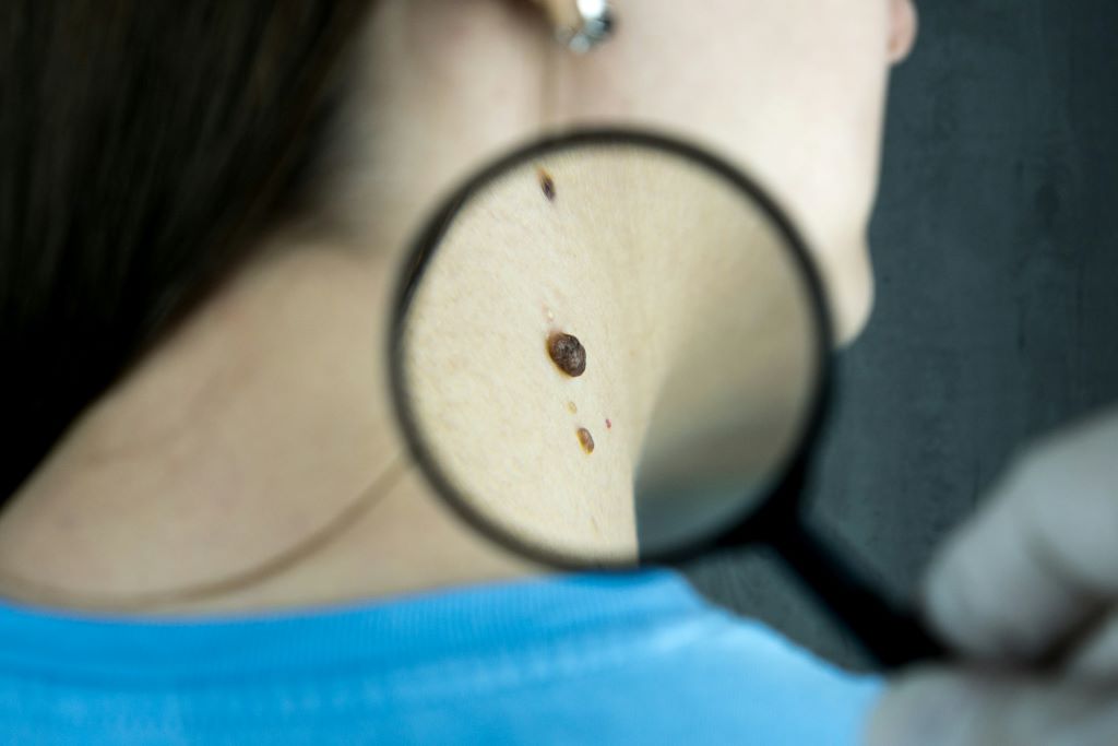
Moles and melanomas share the same cellular origin, forming from a pigment-producing cell called a melanocyte that gives skin its colour. In moles, the cell multiplies to form a cluster then stops growing, leaving a harmless spot. In melanoma, the cell growth continues aggressively.
Moliness is strongly influenced by your genes and having a high mole count is a major risk factor for melanoma. Around a third of melanomas develop from a mole.
The QIMR Berghofer research analysed data from more than 85000 participants of European ancestry discovering 24 new genetic regions that determine the number of moles someone has. This is a five-fold increase on the five areas found in an earlier 2018 study also led by QIMR Berghofer researchers.
All but one of the genetic regions for mole count also play a role in melanoma. The team pinpointed more than 250 key genes in these regions to prioritise for further research.
One of the new genes, SIKE1, regulates immune responses to viral infections. The researchers think it could enable the development of melanomas by malfunctioning and affecting the immune system’s ability to detect and destroy melanocytes that are multiplying abnormally. This could be a promising target for a potential immunotherapy that could possibly prevent early stage melanoma growth.
“I’m really proud to be continuing this long legacy of research. Our study increases understanding of why some people have a lot of moles and why some people develop melanoma so we can better treat and prevent this skin cancer,” Ms Jayasinghe said.
The researchers used the study insights to create a Polygenic Risk Score (PRS) for moliness to predict those who are genetically more likely to have a large number of moles, which could be integrated into melanoma screening tools in future to improve their accuracy in finding those at high risk so they can receive extra monitoring.
The next step is to analyse even larger data sets to find more genetic regions involved in moliness and melanoma. The researchers are also searching for existing drugs that could potentially target the newly identified biological pathways.