Category: Lab Tests and Imaging

New Antenna Upgrade Boosts MRI Image Quality

Photo by Mart Production on Pexels

Magnetic resonance imaging (MRI) is one of medicine’s most powerful diagnostic tools. But certain tissues deep inside the body – including brain regions and delicate structures of the eye and orbit that are of particular relevance for ophthalmology – are difficult to image clearly. The problem is not the scanner itself, but the hardware that sends and receives radio signals. 

Now, researchers at the Max Delbrück Center have developed an advanced materials-based MRI antenna that overcomes these limitations – delivering enhanced images more quickly and that can be used in existing MRI machines. The research, led by Nandita Saha, a doctoral student in the Experimental Ultrahigh Field Magnetic Resonance lab of Professor Thoralf Niendorf, was published in Advanced Materials.

Niendorf and his team worked closely with researchers at Rostock University Medical Center, combining expertise in MRI physics with clinical ophthalmology and translational imaging. The Rostock team is also supporting clinical validation of the technology.

“By using concepts from metamaterials, we were able to guide radiofrequency fields more efficiently and demonstrate how advanced physics can directly improve medical imaging,” says Niendorf, senior author of the paper. “This work shows a pathway toward faster, clearer MRI scans that could benefit patients in many clinical areas.” 

Rethinking MRI hardware with metamaterials

MRI works by sending radiofrequency (RF) signals into the body and detecting how tissues respond inside a strong magnetic field. The stronger the signal response, the better the image. Conventional MRI antennas – also called RF coils – often struggle to collect enough signal from deep or anatomically complex regions. This leads to images that lack detail and prolongs scan times.

The research team addressed this bottleneck by integrating metamaterials directly into the MRI antenna. Metamaterials are engineered structures that interact with electromagnetic waves in ways not found in natural materials. The engineered RF antenna increases signal strength from targeted tissues, improves spatial resolution and image sharpness and enables faster data acquisition. Crucially, the antenna fits into existing MRI systems, avoiding the need for new infrastructure. The team validated the technology by imaging the eye and orbit region in a group of volunteers at 7.0 Tesla.

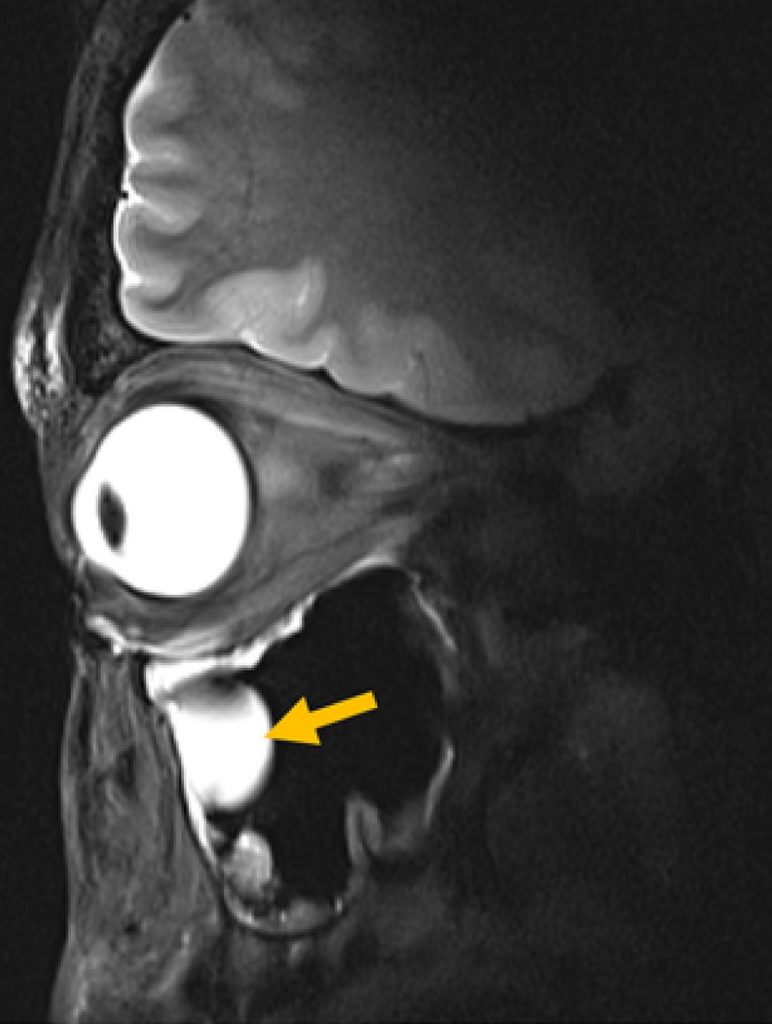
MRI image of an eye, the eye socket and the brain.
© AG Niendorf, Max Delbrück Center

“Our research demonstrates clear relevance for ophthalmological applications as it can facilitate anatomically detailed, high-spatial resolution MRI of the eye,” says Professor Oliver Stachs, a co-author of the paper at University Medicine Rostock. “It offers the potential to open a window into the eye and into (patho)physiological processes that in the past have been largely inaccessible.” 

“Our goal was to rethink MRI hardware from the modern physics of antenna design,” adds Saha. This technology can also be tuned to protect sensitive areas of the body during MRI, for example, to reduce unwanted heating around medical implants, she adds. It could also be used to focus RF energy more effectively for MRI guided therapies for various cancer treatments, such as gentle heating of tumors (hyperthermia) or thermal ablation of tissue. 

Better diagnostics

For patients, MRI scans can be uncomfortable and time-consuming – even more so when images need to be repeated because important details are hard to see. Faster scans mean patients spend less time inside scanners. Clearer images mean doctors can make diagnoses with greater confidence. And because the new antenna is lightweight and compact, it can also be designed to better fit specific parts of the body, improving comfort even further.

The technology could also be adapted to support MRI systems running at magnetic field strengths lower or higher than 7.0T, to image target anatomy other than the eye, orbit or the brain or to track metabolism or drug movement inside the body, says Niendorf. Special MRI scans that use other atoms, such as sodium or fluorine, could also benefit from this technology by producing clearer signals and better images, he adds. 

“Innovations in imaging hardware have the potential to transform diagnostics, and this study is an important step toward next-generation MRI technology,” says Dr Ebba Beller, a co-author of the paper at Rostock University Medical Center.

The researchers are already planning larger studies at multiple hospitals and adapting the design for other organs, such as the heart and kidneys. The collaboration will continue to be strengthened by long-standing reciprocal visiting scientist appointments of Stachs and Niendorf. 

Source: Max Delbrück Center for Molecular Medicine

Study Shows Low-Field MRI Is Feasible for Breast Screening

Mass General Brigham’s evaluation of low-field MRI performance lays potential groundwork for this technology to be a lower-cost, accessible option for breast imaging

Photo by National Cancer Institute on Unsplash

Researchers at Mass General Brigham have demonstrated the technical feasibility of using ultra-low field (ULF) magnetic resonance imaging (MRI) for breast imaging. With further refinement and evaluation, the technology could offer an alternative to existing breast cancer screening methods and may reduce barriers to screening. Results are published in Scientific Reports.

“These results are a very encouraging proof of principle, though larger studies are needed to establish diagnostic performance,” said project principal investigator and co-senior author Matthew Rosen, PhD, an associate professor of Radiology and director of the Low Field MRI laboratory in the Athinoula A. Martinos Center for Biomedical Imaging in the Mass General Brigham Department of Radiology. “They motivate our continued pursuit of safe, comfortable, lower-cost screening approaches that can expand access for patients.”

Current U.S. guidelines recommend screening mammography for women aged 40 to 74 years. Unlike mammography, ULF MRI doesn’t require breast compression, which many patients find uncomfortable. Another benefit of ULF MRI is that it doesn’t use ionizing radiation.

While higher risk patients may receive MRI screening for breast cancer, standard MRI machines are not used in routine breast cancer screening because they are expensive and not widely available. ULF MRI systems cost less than 5% of the price of standard MRI systems and have lower long-term operating costs.

In this study, ULF MRI scans were performed on 14 participants, including 11 women with no history of breast cancer, two women with a prior breast cancer diagnosis, and one woman with a benign mass.

When interpreting the ULF MRI scans, three radiologists could reliably identify essential breast features and distinguish fibroglandular tissue from adipose tissue. The authors note that discrepancies were likely related to the novelty of ULF MRI and may be reduced with additional training and experience.

“This early evidence suggests that ULF MRI can detect essential breast features and some abnormalities without radiation or injected contrast,” said co-first author Neha Koonjoo, PhD, an investigator at the Martinos Center. “These findings point to the potential for ULF MRI as an option that could complement existing screening tools in the future.”

“Even at very low magnetic field, the radiology team was able to make observations about the breast,” said co-principal investigator and co-senior author Kathryn E. Keenan, PhD, from the US National Institute of Standards and Technology. “We attempted this study in hopes that the breast features would be visible, but you don’t always have success. We’re very motivated by this study to continue our work on ultra-low-field MRI for breast screening.”

The researchers note that further study is needed to determine the diagnostic accuracy of ULF MRI for breast cancer screening, including studies in larger cohorts that include patients with benign and malignant lesions. They also emphasize that further refinements in ULF MRI technology are needed to meet clinical resolution standards for breast cancer screening.

“These results will guide the next engineering steps to improve image quality and enable a more comfortable exam and help bring screening to more settings and more patients,” said co-first author Sheng Shen, PhD, of the Martinos Center for Biomedical Imaging.

Source: Mass General Brigham

Point-of-care Rapid Tests can Improve Screening for Latent Tuberculosis

Photo by National Cancer Institute on Unsplash

A new test shows promising results for detecting latent tuberculosis infection in resource-limited settings. This is according to a study from Karolinska Institutet, published in the journal Clinical Infectious Diseases.

“This test can help more people with latent tuberculosis to be detected and receive preventive treatment, especially in rural areas in countries with limited resources,” says last author Lina Davies Forsman, a researcher at the Department of Medicine, Solna, Karolinska Institutet.

Tuberculosis remains one of the world’s deadliest infectious diseases. To reduce the number of new cases, infected individuals with latent infection must be detected and offered preventive treatment to avoid active tuberculosis, which can spread the disease to others.

Currently, latent tuberculosis is often diagnosed using a laboratory test called QuantiFERON-TB Gold Plus. This test involves several steps and can take one to two days before the results are available, as well as requiring advanced laboratory infrastructure and trained personnel. This makes it difficult to carry out tests in areas with a high prevalence of tuberculosis where access to laboratories and trained personnel is limited.

Results within 15 minutes

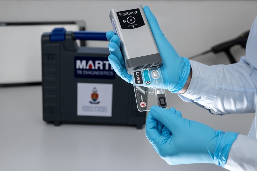
In the new study, researchers from Karolinska Institutet, together with colleagues in Vietnam, have therefore compared this test with another test, TB-Feron. This is a point-of-care test that provides results within 15 minutes and does not require an advanced laboratory or trained personnel.

The study included 345 adult participants in Hanoi, Vietnam, divided into three groups: people with confirmed tuberculosis, people in the same household as people with infectious tuberculosis, and people with no known exposure to tuberculosis. All were tested with both TB-Feron and the established laboratory test QuantiFERON-TB Gold Plus.

The results show that TB-Feron has high sensitivity – 88 percent of individuals with expected positive results were correctly identified. The corresponding figure for QuantiFERON-TB Gold Plus was 92 percent.

However, the specificity, i.e. TB-Feron’s ability to rule out tuberculosis infection in healthy individuals, was moderate at 70 percent. The corresponding figure for QuantiFERON-TB Gold Plus was 96 percent.

Among household contacts, the concordance between TB-Feron and the established test was good, with 92 percent concordance for positive samples.

“It is promising that TB-Feron works so well in an environment with a high disease burden. The test is patient-friendly and easy to use, with rapid same-day results, making it useful in primary care,” says Han Thi Nguyen, pulmonologist and doctoral student at the same department and first author of the study.

The researchers also investigated the reliability of TB-Feron by comparing results from two different groups with laboratory staff. No systematic differences were observed, indicating good reproducibility.

Source: Karolinska Institutet

Simple Method for Early Detection of Chronic Kidney Disease

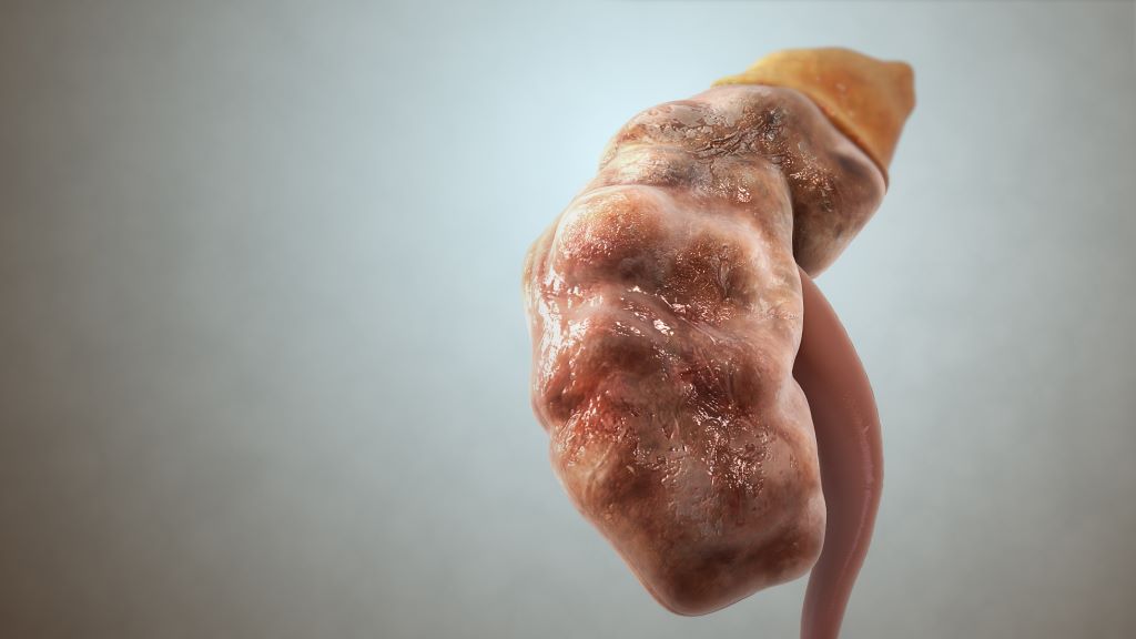
Chronic kidney disease (CKD). Credit: Scientific Animations CC4.0

Subtle abnormalities in kidney function – even within the range considered normal – may help identify people at risk of developing chronic kidney disease. This is shown in a new study from Karolinska Institutet, published in Kidney International. The researchers have therefore developed a web-based tool that could aid in early detection and thus primary prevention.

Chronic kidney disease is a growing global health concern afflicting 10−15% of adults worldwide, and is projected to become one of the top five leading causes of years of life lost by 2040. In the absence of effective screening programmes, patients are often diagnosed late, when more than half of their kidney function has already been lost. 

To address this gap, researchers at Karolinska Institutet have constructed population-based distributions for estimated glomerular filtration rate (eGFR), the most widely used measure of kidney function. The aim is to help doctors identify people at risk, thus enabling early preventive action.

“We were inspired by the growth and weight charts used in paediatrics, which intuitively help clinicians identify children at risk of obesity or undergrowth,” says the first author of the study, Yuanhang Yang, Postdoctoral Researcher at the Department of Clinical Science and Education, Södersjukhuset, Karolinska Institutet.

A web-based calculator

The researchers have made their eGFR distribution charts openly available to healthcare professionals and developed a web-based calculator, developed by PhD student Antoine Creon, that can help assess how a patient’s eGFR compares with population norms for their age.

The study included over 1.1 million adults in the region of Stockholm, covering roughly 80 per cent of the population aged between 40 and 100 years. Nearly seven million eGFR tests collected between 2006 and 2021 were used to construct age- and sex-specific distributions. 

The findings show that departures from the median eGFR for one’s age and sex are associated with worse outcomes. Individuals with an eGFR below the 25th percentile had a markedly higher risk of developing kidney failure requiring dialysis or transplantation. Mortality also displayed a U-shaped relationship; both low and high percentile extremes were linked to increased risk of death. 

Ability to act earlier 

The study also illustrates this lack of awareness in healthcare, according to the researchers. Among those with a seemingly normal eGFR above 60 ml/min/1.73 m², but below the 25th percentile, only one fourth had received additional testing for urinary albumin, which is important for detecting early kidney damage.

“For example, consider a 55-year-old woman with an eGFR of 80. Most clinicians would not react to such a seemingly normal value. However, our charts show that this corresponds to the 10th percentile for women of that age – and that she has a three-fold higher risk of starting dialysis in the future. This signals an opportunity to act earlier,” says Juan Jesús Carrero, Professor at the Department of Medical Epidemiology and Biostatistics, Karolinska Institutet.

Source: Karolinska Institutet

New Optical Tool Lights up IBD Biomarkers

A test that rapidly detects signs of inflammatory bowel disease (IBD) in stool samples could improve future diagnosis and monitoring of the condition, a study suggests.

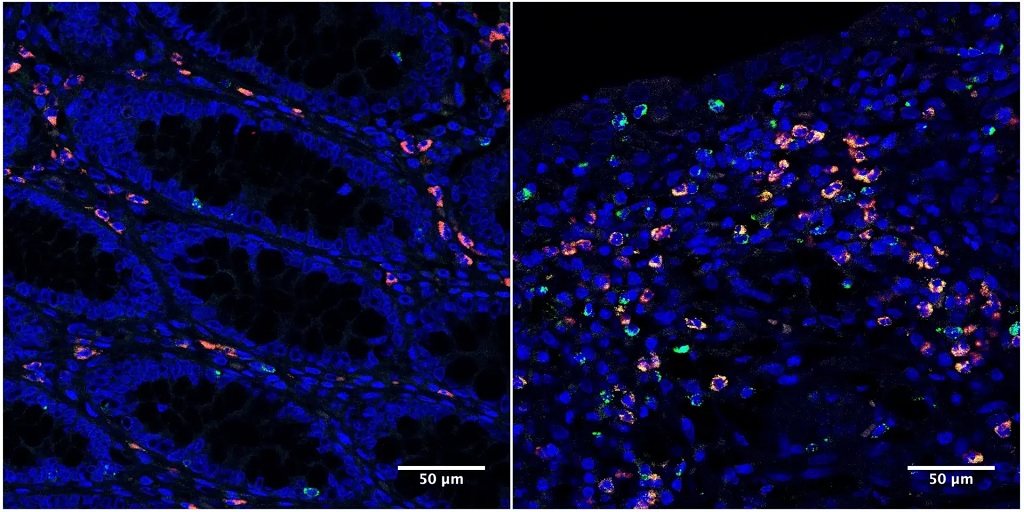
Microscopy images of colon tissue samples from IBD patients under remission (left) and with active disease (right). Granzyme A, indicated by green fluorescence, is elevated in the intestinal tract of IBD patients with active disease. Credit: Emily Thompson.

Scientists have developed a tool to measure the activity of a molecule linked to gut inflammation within faecal samples. The optical tool, known as a luminescent reporter, lights up when it detects the molecule, with higher readouts indicating increased activity and inflammation.

The new technique could boost the accuracy of stool sample tests for IBD, reducing the need for invasive, expensive procedures, experts say.

Gut inflammation

IBD is a chronic illness where the body’s immune system mistakenly attacks the digestive tract, leading to long-lasting inflammation. Diagnosis and monitoring of the condition often rely on colonoscopies, where a small camera is used to examine the gut.

Current IBD stool tests measure general markers of inflammation, such as the protein calprotectin, so a positive result requires further investigation to confirm the source. 

University of Edinburgh researchers studied gut tissue from IBD patients and identified high levels of an enzyme – a molecule that speeds up chemical reactions in cells – called granzyme A (GzmA) in inflamed gut tissue compared with non-inflamed tissues. 

Enzyme activity

GzmA is released by T cells; in IBD, T cells mistakenly see the gut as a threat and become overactive, which can lead to tissue damage and inflammation.

The research team developed a luminescent reporter to measure the activity of GzmA in stool samples. The reporter tool was tested on 150 samples from both IBD and healthy patients. 

Combining the new reporting tool with the current common testing of faecal calprotectin levels was more successful in identifying IBD in patients than using faecal calprotectin scores alone.

Researchers say the ability to identify gut-specific inflammation is a step forward for IBD diagnosis, but caution further research is needed before it can be used in a clinical setting.

Spin-out company

The tool will form part of the assets of a new company in the process of spinning out of the University of Edinburgh, called IDXSense, supported by Edinburgh Innovations, the University’s commercialisation service. 

The technique could also support the development of personalised IBD treatments in the future, with the ability to rapidly and accurately monitor gut inflammation levels in response to different therapies, experts say.

Source: The University of Edinburgh

Diagnostic Breast MRI may be Unnecessary for Some Patients with Early-stage Breast Cancer

Adding breast magnetic resonance imaging (MRI) to a diagnostic mammogram did not reduce five-year cancer recurrence rates for patients with stage I/II hormone receptor (HR)-negative breast cancer, according to researchers at The University of Texas MD Anderson Cancer Center. 

The Phase III Alliance A011104/ACRIN6694 trial found that five-year locoregional recurrence rates were 6.8% in patients who received an MRI as part of a diagnostic work-up and 4.3% in those who did not. These data were presented today at the San Antonio Breast Cancer Symposium (SABCS) by principal investigator Isabelle Bedrosian, MD, professor of Breast Surgical Oncology (Abstract GS2-07).

“We have long assumed that finding more breast cancer on an MRI and removing it with surgery would help lower the chance of a patient’s cancer coming back,” Bedrosian said. “When you look at our findings alongside earlier trials, the message is clear: adding MRI before surgery doesn’t improve results for patients – and may not have to be used as a standard part of the diagnostic process.”

No additional MRI benefit in this group

The trial enrolled 319 patients between 2014 and 2018 with newly diagnosed stage I or II HR-negative breast cancer. These patients were eligible for lumpectomy and did not have germline BRCA1/2 mutations, bilateral breast cancer or a history of prior breast cancer. All patients had undergone diagnostic mammography with or without ultrasound prior to trial enrolment.  Patients were randomly assigned to undergo additional imaging by breast MRI (161 patients) or to receive no further imaging (158 patients).

Not only did breast MRI not impact five-year recurrence rates, but there were also not significant differences between groups for five-year distant recurrence-free survival nor overall survival. 

A small subset of patients with tumour subtypes (HR- HER2+ and HR-HER2-) and those over the age of 50 at diagnosis also showed no benefit to MRI.

Pre-op MRI not finding anything important

Breast MRI is a common part of the diagnostic evaluation because it can reveal cancer that mammography might not detect. However, the evidence that it improves surgical outcomes for patients has been limited.

“We believe the reason MRI did not reduce recurrence rates may be twofold,” Bedrosian said. “It is possible that MRI didn’t uncover many lesions that mammography hadn’t already found, or perhaps identifying and surgically removing those additional lesions was not important to reducing risk of the cancer coming back. It’s possible that in the group that did not receive MRI, radiation and chemotherapy effectively treated the occult areas of disease”. 

Experts are now analysing how often breast MRI identified additional lesions in the trial population to better understand why breast MRI did not impact oncologic outcomes.

Study limitations 

Limitations included that most patients involved in the trial had breast cancer that hadn’t spread to their lymph nodes, which may partly explain why recurrence rates were low overall. Despite being open to women of all ages, the study enrolled mostly older women who may have been less likely to benefit from breast MRI. 

New Scan Could Help Millions with Hard-to-treat High Blood Pressure

A speedy new scan could improve how millions of people with hypertension are treated, suggests a new study

Credit: Pixabay CC0

About a quarter of people with high blood pressure have been estimated to have primary aldosteronism, a problem with their adrenal glands producing too much of the hormone aldosterone, which regulates levels of salt in the body.

This problem is often missed, as the path to diagnosis is complex, involving multiple tests and, to guide treatment, an invasive procedure that is not always reliable.

The new 10-minute scan, developed at University College London and described in a research letter in the New England Journal of Medicine (NEJM), reveals overactivity in adrenal glands that was invisible with conventional tests, showing exactly where too much aldosterone is being made.

This, the researchers say, will make it easier to decide on the best treatment approach – either removal of an adrenal gland that is producing too much aldosterone, or the use of new medications that block aldosterone production, targeting the cause of high blood pressure in many patients.

Professor Bryan Williams, Chair of Medicine at UCL and clinical lead for the study, said: “We have been waiting for a test like this for many decades. This British innovation is going to transform the diagnosis of aldosterone excess as an important and previously hidden cause of hypertension in many of our patients. It offers huge potential to completely change the way we make this diagnosis and enable us to provide better targeted treatment for our patients.”

The over-production of aldosterone, which raises high blood pressure by causing the body to retain too much salt, can result in a condition called primary aldosteronism, which increases the risk of heart disease, stroke and kidney disease. However, many people who do not meet the threshold for this condition are thought to have excess aldosterone raising their blood pressure.

Currently the condition is screened with a blood test and confirmed with a second test*. To decide on treatment, two catheters are inserted in veins on either side of the groin to measure levels of aldosterone on each side of the body. This helps clinicians determine if the problem is only located in one adrenal gland or both – but the test is not always accurate and not often offered as few hospitals have the expertise to perform this complex procedure.

To better detect the condition, researchers at UCL used a PET-CT scan, which creates detailed 3D images of parts of the inside of the body and maps the accumulation of a tiny amount of radioactive tracer injected into a person’s vein.

They built a new tracer compound designed to bind to the aldosterone-producing enzyme, aldosterone synthase. The tracer was highly selectively taken up by the parts of the adrenal gland that were over-producing aldosterone, lighting up these areas on the scan.

In their NEJM research letter, the researchers described how 17 patients were scanned in the world’s first use of this technique at UCLH. The team found the source of over-production of aldosterone in every patient and did not see any side effects.

Professor Williams added: “This is the first time we have been able to visualise this disease. We can see it light up on the scan. The intensity of the signal reflects the level of aldosterone over-production. This might allow us, in future, to more precisely target these over-producing areas.” 

The achievement builds on more than a decade’s work by Professor Erik Arstad (UCL Division of Medicine and UCL Chemistry) and colleagues, who pioneered and patented a new method to make radioactive tracers.

Using this method, they were able to repurpose a drug-like molecule that bound to the aldosterone-producing enzyme for use as a tracer, replacing a single atom with a radioactive version of that atom – meaning this molecule would light up on a PET-CT scan.

Professor Arstad said: “It is very rewarding to be able to bring laboratory innovation into the clinic for the benefit of patients with hard-to-treat hypertension.”

The study was conducted at UCL and UCLH and was funded by the MRC and the NIHR University College London Hospitals Biomedical Research Centre.

The team is now embarking on a phase 2 clinical trial to gather sufficient data for the test to be approved for routine clinical use in the NHS.

In the UK, more than 14 million people are estimated to have high blood pressure (about one in three adults).

*For instance, a salt loading test, where a person increases their intake of salt (sodium), which would be expected to suppress aldosterone levels. If aldosterone levels are still high despite this increase, that confirms a primary hyperaldosteronism diagnosis.

Source: University College London

A Simple Neck Scan Could Detect Men at High Risk of Heart Failure

Right side heart failure. Credit: Scientific Animations CC4.0

A simple neck scan can identify men with double the risk of heart failure, according to a new study led by UCL researchers and funded by the British Heart Foundation and the National Institute for Health and Care Research.

A carotid ultrasound, like the ultrasound for pregnant women, is quick and painless, using a small handheld device moved gently over the neck to scan the arteries underneath.

When around 1600 men over the age of 70 received the scan, it showed the ‘flexibility’ of their carotid arteries – how much they stretch and expand with each heartbeat.

Researchers found that the quarter of men with the least flexible carotid arteries were 2.5 times more likely to develop heart failure than those with the most flexible carotid arteries.

These people could be encouraged by doctors to eat more healthily, do more exercise and take medications, if needed, to help reduce their risk of developing heart failure.

GPs do not currently routinely carry out the cheap and easy scan on healthy patients without symptoms. But, where GP surgeries have the capacity, offering a neck scan to older people to measure the flexibility of their arteries could help them better understand their risk of future heart failure, according to the researchers.

Having relied upon data from the British Regional Heart Study, which began in the 1970s and only involved men, researchers highlight that these findings next need to be looked at in women.

Dr Atinuke Akinmolayan (UCL Primary Care & Population Health), who is now a GP, said: “The carotid ultrasound is a safe, cheap and painless investigation, and our findings suggest it may be able to provide an early warning sign for heart failure.

“More research is needed, especially to see if this works for women, but this is something GPs could look at offering to people over the age of 60, where possible and believed needed.

“A patient who gets an ultrasound result indicating they may be at higher risk of future heart failure could have an important conversation with their doctor about lifestyle changes they could make to lower that risk.”

Doctors tend to scan the two carotid arteries, which run up either side of the neck, when someone has had a stroke or is at risk of a stroke following a transient ischaemic attack, known as a ‘mini-stroke’. A scan can identify carotid artery disease – a build-up of fatty material which can cause a stroke by breaking off and travelling into the brain or by narrowing the arteries and stopping blood reaching the brain.

However, the carotid arteries may be a red flag for heart failure also. This is because, when the carotid arteries become less flexible, they do not expand properly to let blood through. This can raise blood pressure, which forces the heart muscle to work harder. Over time, this can lead to heart failure.

The study, published in the Journal of the American Heart Association, looked at 1631 British men, aged 71 to 92, who had a carotid artery ultrasound between 2010 and 2012 as part of the British Regional Heart Study.

A carotid ultrasound, sometimes called a Doppler scan, takes an average of 15 to 30 minutes for most people, although this can vary.

A small handheld sensor is moved back and forth over the neck, generating sound waves which bounce off the arteries. That provides an echo which changes in frequency when blood flow is reduced in the blood vessels because they are narrowed by built-up fatty material.

The narrowing identified by a carotid scan can then be used to calculate the arteries’ flexibility, after factoring in other measures, including blood pressure. Researchers were able to identify the quarter of men with the least flexible carotid arteries, and the quarter of men whose carotid arteries were most flexible.

They then compared the rates of heart failure in each group  over an average of six years after their neck scans.

Even after considering other causes of heart failure, like age, weight, smoking and whether people had previously suffered a heart attack, the quarter of men with the least flexible carotid arteries had 2.5 times the risk of developing heart failure, compared to the quarter with the most flexible carotid arteries.

In a separate finding, looking at the thickness of people’s carotid arteries rather than their flexibility, the study found that men with thicker carotid arteries were more likely to have a heart attack or die from one.

For every ‘unit’ increase in the thickness of the carotid artery wall – with a unit equalling 0.16 millimetres – the risk of having a heart attack increased by about 29 per cent, even after considering other relevant factors like age and weight.

However the thickness of the carotid arteries was not found to be significantly linked to future heart failure in the study.

There are around 200 000 new cases of heart failure diagnosed every year in the UK.

It occurs when the heart is not pumping blood around the body as well as it should, most commonly when the heart muscle has been damaged – for example, after a heart attack.

Heart failure can cause extreme fatigue, shortness of breath and fainting.

Professor Bryan Williams, chief scientific and medical officer at the British Heart Foundation (BHF), who is also Chair of Medicine at UCL Institute of Cardiovascular Science, said: “The findings of this study are interesting and show that stiffening of arteries is associated with increased risk of heart failure, most likely due to the heart having to work harder against the resistance caused by these stiffer arteries.

“It is an important signal that whenever we detect such changes in the carotid arteries, we should also be thinking of the potential impact on the heart and an increased risk of heart failure – which we have treatment strategies to prevent.”

Source: University College London

New Method Noninvasively Assesses Achilles Tendon in Dancers

Photo by Nihal Demirci on Unsplash

A study in the Journal of Orthopaedic Research uses a noninvasive, nonradioactive imaging-based method to measure the structure and function of the Achilles tendon in professional ballet dancers. The method could potentially be developed to help prevent injuries and improve rehabilitation efforts in athletes, as well as in the general public.

The study involved what is called multi-echo ultrashort echo time (UTE) magnetic resonance imaging (MRI) to assess collagen and other components of the Achilles tendon. These structural UTE MRI assessments were combined with functional assessments of the Achilles tendon based on sheer wave elastography (SWE) ultrasound, which measures tendon stiffness.

Professional dancers tended to have more tendon stiffness compared with non-dancers, consistent with prior observations of a training effect from repeated loading with exercise. UTE MRI measures corresponded with the degree of stiffness from SWE ultrasound.

“These findings highlight the potential of integrating UTE and SWE imaging to investigate tendon structure‐function relationships and adaptations to mechanical loading,” the authors write. “Enhanced structure‐function assessment of tendon health and injury status could improve rehabilitation protocols or injury prevention strategies for athletes, including professional dancers.”

Source: Wiley

When a Limp Isn’t Just a Sprain in Adolescents

A timely X-ray can save young hips

Frog leg lateral view of the hips. Widening of the growth plate (physis) with blurring and irregularity of the femoral neck (metaphysis). Inferior offset of the head in relation to the neck (early slip).

Slipped Capital Femoral Epiphysis (SCFE) is the most common adolescent hip disorder. It occurs when the ball at the top of the thigh bone (femoral head) slips off the neck of the bone through the growth plate (physis). A bit like an ice cream sliding off a cone… Dr Ryno du Plessis, a renowned orthopaedic and joint replacement surgeon in the Western Cape, talks about what it is and why it is often misdiagnosed.

SCFE usually happens during growth spurts in children aged 9 to 16 years and is more common in boys and in children with obesity, endocrine disorders, or other risk factors.

Why is this problem often missed?

AP view of the hips. ‘Melting ice cream sign’: Femoral head (epiphysis) slipping off the femoral neck (metaphysis) though the growth plate (physis) like an ice cream melting from the cone.

Despite its frequency, SCFE is routinely misdiagnosed or diagnosed late – unfortunately, sometimes months after symptoms start. Studies show that over 50% of SCFE cases are not diagnosed at the first medical visit.

Here’s why:

  • Pain felt in the knee or thigh: Physicians often focus on the wrong joint and the hip is never X-rayed
  • Labeled as a groin strain: Adolescents in sports may be diagnosed with muscle strains or ‘growing pains’
  • Symptoms develop gradually: Children may limp without severe pain, leading to delayed concern
  • Physiotherapy prescribed early: Instead of imaging – patients are referred to physio – delaying diagnosis
  • Lack of hip-specific X-rays: It requires a frog-leg lateral X-ray.

Why does delay matter?

The longer the slip is left untreated, the more serious the outcome. Every week or month of delay increases the severity of the deformity, often silently.

Late diagnosis risks:

  • More severe deformity
  • Loss of bloody supply to the femoral head. This is known as avascular necrosis and can lead to pain, limited movement and eventually, hip collapse and osteoarthritis
  • Early-onset hip arthritis
  • Complex surgery

Children diagnosed early often need just one screw to stabilise the hip. Those who are diagnosed late may face major reconstructive surgery, longer recovery, and reduced hip function for life.

Red flags for parents, teachers and coaches

If you notice any of the following signs in a child or teen – especially those who are overweight – take it seriously and ask for a hip X-ray:

  • Limping for more than a week
  • Complaints of pain in the knee, thigh, groin, or buttock
  • Walking with the foot turned outwards
  • Stiffness or loss of motion in the hip
  • Sudden inability to walk or stand after a minor stumble (may indicate an unstable SCFE)

Radiology – diagnostic challenges

Dr Jaco Greyling, a radiologist from SCP Radiology, says SCFE diagnoses can be delayed due to several factors, including

  • Hip X-rays not ordered by the initial healthcare provider (eg, GP or physiotherapist)
  • Only a single anterior-posterior pelvis projection is performed, whereas a frog-leg lateral view must also be specifically requested by the referring physician. Radiologists should ensure the child returns for this view if it was not initially ordered
  • Findings in the pre-slip phase are subtle and may be missed, even by experienced radiologists

He says, ’the recommended imaging is an anterior-posterior pelvic view which shows malalignment and widening of the growth plate and a frog-leg lateral view, the most sensitive for detecting early or subtle slips.’

‘Key radiological signs,’ says Dr Greyling are:

  • Widening of the growth plate
  • Loss of height of the femoral head
  • Loss of alignment of the anatomical lines that intersect with the femoral head
  • ‘Melting ice cream sign’ slipping off the femoral neck at the growth plate (epiphysis).

Follow-up recommendations:

Dr Greyling suggests repeat imaging within two weeks if symptoms persist, and an early referral to a paediatric orthopaedic surgeon and an MRI for patients with risk factors and ongoing pain.

Who’s at risk?

  • Children aged 9-16 years
  • Boys are at greater risk than girls
  • BMI in the overweight/obese range
  • Family history of hip disorders
  • Endocrine disorders: Hypothyroidism, growth hormone treatment, kidney disease

Treatment

Early SCFE is usually treated with in-situ fixation using one or two screws. The goal is to stabilise the rounded end of a long bone to prevent further slippage.

In cases where both hips are at risk (especially in young or overweight patients), pinning of the opposite hip as well is sometimes recommended to prevent it from occurring.

Severe or late cases have a high risk of AVN, which is the death of bone tissue caused by a disruption in its blood supply, leading to pain, stiffness, and potential bone collapse or joint destruction over time and permanent disability.

The take-home message

SCFE is treatable and preventable if recognised early.

If a child has an unexplained limp, especially with thigh or knee pain, don’t assume it’s just a strain. Ask the doctor directly: “Could this be SCFE? Should we get hip X-rays done?”

One simple question. One X-ray. It could save a child’s hip.