The Science and Challenge Behind Replacing MRI Machines
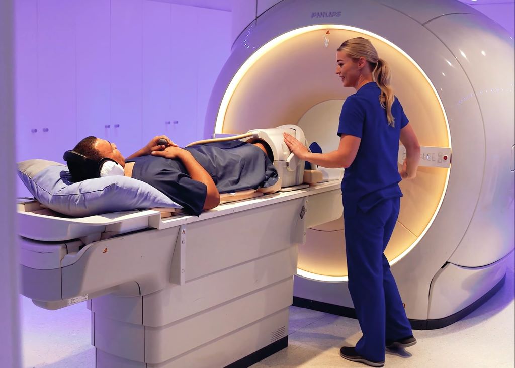
Moving a Magnetic Resonance Imaging (MRI) machine is not as simple as out with the old and in with the new. It is an engineering feat – part physics, part choreography. It is not just a machine, it is an entire system that needs structural support and specialised housing.
Some Quite Interesting (QI) facts about moving an MRI machine
MRI machines are marvels of engineering, with their powerful magnets requiring precise handling and specialised support. The magnet, which is the heart of the MRI, can weigh several tonnes and must remain cold, often near absolute zero, maintained by liquid helium or other cooling methods.
‘When replacing an MRI machine, the process is carefully orchestrated, starting with meticulous planning and structural assessments,’ explains Tinus van Rooyen, Business Project Manager at SCP Radiology. The date of the move is carefully selected to coordinate the team of engineers, crane operators and logistics professionals.
‘It is far more complex than moving almost any other piece of hospital or healthcare equipment. Which is why,’ says van Rooyen, ‘removing our old MRI machine and replacing it is not quite ‘all in a day’s work’. And our practice is doing it across multiple sites over the coming months.’
The new MRI machines contain less than 1% of the scarce and non-renewable resource, helium, than that of a conventional MRI machine, improving operational efficiency and long-term sustainability. ‘The newer MRI systems use sealed magnets that, although having to be kept at a temperature of 4 Kelvin (-269.15° Celsius), require very little helium and no refilling over their lifetime’, explains van Rooyen. ‘The improvements in technology in the new machines also ensure improved image quality.’
What is an MRI machine?
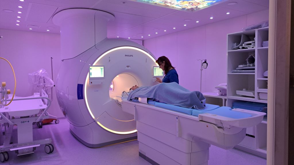
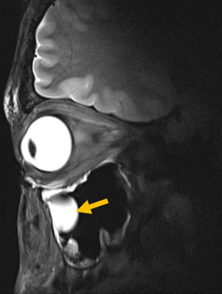
The powerful magnetic field and radio waves create detailed images of the body, enabling radiologists to look at soft tissues like the brain, spine, joints and organs in extraordinary detail and without using any radiation. ‘Everything is controlled by advanced computer systems, which convert signals into detailed scans. If an MRI is listening for whispers from the body, a Faraday cage – using copper or aluminium – shields the room to ensure it’s completely silent, so these can be heard clearly.’
How heavy is an MRI machine?
Heavier than most people expect. A low helium MRI machine weighs in at around 3.3 tonnes (3 300kg) – about the weight of an elephant and is significantly less than the conventional machines. The magnet alone is about 60 000 times stronger than Earth’s magnetic field when it’s fully operational and can weigh several tonnes. It’s unsurprising that installing one is a major engineering exercise.
Why would an MRI machine be replaced – what is the usual lifespan of a machine?
The lifespan of an MRI is approximately 10 – 15 years. Replacing ageing equipment ensures access to the latest technology, including improved image quality and an enhanced patient experience. Machines can also reach End of Support (EOS). ‘This means manufacturers no longer support and maintain the unit and parts are unavailable,’ says van Rooyen. ‘It therefore becomes unreliable to keep it running. Patient care is paramount, as is minimising potential downtime and ensuring continuity of service.’
What happens when a machine is replaced?
The old one needs to be ramped down (gradually reducing the strength of the scanner’s main magnetic field to zero in a controlled way). It is then disconnected and removed.
Installation of new unit is basically a reverse of the ramping down process. Once the unit is in place, the magnet is cooled to a superconducting state, then ramped up by gradually increasing the electrical current in the magnet coils, causing the magnetic field to slowly rise. After the magnet has reached its specified strength, the magnetic field is then aligned to make it as uniform as possible, ahead of the unit being calibrated and tested. The entire process can take up to eight weeks. Careful planning ensures continuity of service, with alternative arrangements in place where necessary.
What are the challenges and logistics during the move?
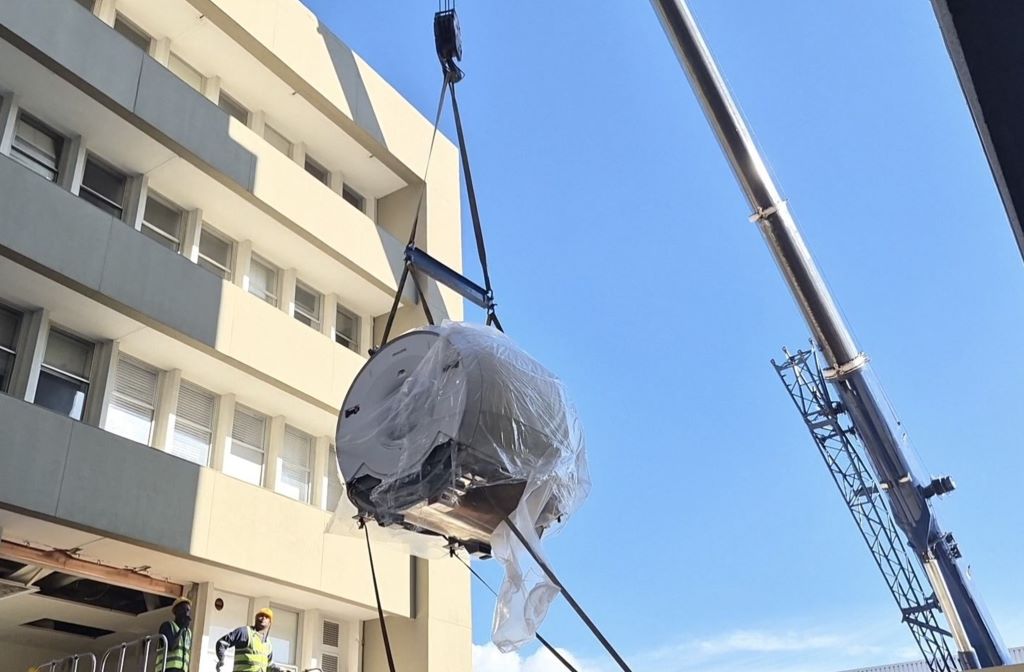
The MRI machine at the SCP Radiology branch at Mediclinic Louis Leipoldt is housed on the first floor. The challenges included building a platform outside the building, moving and lifting the units, with the external wall being removed to create access. The equipment manufacturers oversee the installation but it required a team of riggers to assist with taking out the old unit, then lifting and installing the new unit off the delivery vehicle using a crane, onto a platform and then into the building. Additional contractors are responsible for preparing the room – this includes copper cladding, drywalling, reinforced flooring (if required), painting, joinery, etc.
‘The installation of each MRI unit is unique and depends on a number of factors. In the case of this new one at Louis Leipoldt, we had to partly close off a section of the road for the unloading and lifting. Getting the MRI into the building is a display in itself. Powerful cranes are used to lift the machine, hoisting it through a specially constructed opening. Every step demands precision to avoid damaging the magnet or the building and the installation requires coordinated planning with multiple stakeholders to ensure that the project is executed safely and efficiently,’ explains Heinie Matthysen, SCP’s Facilities Manager.
‘Everything is planned in minute detail by our facilities manager,’ says van Rooyen, ‘however, we also have to factor in the Cape Town weather that has a mind of its own’.
So, you can’t install an MRI in any room?
No, there are key requirements for the machine to work effectively and safely. These include a Faraday cage, which serves two purposes: To keep external radiofrequency signals out (so that they do not interfere with the MRI) and keep MRI radiofrequency signals in (to prevent these signals from affecting other equipment).
Copper (which this cage is made of, although aluminium can also be used) is an excellent conductor of electricity, highly effective at blocking electromagnetic waves, durable and relatively easy to install as sheets or mesh.
‘The room is engineered around the scanner. Without shielding and safety systems, the images would be unreliable and the risks much higher, ‘says van Rooyen.
The three facilities having their MRI machines replaced are:
- SCP Radiology Louis Leipoldt (at Mediclinic Louis Leipoldt in Bellville): 16 April to 3 June
- SCP Radiology Vredenburg (at Life West Coast Private Hospital in Vredenburg):
27 June to 3 August
- SCP Radiology Worcester (at Mediclinic Worcester in Worcester): Timelines TBC