Tag: hip replacement surgery

Is it Safe to Have an MRI After Hip or Knee Replacement Surgery?

A patient with a knee replacement undergoing an MRI where modern technology reduces the distortions in the images.

It is a common concern for patients that metal implants, such as hip or knee replacements, may prevent them from having an MRI scan. In most cases, this is not true. Patients with modern joint replacements can safely undergo MRI, depending on the materials used in the implant. It is important to inform the radiology team about the implant before your scan.

Dr Jean de Villiers, a radiologist and director of SCP Radiology, answers some of the questions most frequently asked by patients, specifically around the process from referral to reporting in radiology imaging.

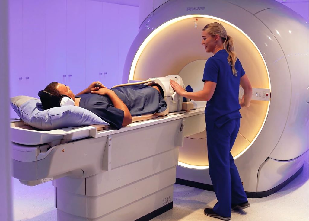
What is Magnetic Resonance Imaging (MRI)?

MRI is a non‑invasive imaging technique that uses powerful magnets and radio waves to create detailed images of the body’s internal structures. Unlike X‑rays or CT scans, MRI does not involve ionising radiation and is used extensively to diagnose a wide range of conditions.

Because MRI uses strong magnetic fields, many patients ask whether it is safe to have an MRI after a hip or knee replacement.

Can you have an MRI after a hip or knee replacement?

Yes, you can have an MRI scan on other parts of the body, as well as on the knee or hip where the implant is. Although some older MRI scanners may not be compatible with certain prostheses, the vast majority of MRI equipment in use today is safe and compatible with modern hip and knee implants.

How safe is MRI if the implant is made of metal?

Most implants are made from titanium or cobalt‑chromium alloys. Although these materials are metallic, they are not significantly affected by the magnetic field of an MRI scanner, nor do they heat up during the scan. Many implants also contain hard plastic components, all of which are designed to be compatible with MRI scanners. They are not attracted to the powerful magnet in the same way as older or highly magnetic materials.

Dr de Villiers explains, “The vast majority of joint replacements used today are MRI‑safe. The key is that we know about them in advance, so we can adjust the scan if needed.”

What is the main challenge with MRI and an implant?

The main challenge is image quality. Metal can sometimes cause image distortion, known as artefact, on MRI images. This may make it more difficult to assess structures close to the implant. However, modern MRI techniques have improved significantly and can often minimise these effects, allowing radiologists to assess surrounding tissues such as muscles and ligaments, and to detect complications such as infection or loosening. MRI is often the best imaging method for evaluating pain or complications after joint replacement surgery.

What happens if MRI does not produce clear diagnostic images?

In some cases, alternative imaging techniques such as CT or ultrasound may be recommended, depending on the clinical question. However, MRI remains safe and highly valuable for many patients with joint prostheses.

Are there implants that prevent you from having an MRI?

Certain implants and devices may be unsafe or require special precautions during MRI, including:

  • Implanted pacemakers
  • Intracranial aneurysm clips
  • Cochlear implants
  • Certain prosthetic devices
  • Implanted drug‑infusion pumps
  • Neurostimulators
  • Bone‑growth stimulators
  • Any other iron‑based metal implants

MRI is also contraindicated in the presence of some internal metallic objects such as bullets or shrapnel, as well as certain surgical clips, pins, plates, screws, metal sutures or wire mesh.

Having a hip or knee replacement does not automatically exclude you from having an MRI scan. With modern implants and appropriate planning, MRI is both a safe and important diagnostic tool. As technology continues to evolve, future developments are expected to further enhance MRI compatibility with hip and knee implants, making it an even more reliable tool for ongoing patient care.

It is crucial for patients to inform their healthcare providers about their joint replacement before undergoing an MRI. This allows the medical team to adjust the MRI settings and take appropriate precautions to ensure both safety and diagnostic accuracy.

New Global Study Estimates that Modern Hip Replacements Could Last at Least 30 Years

New global study using data from the National Joint Registry, estimates that modern hip replacements could last at least 30 years

Photo by DanR. CC BY-NC-SA-2.0

A major international study led by researchers who have used extensive data from the NJR estimates that modern total hip replacements, those using today’s more advanced bearing surfaces, are likely to last over 30 years in 92% of patients. This new finding marks a significant improvement in long term implant longevity and durability, compared with previous generations of medical implant devices.

Published on 26 February 2026, the research represents the largest and most contemporary analysis of hip replacement conducted to date. The study was a global collaboration including data contribution from eight joint registries. The data of just under two million hip replacement procedures were analysed, with the NJR accounting for almost two-thirds of that data. Registry data were combined with evidence from 29 long term clinical studies, across 18 countries.

Data was included from adult patients undergoing primary hip replacement with contemporary bearing surfaces: highly cross‑linked polyethylene (XLPE), ceramic‑on‑XLPE, or third‑ and fourth‑generation ceramic‑on‑ceramic articulations. Only implants that are still in routine clinical use were included, ensuring the study reflects modern practice, rather than historic device performance. Across all registries, cases were followed for a minimum of 10 years, with implant survival tracked until first all‑cause revision. All three material types demonstrated similarly high survivorship.

The results of the study provide patients with reassurance in consideration of the commonly asked question “How long will my hip replacement last?”  It is encouraging to know that modern hip replacements could last decades.

With regard to previous research on implant longevity, a 2019 study into hip replacement longevity which was supported by the NJR, suggested that over half, ie. 58% of hip replacements lasted 25 years, but those estimates were based on some implants made of materials that are no longer widely used. In 2022, another review of NJR data was conducted to enable further understanding of implant longevity, which produced the paper: ‘How long revised and multiply-revised hip replacements last?’ You can read more on that here.

You can read the recent Lancet paper here: Survivorship of modern total hip replacement to 30 years: systematic review, meta-analysis, and extrapolation of global joint registry data – The Lancet

Source: National Joint Registry

Overlooked Factor Key to Good Recovery After Hip Replacement Surgery

Photo by DanR. CC BY-NC-SA-2.0

Hip replacement surgery, or total hip arthroplasty (THA), can lessen pain and improve function in individuals with hip osteoarthritis. Some patients, however, continue to experience long-term physical deficits, including muscle weakness, decreased functional mobility, and increased fall risk, after the procedure. New research published in the Journal of Orthopaedic Research reveals that a patient’s muscle quality before THA may predict their risk of such suboptimal recovery after surgery.

In the study, 10 people undergoing THA underwent imaging tests before surgery. Patients whose imaging results indicated poor muscle quality were more likely to perform poorly on movement tasks after surgery, compared with those with good muscle quality. The severity of patients’ osteoarthritis before surgery (as indicated by the imaging tests performed) was not linked to their functional abilities after surgery.

“The findings from this study indicate that hip muscle quality may be an important predictor of post-operative biomechanical recovery following hip replacement. Muscle quality is often overlooked, and magnetic resonance imaging is needed to visualise muscle composition, which is not routinely collected for hip replacement patients,” said corresponding author Jeannie F. Bailey, PhD, of the University of California, San Francisco. “Future studies will seek to understand possible implications for poor hip muscle quality on long-term functional outcomes.”

Source: Wiley

Study Identifies Hip Implant Materials with Lowest Risk of Revision

Photo by DanR on Flickr

Hip implants with a delta ceramic or oxidised zirconium head and highly crosslinked polyethylene liner or cup had the lowest risk of revision during the 15 years after surgery, a new University of Bristol-led study has found. The research could help hospitals, surgeons and patients to choose what hip implant to use for replacement surgery.

The aim of the study, which appears in PLOS Medicine, was to establish hip implant materials at risk of revision. This would help orthopaedic surgeons, and patients, and to improve shared decision making before surgery by identifying hip implants with the lowest risk of revision.

The researchers analysed the UK’s National Joint Registry (NJR) data from 1 026 481 hip replacement patients carried out in the NHS and private sectors in England and Wales for up to 15 years after initial hip replacement operations (between 2003 to 2019). 

After reviewing hip implants from the NJR data, the research team found the risk of revision following a hip replacement is influenced by the type of material used in the bearing surface. Bearing surfaces are the moving parts of an artificial hip joint that glide against each other during activity.

The data indicated that hip implants with a delta ceramic or oxidised zirconium head and highly crosslinked polyethylene liner or cup had the lowest risk of revision throughout the 15 years following hip replacement surgery.

These findings were confirmed when the research team investigated the specific reasons for revision hip replacements being performed.  The data also showed 20 869 (2%) of hip replacement patients had to undergo revision after the initial surgery.

Senior author Dr Erik Lenguerrand, Senior Lecturer in Medical Statistics and Quantitative Epidemiologist in the Bristol Medical School: Translational Health Sciences (THS), said: “Our research has found the risk of hip replacement revision depends on the hip implant materials used in the original surgery.  The lowest risk of revision are from implants with delta ceramic or oxidised zirconium head and a highly crosslinked polyethylene (HCLPE) liner or cup.

“Further research is needed to find out the association of implant materials with the risk of rehospitalisation, re-operation other than revision, mortality and the cost-effectiveness of these materials.”

Michael Whitehouse, Professor of Trauma and Orthopaedics at Bristol Medical School: THS, and senior clinical lead for the paper, explained: “Our study has used data from one of the largest registries in the world that includes all public and private health care sectors in England and Wales. This means that the data is more generally applicable than that available previously, which was limited by broad groupings of implant types or much smaller study size. It highlights the importance of considering the whole structure that is created when implants are put together to make up a hip replacement rather than focusing on individual components.

“Our findings will help hospitals, surgeons and patients to choose hip implants and combinations of them with the lowest risk of revision following an initial hip replacement operation.”

Tim Wilton, Medical Director of the National Joint Registry (NJR), added: “We are always delighted when the data from the NJR can be used by researchers to produce important research of this kind which gives meaningful analysis to guide surgeons and patients in their decisions. An important value of the NJR data is that it allows researchers a unique insight to assess the long-term performance of different hip implant materials.

“By tracking the combinations of materials used and subsequent revision rates, this research highlights the role of implant material choice in surgical outcomes. This ensures that the materials used can be optimised for longevity and patient health. Surgeons would be well advised to study these findings carefully in relation to the implant choices they make, and to use the information in pre-operative discussions with their patients. As the demand for joint replacements continues to rise, this insight can be invaluable in reducing revision surgery.”

The research was not a randomised controlled trial and therefore it was not possible to control all factors that can influence the risk of revision.

The categorisation of hip implants used as part of hip replacements is often broad in national joint replacement surgery registries and does not fully show differences in revision risks associated within the different types of implant materials grouped together.

Source: University of Bristol

Genetic Risk Score for Hip or Knee Replacements

A Monash University-led research team has developed a risk score based on individuals’ genetic data to predict their likelihood of needing hip or knee replacement surgery for osteoarthritis. The team validated the score’s predictive ability in a study published in Arthritis & Rheumatology.  

The score incorporates 10 genetic sequence variants for predicting a person’s risk of needing knee replacement surgery and 37 genetic sequence variants for predicting the risk of needing hip replacement surgery.  

Among 12093 individuals of European genetic descent aged 70 years or older, 1422 (11.8%) had knee replacements and 1,297 (10.7%) had hip replacements. Participants with high risk scores had a 1.44-times higher odds of knee replacement and a 1.88-times higher odds of hip replacement, compared with those with low risk scores.   

 “Genetic scores, such as the one we developed, do not change over a person’s life. They provide an individual  with further information about their risk of severe osteoarthritis in later life and have the potential to improve prevention of severe knee and hip osteoarthritis by identifying those who may benefit from early intervention,” said senior author Flavia Cicuttini, PhD, of Monash University.  

Source: Wiley