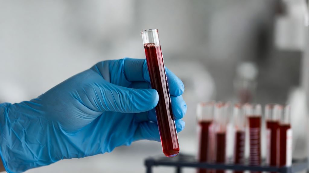
Category: Lab Tests and Imaging

An Early Blood Test can Predict Survival in Patients with Metastatic Prostate Cancer

Credit: Darryl Leja National Human Genome Research Institute National Institutes Of Health

A blood test, performed when metastatic prostate cancer is first diagnosed, can predict which patients are likely to respond to treatment and survive the longest. It can help providers decide which patients should receive standard treatment versus who might stand to benefit from riskier, more aggressive new drug trials. The research, which forms part of a Phase III clinical trial, was just published in JAMA Network Open.

Once prostate cancer has metastasised and is no longer curable, systemic treatments are used to prolong survival as much as possible. Biomarkers that predict how patients will respond could allow for better personalisation of treatments, but they are few and far between.

A new study found that measuring circulating tumour cells (CTCs), rare cancer cells shed from tumours into the blood, is a reliable way to predict later treatment response and survival prospects. CTCs have been studied in prostate cancer before, but only in its later stages.

“No one, until now, has looked at whether CTC counts can be used right at the beginning, when a man first presents with metastatic prostate cancer, to tell us whether he’s going to live a long or short time, or whether or not he will progress with therapies,” said Amir Goldkorn, MD, lead author of the study and associate director of translational sciences at the USC Norris Comprehensive Cancer Center at the Keck School of Medicine of USC.

The research leveraged CellSearch (Menarini, Inc.), an FDA-cleared liquid biopsy technology at the Norris Comprehensive Cancer Center, to detect and measure CTCs in blood samples. Patients with more CTCs had shorter median survival lengths and a greater risk of death during the study period. Those with more CTCs also had less “progression-free survival,” which refers to the length of time when a patient’s disease is controlled by treatment without getting worse.

“You couldn’t tell these men apart when they walked through the door,” said Goldkorn, who is also a professor of medicine at the Keck School of Medicine. “All of their other variables and prognostic factors were seemingly the same, and yet they had very, very different outcomes over time.”

The researchers say that the CellSearch blood test, which is already widely available from commercial providers, can help quickly identify patients who are unlikely to respond to standard treatment options. Those men could benefit from a more intensive approach to therapy, including clinical trials of new drugs that may have more side effects but could improve survival in these high-risk patients.

Counting CTCs

The research was part of a phase 3 clinical trial of the NCI-funded SWOG Cancer Research Network, a group of more than 1300 institutions around the country that collaborate to study various cancers. Baseline blood samples from 503 patients with metastatic prostate cancer, who were participating in a new drug trial, were sent to the Keck School of Medicine team for analysis.

To analyze the blood samples, the researchers used the CellSearch platform at the Norris Comprehensive Cancer Center’s Liquid Biopsy Research Core, a facility that Goldkorn founded and directs. CellSearch uses immunomagnetic beads, antibodies attached to small magnetic particles, which bind to CTCs in the blood and pull them out to be detected and counted by specialised equipment.

Patients with five or more CTCs in their blood sample had the worst outcomes. Compared to patients with zero CTCs, they were 3.22 times as likely to die during the study period and 2.46 times as likely to have their cancer progress. They were only 0.26 times as likely to achieve a complete prostate-specific antigen (PSA) response, meaning they responded poorly to treatment.

Men with five or more CTCs had a median survival length of 27.9 months following the blood test, compared to 56.2 months for men with one to four CTCs and at least 78 months for men with zero CTCs. (Many patients in the latter group survived past the date of publication, so the median survival length could not yet be calculated.)

The bottom line: more CTCs meant that patients survived for less time, progressed much more quickly and were unlikely to respond to standard treatments.

Candidates for clinical trials

The new study shows that measuring CTC counts at the start of therapy can predict long-term survival rates, even in men who go on to receive many treatments for metastatic prostate cancer over a years-long period. That means the test can help identify men early on for trials of new and potentially more aggressive therapies.

“We want to enrich these clinical trials with men who need all that extra help – who really would benefit from three drugs versus just two, or from being on a new chemotherapy drug, even though it may have more side effects,” Goldkorn said.

Goldkorn and his team are now testing a new blood test that measures not just CTC counts, but also the molecular composition of CTCs and tumour DNA circulating in the blood, as well as other factors. Their goal is to create biomarkers with even more predictive power, which may ultimately help match patients with specific treatment options.

Source: Keck School of Medicine of USC

A Potential Stool Test for Endometriosis also Suggests an IBD Link

Photo by Sora Shimazaki on Pexels

Promising findings by researchers at Baylor College of Medicine and collaborating institutions could lead to the development of a non-invasive stool test and a new therapy for endometriosis, a painful condition that affects nearly 200 million women worldwide. The study appeared in the journal Med.

“Endometriosis develops when lining inside the womb grows outside its normal location, for instance attached to surrounding intestine or the membrane lining the abdominal cavity. This typically causes bleeding, pain, inflammation and infertility,” said corresponding author Dr Rama Kommagani, associate professor in the Department of Pathology and Immunology at Baylor. “Generally, it takes approximately seven years to detect endometriosis and is often diagnosed incorrectly as a bowel condition. Thus, delayed diagnosis, together with the current use of invasive diagnostic procedures and ineffective treatments underscore the need for improvements in the management of endometriosis.”

“Our previous studies in mice have shown that the microbiome, the communities of bacteria living in the body, or their metabolites, the products they produce, can contribute to endometriosis progression,” Kommagani said. “In the current study, we took a closer look at the role of the microbiome in endometriosis by comparing the bacteria and metabolites present in stools of women with the condition with those of healthy women. We discovered significant differences between them.”

The findings suggested that stool metabolites found in women with endometriosis could be the basis for a non-invasive diagnostic test as well as a potential strategy to reduce disease progression.

The researchers discovered a combination of bacterial metabolites that is unique to endometriosis. Among them is the metabolite called 4-hydroxyindole. “This compound is produced by ‘good bacteria,’ but there is less of it in women with endometriosis than in women without the condition,” said first author Dr Chandni Talwar, postdoctoral associate in Kommagani’s lab.

“These findings are very exciting,” Talwar said. “There are studies in animal models of the disease that have shown specific bacterial metabolite signatures associated with endometriosis. Our study is the first to discover a unique metabolite profile linked to human endometriosis, which brings us closer to better understanding the human condition and potentially identifying better ways to manage it.”

Furthermore, extensive studies also showed that administering 4-hydroxyindole to animal models of the disease prevented the initiation and progression of endometriosis-associated inflammation and pain. 

“Interestingly, our findings also may have implications for another condition. The metabolite profile we identified in endometriosis is similar to that observed in inflammatory bowel disease (IBD), revealing intriguing connections between these two conditions,” Kommagani said. “Our findings support a role for the microbiome in endometriosis and IBD.”

The researchers are continuing their work toward the development of a non-invasive stool test for endometriosis. They are also conducting the necessary studies to evaluate the safety and efficacy of 4-hydroxyindole as a potential treatment for this condition.

Source: Baylor College of Medicine

Most Accurate Ultrasound Test Could Detect 96% of Women with Ovarian Cancer

Photo by Mart Production on Pexels

An ultrasound test that detected 96% of ovarian cancers in postmenopausal women should replace current standard of care test in the UK according to a new study.

In a paper published in Lancet Oncology, research led by Professor Sudha Sundar from the University of Birmingham compared all currently available tests to diagnose ovarian cancer in postmenopausal women head-to-head in a high-quality diagnostic test accuracy study.

Of the six diagnostic tests investigated, the IOTA ADNEX model which looks at ultrasound features (how the lump looked like on ultrasound) had the best accuracy of all and could detect up to 96% of women with ovarian cancer.

The ultrasound test outperforms the current standard of care in the UK significantly and so the researchers recommend that the IOTA ultrasound ADNEX model should replace the current standard of care test called risk of malignancy (RMI1) test in the UK which identifies 83% of ovarian cancers.

Sudha Sundar, Professor of Gynaecological Cancer at the University of Birmingham and consultant in gynaecological cancer surgery at Sandwell and West Birmingham NHS Trust said:

“This is the first time that a head-to-head study of all available ovarian cancer tests have been done in the same population. Here we studied their use with symptomatic, postmenopausal women who are most at risk of this cancer. Our trial found that the IOTA ADNEX ultrasound protocol had highest sensitivity for detecting ovarian cancer compared to the standard of care and other test.

“The ultrasound test also performs well when delivered by a trained sonographer who have received specific training and certification and quality assurance, and as the vast majority of ultrasound scans are performed by sonographers it is important that a new standard is able to be delivered by as many clinical professionals as possible.

“We found that the higher sensitivity of the IOTA ADNEX model is likely to lead to some women who don’t have cancer also being flagged up as having a higher risk of cancer. We however did discuss this extensively with patients, cancer charity Target ovarian cancer and NHS experts who all agreed that in postmenopausal women who are at higher risk of ovarian cancer, picking up more women with cancer would benefit women overall.”

The research team note that the IOTA ADNEX model achieved 96% accuracy when delivered by NHS sonographers who were appropriately trained and received quality assurance. As most scans worldwide are carried out by sonographers rather than gynaecologists, introductory free online resources have been created by the researchers for NHS staff to undergo the specialist ultrasound training and get certification and quality assurance.

Source: University of Birmingham

New Images of RSV may Expose Weak Points in the Stubborn Virus

Photo by Andrea Piacquadio on Pexels

The complex shape of respiratory syncytial virus is one hurdle limiting the development of treatments for an infection that leads to hospitalisation or worse. New images of the virus published in Nature from researchers at the University of Wisconsin–Madison may hold the key to preventing or slowing RSV infections.

RSV is of greatest concern in young children, the elderly and adults at high risk for respiratory complications. Yet unlike the flu and other common, communicable respiratory illnesses that annually sweep through schools, there are few options for fighting RSV. In the US, prophylactic treatments are available for young children, and existing vaccines are approved only for pregnant women and the elderly.

The virus’s structure, which consists of tiny, bending filaments, have eluded researchers. This has made it difficult to identify key drug targets, including viral components that are conserved across related viruses.

RSV F proteins, shown in this image created by University of Wisconsin–Madison researchers using a technique called cryo-electron tomography, may make RSV more potent by keeping it from infecting cells prematurely. Image by Wright Lab, UW–Madison

“There are a number of viruses related to RSV that are also significant human pathogens, including measles,” says Elizabeth Wright, a UW–Madison biochemistry professor. “What we know about related viruses gives us clues about RSV protein structures, but to identify drug targets we need a closer look at RSV proteins that are intimately associated with the membranes of host cells.”

Using an imaging technique called cryo-electron tomography, Wright and her team have now revealed details of molecules and structures essential to RSV’s form and function. They published their findings recently in Nature.

Cryo-ET freezes viral particles or other molecules at ultracold temperatures, stopping biological processes in action. This allows researchers to examine the structures of organisms, cells and organelles, and viruses and capture small-scale images of structures frozen in time. Flash-freeze many RSV particles, and cryo-ET imaging will capture (nearly) all the virus’s possible configurations from many different angles. These 2D images are combined to produce a representation of the virus’s 3D structures at high resolutions – even at the level of individual atoms.

Wright’s recent study produced high-resolution images detailing the structure of two RSV proteins, RSV M protein and RSV F protein, that are crucial to the interaction between the virus and the host cell membrane. Both proteins are also present in related viruses.

RSV M protein interacts with host cell membranes, holding together the virus’s filamentous structure and coordinating viral components and other proteins – including RSV F proteins. RSV F proteins sit on the viral surface, ready to engage with host cell receptors and regulate the virus’s fusion and entry into the host cell. The scientists’ images reveal that in RSV, two F proteins come together to form a more stable unit. Wright says that this association may prevent the F proteins from prematurely infecting the host cell.

“Our primary findings reveal structural details that allow us to better understand not only how the protein regulates assembly of viral particles, but also the coordination of proteins that enable the virus to be infectious,” says Wright.

The scientists believe that F protein pairs may be a key to destabilising the virus before it is ready to infect its next host, making pairs of F proteins a possible target for future drug development. They will continue to explore how RSV proteins interact with each other to cause infection.

Source: University of Wisconsin-Madison

Hand-held Medical Scanner could Transform Cancer and Arthritis Diagnosis

PAT images of wrist vasculature acquired in high-resolution scan mode. Wrist region, (i) x-y and (ii) x-z depth-to-colour encoded MIPs, (iii) x-z and (iv) y-z greyscale MIP slices of regions indicated by dashed red and blue rectangles in (i) showing fine dermal microvasculature (DM), radial artery (RA) and large wrist veins. Inset: x-z greyscale MIP showing cross-sectional view of the radial artery and adjacent veins in the plane indicated by the dashed yellow line in (iv). Huynh et al., Nature Communications, 2024.

A new hand-held scanner developed by UCL researchers and tested in a series of clinical trials on UCLH patients can generate highly detailed 3D photoacoustic images in just seconds, paving the way for their use in a clinical setting for the first time and offering the potential for earlier disease diagnosis.

In the study, published in Nature Biomedical Engineering, the UCL and UCLH team show their technology can deliver photoacoustic tomography (PAT) imaging scans to doctors in real time, providing them with accurate and intricate images of blood vessels, helping inform patient care.

Photoacoustic tomography imaging uses laser-generated ultrasound waves to visualise subtle changes (an early marker of disease) in the sub-millimetre-scale veins and arteries up to 15mm deep in human tissues.

However, up until now, existing PAT technology has been too slow to produce high-enough quality 3D images for use by clinicians.

The older PAT scanners took more than five minutes to take an image – by reducing that time to a few seconds or less, image quality is much improved and far more suitable for people who are frail or poorly.

The researchers say the new scanner could help to diagnose cancer, cardiovascular disease and arthritis in three to five years’ time, subject to further testing.

In this study, the team tested the scanner during pre-clinical tests on 10 UCLH patients with type-2 diabetes, rheumatoid arthritis or breast cancer, along with seven healthy volunteers. They also compared the PAT scans to regular clinical scans taken at UCLH. Larger scale trials of the device are ongoing at UCLH and UCL.

In three patients with type-2 diabetes, the scanner was able to produce detailed 3D images of the microvasculature in the feet, highlighting deformities and structural changes in the vessels. The scanner was also used to visualise the skin inflammation linked to breast cancer.

UCLH consultant radiologist Andrew Plumb, a senior author of the study and Chief Investigator of the clinical PAT studies, said: “One of the complications often suffered by people with diabetes is low blood flow in the extremities, such as the feet and lower legs, due to damage to the tiny blood vessels in these areas. But until now we haven’t been able to see exactly what is happening to cause this damage or characterise how it develops.

“In one of our patients, we could see smooth, uniform vessels in the left foot and deformed, squiggly vessels in the same region of the right foot, indicative of problems that may lead to tissue damage in future. Photoacoustic imaging could give us much more detailed information to facilitate early diagnosis, as well as better understand disease progression more generally.” Dr Plumb is also Associate Professor of Medical Imaging at UCL.

Patients were identified and recruited from a number of clinics at UCLH, including consultant rheumatologist Madhura Castelino, consultant interventional radiologist Conrad von Stempel and research staff Katerina Soteriou and Antonia Yeung who co-ordinated safe, timely scanning at UCLH and UCL on the new PAT scanner.

UCL Professor of Biomedical Photoacoustics Paul Beard, corresponding author, said: “We’ve come a long way with photoacoustic imaging in recent years, but there were still barriers to using it in the clinic.

“The breakthrough in this study is the acceleration in the time it takes to acquire images, which is between 100 and 1000 times faster than previous scanners.

“This speed avoids motion-induced blurring, providing highly-detailed images of a quality that no other scanner can provide. It also means that rather than taking five minutes or longer, images can be acquired in real time, making it possible to visualise dynamic physiological events.

“These technical advances make the system suitable for clinical use for the first time, allowing us to look at aspects of human biology and disease that we haven’t been able to before.

“Now more research is needed with larger groups of patients to confirm our findings.”

Professor Beard added that a key potential use for the new scanner was to assess inflammatory arthritis, which requires scanning all 20 finger joints in both hands. With the new scanner, this can be done in a few minutes – older PAT scanners take nearly an hour, which is too long for elderly, frail patients, he said.

Source: University College London Hospitals

Over Half of Iron Deficiency Cases Unresolved After Three Years

Photo by National Cancer Institute on Unsplash

Over half of people with iron deficiency were found to still have low iron levels three years after diagnosis, and among patients whose condition was effectively treated within that timeframe, they faced longer-than-expected delays, pointing to substantial gaps in appropriate recognition and efficient treatment of the condition, according to a study published in Blood Advances.

Iron deficiency is common, and affecting up to 40% of adolescents and young women. Previous work reported that up to 70% of cases go undiagnosed in high-risk populations, such as those with bleeding disorders, issues with malabsorption, or women who menstruate.

“Iron deficiency is probably a bigger problem than we realise. I’ve seen a lot of cases where people don’t have anaemia, but they are walking around with very little to no iron in their body and it can have a big impact on how people feel in their day-to-day life,” said Jacob Cogan, MD, assistant professor of medicine at the University of Minnesota and the study’s lead author. “Iron deficiency can be challenging to diagnose, but it’s easy to treat. Our findings underscore the need for a more coordinated effort to recognise and treat iron deficiency to help improve quality of life.”

If untreated, low iron stores can lead to mood changes, fatigue, hair loss, exercise intolerance, and eventually anaemia. The condition is generally first treated with oral iron supplementation, and if low iron levels persist after a few months or the patient reports side effects, intravenous (IV) iron is started.

For this study, the researchers retrospectively analysed medical records from one of Minnesota’s largest health system database and identified 13 084 adults with a laboratory diagnosis of iron deficiency (defined as a ferritin value of 25ng/mL, with and without anaemia) between 2010 and 2020 who had available follow-up data for three years.

In the study, iron deficiency was d or less. Patients had to have at least two ferritin values – one initial value and at least one more within the three-year study period. Adequate treatment and resolution was defined as a subsequent ferritin value of at least 50ng/mL. Most patients received some form of treatment, consistent across sex.

Of the 13,084 patients included in the study, 5,485 (42%) patients had normal iron levels within three years of diagnosis, while 7,599 (58%) had persisting iron deficiency based on low ferritin levels. Only 7% of patients had their iron levels return back to normal within the first year of diagnosis.

Factors associated with a higher likelihood of getting iron levels back to normal included older age (age 60 and up), male sex, Medicare insurance, and treatment with IV iron alone. Additionally, compared with patients who were still iron deficient, those whose condition was resolved had more follow-up blood work to check ferritin values (six vs four ferritin tests). Of note, younger patients, females, and Black individuals were most likely to remain iron deficient or experience longer lags in getting their iron stores back to a healthy level.

Even among patients whose iron levels were restored to normal during the study duration, it took nearly two years (the median time to resolution was 1.9 years), which researchers say is longer than expected and signals missed opportunities to more effectively manage the condition. While there was no data to look at whether anaemia iron deficiency was more apt to be treated, Dr Cogan says it’s reasonable to think this might be the case as iron deficiency without anaemia is harder to recognise.

“Two years is too long and well beyond the timeframe within which iron deficiency should be able to be sufficiently treated and resolved [with oral or IV treatments],” said Dr Cogan. “The numbers are pretty striking and suggest a need to put systems in place to better identify patients and treat them more efficiently.”

As with trends showing persisting iron deficiency, Dr Cogan attributes the delays in resolution to the diagnosis either being missed or not treated to resolution. He added that there is a clear need for education about non-anaemic iron deficiency and who is at high risk, more universal agreement on the best ferritin cut off for diagnosis, and efforts to create an iron deficiency clinic or pathway to “assess and treat patients more efficiently and get people feeling better faster.”

The study was limited by its reliance on EMR data and retrospective nature, which prevented researchers from determining why ferritin tests were ordered for patients or the cause of their iron deficiency.

Source: American Society of Hematology

Investigating New Diagnostic Tools for Early Diagnosis of Endometriosis

Photo by Andrea Piacquadio on Pexels

Endometriosis is a common, burdensome, chronic disease that affects 190 million women worldwide, and may cause life-altering consequences such as chronic pain, infertility and quality of life. Early diagnosis remains a major clinical and public health challenge. The average time to diagnose endometriosis is seven years after the onset of symptoms, which include abdominal pain and cramping before, during and after menstruation, among others.

In a commentary published in The Journal of Reproductive Medicine, Gynaecology & Obstetrics, researchers from Florida Atlantic University’s Schmidt College of Medicine and collaborators, conducted a PubMed search to identify promising approaches for early diagnosis of endometriosis.

“Currently, diagnosing endometriosis involves a thorough review of the patient’s medical history and physical examination,” said first author Panagiota “Yiota” Kitsantas, PhD, professor and chair of the Department of Population Health and Social Medicine, FAU Schmidt College of Medicine. “The most commonly used and accurate diagnostic methods are pelvic exams, abdominal ultrasound, MRI and laparoscopy. Laparoscopic surgery is considered the gold standard for diagnosing endometriosis by gynaecologists, but it can be expensive and carries potential risks of surgical complications. Moreover, the accuracy of laparoscopy can vary based on the surgeon’s experience and the stage of the disease.”

The authors say the ideal test for early diagnosis of endometriosis would be to use symptom-based criteria to determine who should undergo testing and then set optimal cut-points to maximise sensitivity and specificity. A test with high predictive value would accurately confirm endometriosis if positive and exclude it if negative. Although less ideal tests may not provide definitive results, they can be useful in reducing the number of patients who need to proceed to more invasive procedures, like laparoscopy.

Endometriosis involves hormonal imbalances that trigger angiogenesis, apoptosis, immune responses, and inflammation. Diagnostic tools for endometriosis have been developed to detect biomarkers, such as mRNA fragments in blood and saliva, but these have shown low accuracy.

“Non-invasive methods like MRI and transvaginal ultrasound are only effective for advanced stages of endometriosis,” said co-author Charles H. Hennekens, MD, professor at FAU Schmidt College of Medicine. “Recent research has focused on a novel noninvasive method of detecting myoelectric activity in the gastrointestinal tract as a potential diagnostic tool. Electroviscerography or EVG could detect unique myoelectric patterns associated with endometriosis, though this approach is promising but unproven.”

Currently, there is no FDA-approved non-invasive test for endometriosis, and further analytic studies leading to peer-reviewed publications are needed to refine these emerging technologies and establish effective diagnostic criteria.

“Early diagnosis of endometriosis remains a challenge, with a succession of promising approaches ultimately not bearing fruit thus far,” said Kitsantas. “Once new technologies such as EVG are more fully evaluated, they may give clinicians the post-test certainty they need to transition from symptom-based to diagnosis-based treatment.”

Source: Florida Atlantic University

Radiology Helps Treat Chronic Pain

Dr Winter performing a CT-guided interventional procedure. Photo: Supploed

Radiology encompasses more than just imaging. It is a medical field that uses various imaging techniques to diagnose conditions, guide minimally invasive procedures and, much to the relief of agonised patients, treat chronic pain.

‘Traditionally, radiology is known as a modality where causes of pain are only diagnosed’, says Dr Arthur Winter, a radiologist at SCP Radiology. ‘Interventional radiology has changed this. It is a rapidly developing branch of radiology involving minimally invasive procedures.  Pain management procedures are becoming a daily part of busy radiology departments.’

Simply put, interventional radiologists can use precisely targeted injections to intervene in the body’s perception of pain.

Understanding pain

Pain is a signal from the nervous system to let you know that something is wrong in your body. It is transmitted in a complex interaction between specialised nerves, the spinal cord and the brain. It can take many forms, be localised to one part of the body or appear to come from all over.

Pain can be acute or chronic

Harvard Medical School gives an overview of the difference between the two. ‘Most acute pain comes from damage to body tissues. It results from physical trauma such as a sports or exercise injury, a broken bone, a medical procedure or an accident like stubbing your toe, cutting a finger or bumping into something. The pain can feel sharp, aching or throbbing and often heals within a few days to a few weeks.’

In comparison, chronic pain lasts at least two to three months, often long after you have recovered from the injury or illness and may even become permanent. It could also be a result of lifestyle diseases. Symptoms and severity vary and may include a dull ache, shooting, burning, stabbing or electric shock-like pain and sensations like tingling and numbness. Chronic pain can be debilitating and affect your ability to perform activities of daily living.

Interventional pain management

Although some acute pain can be managed with interventions, it is patients with chronic pain that truly benefit. ‘These patients often use high doses of opioid painkillers that may cause nausea, constipation, anorexia and addiction. Other painkillers may also irritate the stomach lining and cause kidney problems,’ says Dr Winter.

An alternative that interventional pain management offers, involves injections called nerve blocks that target very specific nerves.

‘Most of these interventions prevent nerve impulses or pain signals from being transmitted, using long-acting local anaesthetics. The effect is usually temporary but the addition of cortisone – or steroids – often brings longer-lasting relief. In some cases, it could be appropriate to follow the temporary block with neurolysis, which is a permanent disruption or destruction of the target nerves.’

Although nerve blocks and other long-acting pain injections have been done for years, the scope of procedures is evolving fast. The involvement of radiologists has also grown.

Dr Winter explains. ‘Pain management has traditionally been the responsibility of clinicians and anaesthetists. During nerve block procedures, they were typically guided by their knowledge of anatomy or a continuous X-ray technique called fluoroscopy. As ultrasound became more widely available, many anaesthetists learned to do these procedures under ultrasound guidance.

‘These specialists still provide these treatments but, thanks to the availability of specialised imaging equipment, radiologists now have the tools and skill to do procedures under sophisticated image guidance. With CT guidance, some procedures can be performed with great accuracy while avoiding blood vessels and non-target organs,’ says Dr Winter.

‘A lower dose of medication is also needed if the needle is placed accurately next to the target nerves. It is therefore not surprising that this is increasingly becoming a responsibility of interventional radiologists.’

Other procedures where radiologists are involved include targeted Botox injections to treat the symptoms of Piriformis syndrome, epidural cortisone injections for inflammation in the spine and a procedure called epidural blood patch. This is to seal spinal fluid leaks that cause low-pressure headaches.

In conclusion, Dr Winter says chronic pain may cause poor quality of life and depression, often seen in patients with underlying cancer. ‘It is especially these patients who should be considered for interventions. There are, for example, very effective procedures to manage pain caused by pancreatic and pelvic cancers.

‘Specialists like oncologists and neurologists recognise the value of interventional radiology in pain management and work closely with us to support their patients. It is a growing branch of radiology that offers a minimally invasive solution and it’s quite rewarding to see patients regain some quality of life.’

Routine Bloods can Improve Cancer Screening in Patients with Abdominal Symptoms

Risk of cancer by specific site based on blood test abnormalities in symptomatic patients can help guide referral strategies

Photo by National Cancer Institute on Unsplash

Incorporating information from common blood tests can enhance cancer risk assessment in patients with abdominal symptoms, according to a study publishing July 30th in the open-access journal PLOS Medicine by Meena Rafiq from University College London, UK, and colleagues.

Early cancer detection is key to successful treatment. However, many undiagnosed cancer patients present to their primary care provider with non-specific symptoms that can be a result of several other benign causes, making it difficult to determine who warrants additional diagnostic testing or referral. Most guidelines focus on “alarm” symptoms specific to a given type of cancer to guide referrals. There is limited guidance on non-specific symptoms to guide cancer assessment and referral decisions across different cancer types.

In this study, researchers used data from the UK Clinical Practice Research Datalink to identify more than 470 000 patients aged 30 years or older who had visited a general practitioner due to abdominal pain or bloating. Within a year of that visit, approximately 9000 patients with abdominal pain and 1000 patients with bloating were diagnosed with cancer. The researchers looked at 19 abnormal blood test results collected during the initial primary care visit to see if they could predict who was more likely to be diagnosed with cancer.

Several blood abnormalities were predictive of cancer risk across sex and age groups. For example, in patients aged 30–59 years with abdominal symptoms, anaemia, low albumin, raised platelets, abnormal ferritin, and increased inflammatory markers strongly predicted a risk of undiagnosed cancer. Among older patients (aged 60 years and above), the presence of abdominal pain or bloating alone was enough to warrant a cancer referral.

The study also showed which types of cancer were most common based on age, sex, and blood test abnormality. For example, among women aged 50–59 years with anaemia and abdominal bloating, the most common types of cancer were bowel and ovarian cancer. This level of granularity can help guide providers on which diagnostic strategies to prioritise.

The study shows that common, routine blood test results can provide additional context in patients with non-specific abdominal symptoms to improve cancer risk assessment and identify patients who warrant additional testing and/or referral to a specialist.

The authors add, “Using existing blood tests can be an effective and affordable way to improve early diagnosis of cancer in people who see their GP with vague symptoms. Our study identified several commonly used GP blood tests where abnormal results increase a patient’s risk of having cancer and these can be used to diagnose cancer earlier.”

Provided by PLOS

“We Were the First Ones to Do It”: Innovative SA Study Takes TB Testing to People’s Homes

Tuberculosis bacteria. Credit: CDC

By Tiyese Jeranji

Most tuberculosis (TB) tests still require a trip to the clinic. Now, new technology has made it possible to test people at home. This could be a big deal for South Africa, where much TB goes undiagnosed. We unpack the findings and implications of a recent study into such TB home testing.

One of the biggest challenges in combatting TB in South Africa is that many people who fall ill with the disease are diagnosed late, or not diagnosed at all.

The World Health Organization (WHO) estimates that 280 000 people fell ill with TB in the country in 2022. Of these, roughly 66 000 were not diagnosed, and accordingly also not treated. Apart from the damage to the health of the people who are not diagnosed and treated, this also has implications for the further spread of TB since untreated TB is often infectious TB – people become non-infectious within a few weeks of starting TB treatment.

Typically, people who fall ill with TB only get diagnosed once they turn up at clinics with TB symptoms – this is called passive case-finding. In recent years, there has been a growing recognition that passive case-finding alone is not good enough if we want to diagnose more people more quickly. As a result, many people in South Africa considered to be at high risk of TB are now offered TB tests whether or not they have symptoms – an approach called targeted universal testing. Screening for TB using new mobile X-ray technology has also been piloted in the country.

Now, in the latest such active case-finding innovation, researchers have been offering people TB tests in the comfort of their own homes.

Dr Andrew Medina-Marino, a senior investigator at the Desmond Tutu Health Foundation (DTHF), tells Spotlight no one in the world was testing for TB at home until they recently started doing so at the DTHF’s new research site in the Eastern Cape.

The testing is done using a molecular testing device, roughly the size of a two litre Coke bottle, called the GeneXpert Edge. The GeneXpert Edge is a portable version of the GeneXpert machines that have been used in labs across the country to diagnose TB for over a decade.

The GeneXpert Edge is a standardised testing device that detects TB DNA in sputum. (Photo: Nasief Manie/Spotlight)

One challenge with the device was that it needed to be plugged into a power outlet in a wall and not all homes in the area have power. “So what we did is, we hooked up a car-like battery to the device and we were able to take it into people’s homes,” says Medina-Marino.

‘Acceptable and feasible’

A study lead by Medina-Marino, and recently published in Open Forum Infectious Diseases, set out to determine the acceptability and feasibility of in-home testing of household contacts of people with TB.

The study was conducted among 84 households in Duncan Village, a township in the Buffalo City Metropolitan Municipality in the Eastern Cape. The Metro had an estimated TB incidence of 876 cases per 100 000 population in 2019, according to the National Institute for Communicable Diseases. This number is much higher than the latest WHO estimate of 468  per 100 000 for South Africa as a whole.

From July 2018 to May 2019, people diagnosed with pulmonary TB were recruited from six government health clinics in the area. They were asked for permission to visit their homes to screen their household contacts for TB. Household contacts were verbally assessed for signs or symptoms of TB, including night sweats, weight loss, persistent cough and a fever.

Households where people had any signs or symptoms of TB were randomised to either be referred to a local clinic for TB testing or tested immediately in their home. Of the eighty-four randomised households, 51 household contacts were offered in-home testing. Everyone accepted the offer for in-home testing.

For the test with the GeneXpert Edge, Medina-Marino says household contacts had to produce a sputum sample. About 47% (24/51) were able to produce sputum. This was then mixed with a reagent containing the required components for a polymerase chain reaction test. This solution was then loaded into a disposable cartridge/test module and inserted into the Edge device. Results were available in about 90 minutes. Anyone who received a positive test result in their home were immediately referred to a clinic for TB treatment.

Regarding the 47 household contacts referred for testing at the clinic, only 15% (7 people) presented for clinic-based TB evaluation, 6 were tested, and 4 out of 6 returned for their results.

Ultimately, the study found that in-home testing of household contacts for TB was acceptable and feasible.

“It’s feasible. If you compare the rate of uptake of treatment versus the rate of uptake for testing, it looks like it’s performing much better when you do home based testing versus referral for testing at the clinic,” says Medina-Marino.

Risk of stigma?

Similar to when HIV home-based testing studies were carried out, Medina-Marino says prior to their study, community members expressed concerns about stigmatising houses that were visited. “[A] lot of people were saying: ‘If you go to people’s houses, you’re going to stigmatise the household.’”

But what they actually found was that people didn’t feel stigmatised. Household contacts of people with TB felt that coming to the house to test people brought a sense of security in the home. He adds that it was easy for people to believe the results because everything was done in front of them.

In instances where people didn’t have TB, Medina-Marino says household contacts were comforted that they didn’t have to be scared of the person tested. In instances where people did have TB, he says the attitude of household contacts was supportive to start treatment.

How the test compares to other tests

Apart from testing for TB, the GeneXpert Edge can also detect whether someone’s TB is resistant to rifampicin. This is one of the medicines in the standard four-drug combination used to treat TB.

Unlike the latest lab-base GeneXpert tests, the GeneXpert Edge does not detect resistance to any TB medicines other than rifampacin. “It is hard to fit the probes needed to detect other forms of resistance into the cartridge,” says study co-author Professor Grant Theron, head of the Clinical Mycobacteriology and Epidemiology Research group at Stellenbosch University’s Molecular Biology and Human Genetics Unit.

Theron notes that the sensitivity and specificity of GeneXpert Edge is similar to that of lab-based GeneXpert machines if the tests are done on specimens from the same type of patient and the same test cartridge. (High sensitivity means the likelihood of false negatives is low wile high specificity means the likelihood of false positives is low.)

Performance may however differ because of differences between people who test at home and people who test at the clinic. Theron explains that in their study they tested people who did not yet feel sick enough to go to get tested at the clinic. People who are sicker, and who are accordingly more likely to go to the clinic, are likely to have more pathogen in their sputum samples and be easier to diagnose.

‘A breakthrough for TB’

Home-based tests is a significant breakthrough in TB because of its crucial role in detecting cases early and enabling timely tracing and testing of household contacts, says Dr Ntokozo Mzimela, a lecturer in integrated pathology in the Faculty of Health Sciences at Nelson Mandela University.

She tells Spotlight it also offers several advantages over clinic-based tests. “They are highly accessible, facilitate mass testing, reduce the risk of disease transmission, and address patient reluctance by allowing testing in the comfort and privacy of one’s home.”

Mzimela adds the GeneXpert Edge and portable X-ray screening serve complementary roles in TB diagnosis. “While the X-ray reveals lung abnormalities, the Edge confirms the presence of TB bacteria. Both tools are essential and should be used in conjunction to provide comprehensive diagnostic insights and ensure accurate and timely treatment for patients,” she says.

Professor Keertan Dheda agrees that home-based testing could link up neatly with portable X-ray, but adds it is still too early to determine where home-based TB testing will fit into the country’s TB testing programme. Dheda heads up the Division of Pulmonology at Groote Schuur Hospital and the University of Cape Town.

“We don’t yet know whether testing everyone is the right approach or whether reflex testing based on chest x-ray abnormalities is the right approach,” Dheda says. “Now that feasibility has been established, it means that more studies can be undertaken, and operational research can be commenced.”

Further studies are already underway, Medina-Marino tells Spotlight.

He says the study in Duncan Village found that about 60% of household contacts who had TB symptoms could not cough up a sputum sample. His team therefore decided to combine in-home testing with an oral swab.

“So in the study that we’re doing now in households, we found an additional 12 people who cannot produce sputum but on their swab test, they showed a positive swab result. Tongue swabs increase yield of case finding among those unable to produce sputum,” he says.

Republished from Spotlight under a Creative Commons licence.

Read the original article