Category: Lab Tests and Imaging

New Brain Imaging Technique Can Detect Early Frontotemporal Dementia

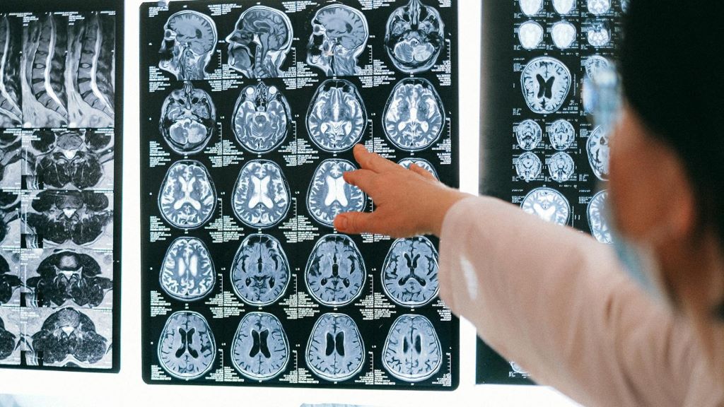
Photo by Anna Shvets

A new international study led by researchers at Karolinska Institutet demonstrates that it is possible to detect subtle changes in the brain and identify early signs of hereditary frontotemporal dementia using advanced brain imaging techniques. The study has recently been published in Molecular Psychiatry.

Frontotemporal dementia, or FTD, is a neurodegenerative disease that often affects people in middle age and is a common cause of dementia before the age of 65. The disease is particularly difficult to diagnose in its early stages, as the earliest symptoms are behavioural changes and may resemble primary psychiatric disease and symptoms later on can resemble conditions such as Alzheimer’s disease and Parkinson’s disease. In about a third of cases, frontotemporal dementia is hereditary, making families with known mutations an important resource for research.

New type of MRI technique

In the current study, researchers from Karolinska Institutet, together with an international research network, examined the brain’s microstructure in over 700 individuals – both carriers of FTD mutations and control subjects. The researchers used a new type of MRI technique that measures how water molecules spread within the grey matter of the brain, where greater diffusion indicates microstructural damage to brain tissue. In this way, the technique can reveal early damage in the cerebral cortex before the brain begins to shrink, known as brain atrophy, or cognitive problems arise.

The results revealed that the new method is more sensitive than the established imaging technique that measures the thickness of the cerebral cortex. Among individuals with a mutation in the C9orf72 gene, the researchers could detect changes in the brain even before any clinical symptoms appeared. For mutations in the MAPT gene, changes were observed at mild symptom stages, whereas for carriers of GRN mutations, alterations emerged only at more advanced stages.

Identifying individuals at risk

“Our findings show that changes in the brain’s microstructure can be detected before visible brain atrophy, and these changes are closely linked to how the disease develops,” explains corresponding author Elena Rodriguez-Vieitez, researcher at the Department of Neurobiology, Care Sciences and Society, Karolinska Institutet.

“This could be valuable for identifying individuals at risk and for evaluating new therapies in clinical trials.”

The researchers also followed the participants over time and showed that a greater spread of water molecules in brain tissue at the start of the study was linked to a faster decline in behaviour and cognitive ability. This was true for all three mutation types.

“Our results suggest that measurements of the brain’s microstructure could become an important tool for identifying individuals at risk of frontotemporal dementia and for monitoring disease progression in clinical trials,” says Caroline Graff, professor at the same department and last author of the study.

Source: Karolinska Institutet

New Laser System Measures Scalp and Brain Blood Flow

This optical measurement could offer an affordable and scalable way to diagnose stroke, brain injury and other conditions

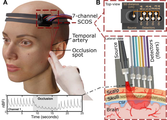
Experimental arrangement of the SCOS system for measuring cerebral blood dynamics during superficial temporal artery (STA) occlusion. (a) 3D visualization of the SCOS device positioned over the temple region and the occlusion site near the ear bone. (b) Top and lateral views of the device, illustrating different detecting channels for sensing the scalp, skull, and brain layers. Credit: Liu et al., APL Bioengineering, 2025

Measuring blood flow in the brain is critical for responding to a range of neurological problems, including stroke, traumatic brain injury (TBI) and vascular dementia. But existing techniques, including magnetic resonance imaging and computed tomography, are expensive and therefore not widely available.

Researchers from the USC Neurorestoration Center and the California Institute of Technology (Caltech) have built a simple, noninvasive alternative. The device takes a technique currently used in animal studies known as speckle contrast optical spectroscopy (SCOS) and adapts it for potential clinical use in humans. It works by capturing images of scattered laser light with an affordable, high-resolution camera.

“It’s really that simple. Tiny blood cells pass through a laser beam, and the way the light scatters allows us to measure blood flow and volume in the brain,” said Charles Liu, MD, PhD, professor of clinical neurological surgery, urology and surgery at the Keck School of Medicine of USC, director of the USC Neurorestoration Center and co-senior author of the new research.

The device has already been tested with humans in small proof of concept studies demonstrating the tool’s utility for assessing stroke risk and detecting brain injury. In the current study, published in APL Bioengineering, Liu and his team sought to confirm that SCOS is truly measuring blood flow in the brain, rather than in the scalp, which also contains many blood vessels. The question has long plagued researchers who use light-based technology to visualize the brain.

Liu’s team took an innovative approach: By temporarily blocking blood flow to the scalp, they confirmed that SCOS readings were indeed measuring signals from blood vessels in the brain. Readings from 20 participants showed that positioning the detector at least 2.3cm away from the laser source provided the clearest measurement of brain blood flow. The study, funded in part by the National Institutes of Health, the Alfred Mann Foundation and the USC Neurorestoration Center, was just published in the journal APL Bioengineering.

“For the first time in humans, this experimental evidence shows that a laser speckle optical device can probe beyond the scalp layers to access cerebral signals,” said Simon Mahler, PhD, who is now an assistant professor in the Department of Biomedical Engineering at the Stevens Institute of Technology and one of the paper’s coauthors. “This is an important step toward using SCOS to non-invasively measure blood flow in the brain.”

Tracking brain blood flow

For years, researchers measuring brain signals with light-based technology, such as lasers and fibre optics, have used statistical simulations to estimate which signals originate in the brain versus the scalp. The USC Neurorestoration Center team found a direct way to test the difference, thanks to a collaboration between surgeons, engineers and neurologists.

“I perform surgeries to increase blood flow in the brain, and many of these involve temporarily stopping blood flow in the scalp,” said Jonathan Russin, MD, now professor and chief of neurosurgery at the University of Vermont, who continues to collaborate with the USC Neurorestoration Center. “That gave us a simple way to test the technology – by creating a change that affected only the scalp’s circulation while leaving the brain’s blood flow untouched.”

In 20 participants, the researchers temporarily stopped blood flow to the scalp, then collected a series of SCOS readings. By gradually moving the detector further from the head, they captured signals reaching progressively deeper towards the brain. They found that positioning the detector 2.3cm from the head allowed them to measure brain blood flow while minimising interference from the scalp.

The findings confirm the utility of SCOS for non-invasively detecting brain blood flow and provide important guidance for other researchers working with light-based technology, Liu said.

Bringing SCOS to patients

Beyond advancing research, the study helps confirm the clinical potential of SCOS for detecting and responding to stroke, brain injury and dementia. Because all of the team’s research has been done with humans, the tool is poised for rapid translation from the lab to the clinic.

“We look directly at humans in essentially the same way the tool will be applied, so there’s nothing lost in translation,” Liu said. “We are never more than one step away from the problem we’re trying to solve.”

The technique is already being used by some of the team’s collaborators to help diagnose stroke and TBI. Next, the researchers will continue to refine the technology and software, working to improve the resolution of images and the quality of data extracted from readings.

“With the knowledge that we’re now measuring exactly what we intend to measure, we’re also going to expand our testing of this technique with patients in clinical settings,” Liu said.

Source: Keck School of Medicine of USC

Faster MRI Scans Offer New Hope for Dementia Diagnosis

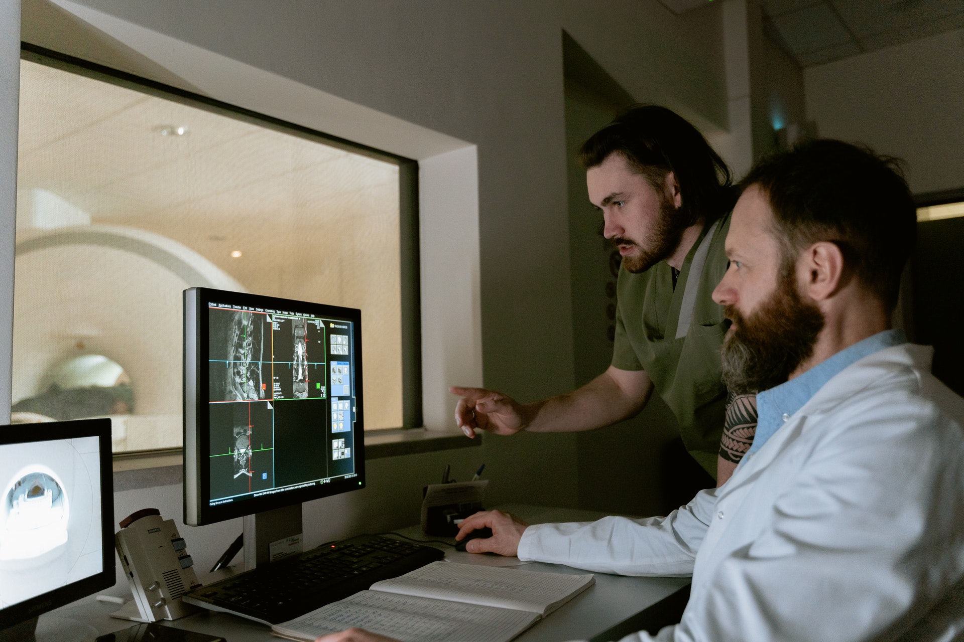
Photo by Mart Production on Pexels

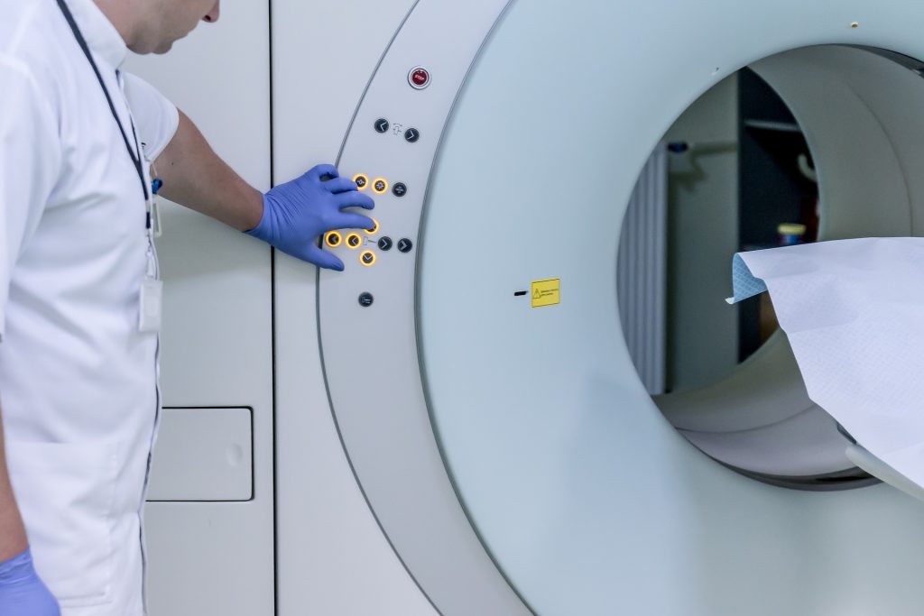
The time to carry out diagnostic MRI scans for dementia can be cut to one third of their standard length, according to a new study led by UCL researchers.

The findings, published in Alzheimer’s & Dementia, have been described as a step towards ending ‘the postcode lottery in dementia diagnosis’. Shorter scans would be easier and more comfortable for patients and also enable more people to be scanned at a lower cost. The team behind the study say this could at least double the number of dementia scans able to be done in one day.

Senior author Professor Nick Fox, Director of the UCL Dementia Research Centre at the UCL Queen Square Institute of Neurology, said: “As more treatments that can slow or change the course of dementia are being developed, it’s important to make sure MRI scans are available to everyone. This is because people living with dementia often need an MRI scan as part of their diagnosis before they can access these treatments.

“To help make this possible, our team carried out the first study looking at how new imaging techniques – called parallel imaging – could speed up MRI scans in clinics. Their goal is to move closer to a future where every person with dementia can get a diagnosis through a scan.”

MRI scans often play a key role in an accurate dementia diagnosis, including ruling out other causes of symptoms and assisting in diagnosing the type of dementia. Emerging disease-modifying treatments such as lecanemab and donanemab also require an MRI scan before starting treatment and for safety monitoring during the course of treatment. Reducing the cost of scanning would contribute to lowering the total cost of delivering for such treatments. 

The ADMIRA study (Accelerated Magnetic Resonance Imaging for Alzheimer’s disease), part funded by Alzheimer’s Society’s Heather Corrie Impact Fund, aimed to understand the reliability of fast MRI scans compared to standard-of-care clinical scans. The neurologists on the study were joined by co-authors from the UCL Hawkes Institute and the UCL Advanced Research Computing Centre in the faculty of Engineering.

The research team scanned 92 people in an outpatient setting where an MRI brain scan was planned as part of their routine clinical assessment. The accelerated scans were carried out and enhanced to increase the quality of the image using new scanning methods. Three neuroradiologists examined these scans, and weren’t aware if they were looking at fast or standard-of-care scans.

Co-author Professor Geoff Parker (UCL Hawkes Institute and UCL Medical Physics and Biomedical Engineering) said: “Our research has taken advantage of recent breakthroughs in scanner technology. Our task was to work out just how fast we could scan while maintaining image quality good enough for diagnosis.”

The team found that the quicker scans reduced time in the scanner by 63% and they were as reliable as the standard-of-care scans for diagnosis and visual ratings.

First author Dr Miguel Rosa-Grilo (UCL Queen Square Institute of Neurology) said: “We were confident that the new scan would prove non-inferior to the standard scan, given the high image quality – but it was remarkable how well it performed.”

Richard Oakley, Associate Director of Research and Innovation at Alzheimer’s Society, said: “Dementia is the UK’s biggest killer, but one in three people living with the condition haven’t had a diagnosis. An early and accurate diagnosis isn’t just a label, it’s the first step to getting vital care, support and treatment.

“While MRIs aren’t the only way to diagnosis dementia, very few people with concerns about their cognitive health are offered one as part of the diagnosis process, mainly because they are expensive and not widely available. These faster MRIs, which take less than half the time of standard scans, could help end this postcode lottery in dementia diagnosis, cut costs and potentially give more people access to them.

“MRI scans can be an uncomfortable and daunting experience for patients, so anything we can do to make it an easier process is really positive.

“So far, this shortened MRI scan has been tested at one specialist centre with one type of MRI scanner, so more research is needed to make sure this works across different types of scanners and a diverse range of people. We’re hugely encouraged by this progress and eager to see how it continues.”

The team will now build on their early results by making sure the approach works across different types of MRI machines, so it can benefit as many hospitals and clinics as possible.

Source: University College London

New One-hour, Low-cost HPV Test Could Transform Cervical Cancer Screening

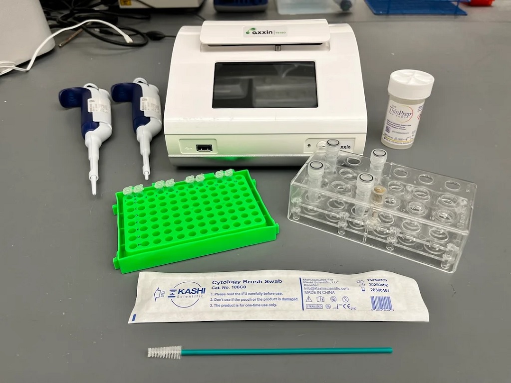
Materials used to run the HPV LAMP assay. A cytology brush is used to collect a cervicovaginal swab sample into ThinPrep buffer. Samples are lysed in screw-on tubes and lysate is added to LAMP reagents in PCR tubes. The assay is run on the Axxin T8-ISO heater/fluorimeter.

A team of researchers led by Rice University, in collaboration with colleagues in Mozambique and the US, has developed a simple, affordable human papillomavirus (HPV) test that delivers results in less than an hour with no specialised laboratory required. The breakthrough could provide an option for women in low-resource settings to be screened and treated for cervical cancer in a single clinic visit, a step that global health experts say could save countless lives. The research was recently published in Nature Communications.

Cervical cancer is considered easily preventable, yet it remains one of the deadliest cancers for women worldwide. According to the World Health Organization (WHO), each year more than 350 000 women die from the disease, and nearly 90% of those deaths occur in low- and middle-income countries where access to regular cervical cancer screening is limited. Persistent infection with high-risk types of HPV causes nearly all cases of cervical cancer. While vaccines are helping reduce HPV infections globally, most women at risk today are adults who did not get the vaccine in childhood. For them, regular and reliable screening is the only path to early detection and lifesaving treatment.

“Cervical cancer is almost entirely preventable, yet it still claims hundreds of thousands of lives each year,” said first author Maria Barra, a bioengineering graduate student at Rice. “Our goal was to build a test accurate enough to guide treatment, fast enough to use during a clinic visit and inexpensive enough to scale. This assay meets all three goals.”

The WHO recommends HPV DNA testing as the gold standard for cervical cancer screening, but existing HPV DNA tests often require expensive lab equipment and trained laboratory technicians – barriers that make widespread use in low-resource settings unattainable. As a result, many women are not screened for cervical cancer. Even where screening programs exist, results may take days or weeks to return. Patients leave to await results. However, where care facilities are remote, few in number and difficult to access, patients are often unable to return for treatment, leaving precancerous lesions to progress unchecked. A faster test without reliance on a lab could provide results and prompt treatment during the same patient visit.

“This is the kind of pragmatic innovation we focus on when engineering for global health – fewer steps, lower cost, higher impact,” said Rebecca Richards-Kortum, Professor of Bioengineering and co-director of the Rice360 Institute for Global Health Technologies at Rice. “Our data show you can bring lab-grade molecular screening to almost any setting without sacrificing reliability. Providing accurate results quickly enables clinicians to start treatment without delay.”

The new test uses a method called loop-mediated isothermal amplification (LAMP), which simplifies DNA detection by running at a single temperature. Instead of requiring DNA extraction – a complicated step in many existing tests – this process is extraction-free. A swab sample is chemically lysed, added directly to the LAMP reagents and incubated for about 45 minutes in a portable heater then read by fluorescence.

The test detects three of the most dangerous HPV types (HPV16, HPV18 and HPV45), which together cause about 75% of all cervical cancers. It also includes a built-in cellular control to ensure that the sample was collected properly.

In clinical studies, the test showed 100% agreement with the reference standard in 38 samples from Houston and 93% agreement in 191 samples from Maputo, Mozambique. The cost of the test is projected to be less than $8 each, and the portable device it runs on is battery-operated, making it ideal for clinics without consistent electricity.

“High mortality rates from cancer are closely associated with delays in diagnoses and limited access to early treatment,” said Cesaltina Lorenzoni, head of the National Cancer Control Program at the Mozambican Ministry of Health, director of science and teaching at Maputo Central Hospital and professor of pathology at the Eduardo Mondlane University Faculty of Medicine. “Point-of-care technologies that can aid clinicians in identifying cancer and guide treatment options in a single patient visit could be lifesaving in clinical settings in Maputo. This assay performed very well in our clinical setting and holds promise of delivering the kind of rapid, specific, cost-effective cancer detection that would meaningfully improve outcomes for women in our country.”

The WHO has set ambitious targets to screen 70% of women worldwide by 2030 as part of its public health campaign to eliminate cervical cancer. Meeting that goal will require screening millions of women in various global settings that lack advanced lab equipment or resources.

By cutting out expensive instruments, minimising sample handling and delivering rapid, accurate results, the LAMP assay represents a significant step toward realistically achieving the WHO goal. Critically, it opens the door to “screen-and-treat” strategies, where if a positive result is found, the patient can be treated on the same medical visit, reducing treatment delays and loss to follow-ups.

The team is currently working to expand the test to cover additional high-risk HPV types and is also working on lyophilised (freeze-dried) reagents that don’t require refrigeration, further increasing the test’s usability in rural or resource-limited areas. The team also plans to conduct usability studies with frontline health workers to refine the design before larger clinical rollouts.

“Our goal is a complete, field-ready kit that community clinics can use anywhere,” Richards-Kortum said. “If we can help health systems move to same-day screen-and-treat, we can move towards a future where cervical cancer can be eliminated globally.”

Source: Rice University

Rapid Diagnostics Test Can Detect Asymptomatic Malaria Cases

Researchers have adapted a rapid diagnostic technology that is able to identify undetected cases of malaria, helping tackle the spread of disease.

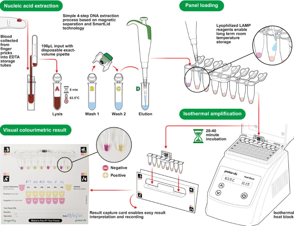
A diagram showing how the Dragonfly technology works (Credit: ProtonDx)

A team of scientists from Imperial College London, the MRC Unit The Gambia, the Clinical Research Unit of Nanoro in Burkina Faso, ProtonDx Ltd, and the NIHR Global Health Research Group have developed and validated a low-cost, point-of-care diagnostic that can rapidly detect low levels of malaria from a finger prick.

The test, called Dragonfly, relies on technology originally created at Imperial and its spinout ProtonDx. The technology allows users to diagnose malaria with high accuracy, without the need for extensive laboratory equipment or infrastructure. Results can be delivered in as little as 45 minutes, and the test is sensitive enough to detect even the lowest levels of malaria parasites in the blood – meaning that people without symptoms of malaria can still be identified.

Malaria is one of the leading causes of preventable deaths worldwide, with around 95% of all deaths occurring in Africa. Asymptomatic infections are a major driver of ongoing transmission, as individuals who carry the disease without showing symptoms do not seek medical treatment. Mosquitos feeding on blood from people without malaria symptoms can still deliver the malaria parasite to other people when they take their next blood meal. The new technology offers hope for combatting this potential spread of infection, by offering a way to identify previously undetectable malaria cases rapidly and on the ground in countries which are most affected by malaria.

The findings, published in Nature Communications, have significant global health implications as this field-deployable molecular diagnostic method offers a sensitive, scalable solution to support test-and-treat strategies for malaria elimination across Africa.

Professor Aubrey Cunnington, from Imperial’s Department of Infectious Disease and Co-Lead of the NIHR Global Health Research Group with Professor Halidou Tinto (from IRSS, Burkina Faso), said: “This is the first time that a diagnostic test for use outside of a laboratory setting has proven sensitive enough to detect low level malaria parasite infections in people who don’t have any symptoms.

“These people are the main source of malaria transmission, and in countries trying to eliminate malaria, there has long been interest in trying to detect these asymptomatically infected people with a screening test performed in their communities, and then giving treatment to those who are positive.

“Until now, no test has been able to detect enough of these infected people to make this a viable proposition, but the Dragonfly test now makes this possible.”

Detecting the undetectable

By collaboratively working as part of the NIHR Global Health Research Group, scientists were able to develop and test this new technology with the help of researchers in the regions affected most by malaria.

Almost 700 blood samples were collected from the community in The Gambia and Burkina Faso to assess the Dragonfly test’s accuracy against gold standard PCR testing and other common methods of testing, including expert microscopy and rapid diagnostic test (e.g., lateral flow immunoassay).

It was found that the Dragonfly tool could detect >95% of all malaria parasite infections, including 95% detection of those where the numbers of parasites were too low to be detected by looking at blood under a microscope.

Although Dragonfly is currently used as a research-used-only device, important progress is being made to understand the potential cost of a final manufactured version – especially when deployed at scale – a critical factor for effective deployment in sub-Saharan Africa. The team is already working closely with the Africa Centres for Disease Control and Prevention to explore opportunities with local manufacturers in the region, ensuring that production and scale-up can be rooted in local capacity. Future studies will also need to assess the robustness of the tool in community settings which are less connected to laboratory facilities.

Dr Jesus Rodriguez-Manzano, last author and technology development lead, from the Department of Infectious Disease, said “This research would not have been possible without the collaborative nature and all the organisations who took part in this study. The technology delivered through this work represents a game changer for malaria control efforts.”

The testing equipment

In the Dragonfly testing process, a capillary blood sample obtained from a simple finger prick is processed in around 10 minutes, without the need for specialised laboratory equipment, to extract high-purity nucleic acids from malaria parasites. The prepared sample is then placed into a detection panel, which is inserted into a portable heater.

After a 30-minute incubation at a constant temperature, results can be read visually using a colour chart: a pink reaction indicates a negative result, while a yellow reaction confirms malaria infection.

The Dragonfly can be manufactured at a fraction of the cost of other platforms, is compact enough to fit into a backpack, and can operate on batteries, an important feature for bringing the tool directly to communities without requiring additional specialised equipment. Testing can be carried out by most people without extensive training, meaning that healthcare providers or scientists do not need to be present for its use.

Source: Imperial College London

Missing First Mammogram Raises Breast Cancer Death Risk

Photo by National Cancer Institute on Unsplash

Women who miss their first mammogram run a higher risk of being diagnosed with advanced breast cancer and dying from the disease. This is shown in a new study from Karolinska Institutet published in The BMJ.

Since the early 1990s, women in Sweden have been offered regular mammograms, which has contributed to a decrease in breast cancer mortality. Despite this, a significant proportion choose not to attend their first examination. The researchers behind the new study wanted to investigate the long-term consequences of this. 

The study is based on data from the Swedish mammography screening program and national health registries, and covers almost 433 000 women in Stockholm between 1991 and 2020, with follow-up for up to 25 years. 

The results show that 32% of all women who were invited to their first screening declined. These women were also less likely to participate in future examinations, which often led to a later diagnosis and poorer prognosis.

“Skipping the first mammogram is a strong indicator of who is at risk of late detection and higher mortality. Our results show that missing the first mammogram is not just a one-time choice, but often marks the beginning of a long-term pattern of not attending check-ups,” says the study’s first author, Ziyan Ma, a doctoral student at the Department of Medical Epidemiology and Biostatistics, Karolinska Institutet.

Were detected at a more advanced stage

When women who skipped their first screening were later diagnosed with breast cancer, the disease was more often detected at a more advanced stage. The risk of developing stage III cancer was approximately 1.5 times higher, and for stage IV, the risk was as much as 3.6 times higher compared to those who participated in the first mammogram. Over a 25-year follow-up period, almost 1 percent of those who did not participate had died of breast cancer, compared with 0.7 percent among the participants – a difference that corresponds to a 40 percent higher risk of dying from the disease. 

However, the total proportion of women who developed breast cancer was almost the same in both groups, approximately 7.7%. According to the researchers, this shows that the increased mortality is mainly due to delayed detection rather than more cases of the disease.

“Family history is a well-known, unchangeable risk factor for breast cancer. Our study shows that missing the very first screening examination carries a similar mortality risk – but unlike family history, this is a behaviour that we can change. Since over 30 percent of women skip their first screening, increased participation could save many lives. Since this group can be identified early, decades before deaths occur, healthcare providers have a chance to intervene with reminders or support to encourage participation, says the study’s last author, Kamila Czene, professor at the Department of Medical Epidemiology and Biostatistics, Karolinska Institutet

Source: Karolinska Institutet

At-home Melanoma Testing with Skin Patch Test

A microneedle patch captures cancer biomarkers in the top-most layer of skin to detect melanoma in animal tissue samples

The newly designed ExoPatch being removed from a sample of mouse skin successfully distinguished melanoma from healthy skin in mice. A gel coating the microneedles picks up cancer indicators from the top-most layer of the skin. Dissolving the gel releases exosomes into a solution, which is then used on a two-lined test strip, similar to an at-home COVID-19 test. Image credit: Jeremy Little, Michigan Engineering.

Melanoma testing could one day be done at home with a skin patch and test strip with two lines, similar to COVID-19 home tests, according to University of Michigan researchers. Developed with funding from the National Institutes of Health, the new silicone patch with star-shaped microneedles, called the ExoPatch, distinguished melanoma from healthy skin in mice.

The patch and test move toward rapid at-home melanoma testing, helping patients catch the most aggressive form of skin cancer early without a biopsy or blood draw.

“The star-shaped needles make puncture easier and less painful, but they are so small that they only go through the top-most layer of the skin, the epidermis, and do not draw blood,” said Sunitha Nagrath, the Dwight F. Benton Professor of Chemical Engineering at U-M and co-corresponding author of the study published in Biosensors and Bioelectronics.

The ExoPatch microneedles, at just 0.6mm long with a width of less than 100 nm (0.0001 mm) at the tip, are coated with a gel that picks up exosomes, tiny packages released by cells, from the interstitial fluid that fills the spaces between cells in the epidermis.

Once thought to be trash ejected from cells for cleanup, exosomes actually contain DNA and RNA fragments that cells use to communicate with each other. Cancer cell exosomes can help tumours spread by preparing tissues to accept tumour cells before arrival, and detecting them can catch cancer earlier than past methods.

The gel that coats the ExoPatch contains a protein called Annexin V that attracts and sticks exosomes to the microneedles’ surface. Once removed from the skin, placing the patch in an acid dissolves the gel, which releases the exosomes into a solution. After dipping a test strip into the solution, two lines form if the sample contains melanoma exosomes, and one line forms for a negative test – the same way an at-home COVID-19 test strip works.

“A fair-skinned person with moles must go to the doctor about every six months to send off a biopsy to see if they’re malignant or benign. With this test, they could instead test at home, get the results right away and follow up with a dermatologist for a positive result,” Nagrath said.

As a first step in the proof-of-concept study, the researchers tested the ExoPatch on a tissue sample of pig skin, which closely resembles human skin in thickness and composition. Using a microscope, they found the microneedles penetrated about 350 to 600nm into the skin. For scale, the epidermis on the human forearm is about 18 300nm thick.

To test whether the ExoPatch could capture melanoma exosomes from skin tissue, the research team tested tissue samples of mouse skin, half from healthy mice and half from mice injected with a fragment of a human melanoma tumour. After a 15-minute application, the ExoPatch was placed under a powerful microscope.

“When looking at microscopy images, I was happy to see how nicely the exosomes adhered to the microneedles and were within the 30 to 150 nanometre size range we expect,” said Scott Smith, U-M doctoral student of chemical engineering and co-lead author of the study.

After confirming the exosomes stuck to the ExoPatch, the researchers dissolved the gel and ran the sample through the test strips. The test successfully distinguished between melanoma and healthy tissues with a 3.5-fold darker line in melanoma samples.

The ExoPatch isolated 11.5 times more exosomal protein from melanoma tissue samples compared to healthy tissue, showing it can specifically target cancerous exosomes.

A pilot study in humans followed by a series of clinical trials will be the next steps to move the technology toward use. Beyond melanoma, the ExoPatch gel coating could be modified to detect exosomes released by other cancers with a solid tumour, including lung, breast, colon, prostate and brain cancer.

“This is the first patch designed to capture disease-specific exosomes from fluid under the skin. The potential applications are huge,” said Nagrath.

Source: University of Michigan

Researchers Pioneer New Breathing Test to Detect Lung Disease Earlier

Credit: Scientific Animations CC4.0

A new project led by Oxford University aims to develop a novel breathing test that could detect asthma and COPD earlier, more accurately, and closer to home – reducing pressure on the NHS and improving outcomes for patients.

This work is included in a portfolio of research funded by the Engineering and Physical Sciences Research Council (EPSRC), part of UK Research and Innovation, to make diagnostic testing more accessible by designing simple, affordable tools that can be used in everyday settings. The project, called ACCESS (A Community-based diagnostiC for early airwayS disease), focuses on airway diseases, and will receive £1.3 million over three years.

Chronic respiratory diseases such as asthma and chronic obstructive pulmonary disease (COPD) affect more than half a billion people worldwide and cause over four million deaths every year. In the UK, these conditions hit disadvantaged communities hardest, with people in areas of socioeconomic deprivation facing particular challenges in accessing hospital-based diagnostic care.

A volunteer undertaking a computed cardiopulmonography based breath test. (The volunteer is Dr Nick Smith, a member of the research team developing the CCP technology). Credit: Department of Chemistry, University of Oxford.

Currently, asthma and COPD diagnosis relies on a test called spirometry. This measures how much air a person can forcefully breathe out, but it is difficult for many patients to perform and often fails to detect disease in its earliest stages. By the time spirometry picks up abnormalities, damage to the lungs is usually irreversible, resulting in late diagnoses, missed chances for early treatment, and poorer outcomes.

The project is enabled by the strong ethos for collaborative and innovative research in this University and will be delivered by a multi-disciplinary team, working across university departments and the NHS.

Project lead Professor Grant Ritchie

To address these issues, Oxford researchers have developed a new test, known as computed cardiopulmonography (CCP), that could transform the way lung diseases are diagnosed. CCP is performed while a patient breathes normally for 12 minutes through a mouthpiece linked to a highly accurate gas analyser. Using cutting-edge laser technology and advanced mathematical modelling, the test captures a detailed picture of how evenly air flows through the lungs. This “fingerprint” of lung function changes early in the course of disease, making CCP a powerful tool for spotting subtle problems that spirometry can miss.

Early studies in people with asthma, COPD, and even in otherwise healthy smokers suggest CCP is a highly sensitive marker of small airways damage. But while the results are promising, the test currently takes too long and requires specialist gas supplies, restricting its use to hospitals and research labs.

The goal of the ACCESS project is to adapt CCP for community use, for instance in GP surgeries, pharmacies, and community diagnostic hubs. Specifically, the research team will work to reduce the time duration and volume of gas needed for each test, and speed up the data analysis so that results can be shared with the patient during the same appointment. Towards the end of the project, CCP will be trialled in a community diagnostic centre to gather feedback from patients and healthcare professionals.

The long-term goal is to support earlier diagnosis and treatment, helping reduce hospital visits and tackle health inequalities. By enabling earlier intervention and delivering care closer to home, CCP has the potential to improve outcomes for millions of people living with lung disease.

Source: Oxford University

Simple Blood Test Can Predict Risk of Severe Liver Disease

Photo by National Cancer Institute on Unsplash

A new study from Karolinska Institutet, published in The BMJ, shows how a simple blood analysis can predict the risk of developing severe liver disease. The method may already start to be applied in primary care to enable the earlier detection of cirrhosis and cancer of the liver.

“These are diseases that are growing increasingly common and that have a poor prognosis if detected late,” says Rickard Strandberg, affiliated researcher at Karolinska Institutet’s Department of Medicine, Huddinge, who has developed the test with his departmental colleague Hannes Hagström. “Our method can predict the risk of severe liver disease within 10 years and is based on three simple routine blood tests.” 

For the study, the researchers at Karolinska Institutet and their colleagues in Finland evaluated how well the method can estimate the risk of severe liver disease. The model, which is called CORE, was produced with advanced statistical methods and is based on five factors: age, sex and levels of three common liver enzymes (AST, ALT and GGT), which are commonly measured during regular health checks. 

A web-based calculator 

Their aim has been to produce a tool that is easy to use in primary care, where most patients first seek medical attention. A web-based calculator is already available for doctors and nurses at www.core-model.com

“This is an important step towards being able to offer early screening for liver disease in primary care,” says principal investigator Hannes Hagström, adjunct professor at the Department of Medicine, Huddinge, and senior consultant at Karolinska University Hospital. “Drug treatment is now available, soon hopefully also in Sweden, for treating people at a high risk of developing liver diseases such as cirrhosis or liver cancer.” 

The study is based on data from over 480 000 people in Stockholm who underwent health checks between 1985 and 1996. On following the participants for up to 30 years, the researchers could see that some 1.5% developed severe liver disease, such as liver cirrhosis and liver cancer, or required a liver transplant. 

Highly accurate risk prediction 

The CORE model proved highly accurate and was able to differentiate between people who either did or did not develop the disease in 88 per cent of cases, which is an improvement on the currently recommended FIB-4 method. 

“Primary care hasn’t had the tools to detect the risk of severe liver disease in time,” says Professor Hagström. “FIB-4 is not suited for the general population and is less effective at predicting the future risk of severe liver disease.” 

The model was also tested on two other population groups in Finland and the UK, where it again demonstrated a high accuracy in predicting this risk. The researchers make the point, however, that it needs to be further tested on groups at especially high risk, such as people with type 2 diabetes or obesity. They also recognise a need to integrate the model into medical records systems to facilitate its clinical use. 

Source: Karolinska Institutet

Pocket Ultrasound Reduces Hospital Stays for Patients with Shortness of Breath

Credit: Rutgers Health/RWJBarnabas Health

When hospitalised patients struggle to breathe, doctors typically reach for their stethoscopes, but results from a clinical study in JAMA Network Open suggest they should diagnose the problem with portable ultrasounds instead.

The study, by Rutgers Universitty and RWJBarnabas Health, found initial exams with portable ultrasounds led to better diagnoses, shorter hospital stays and big cost savings. However, the findings revealed a need for additional training and workflow integration to help clinicians transition from traditional tools to this promising new technology.

“The study clearly shows that ultrasound is the superior diagnostic technology, even for long-time stethoscope users who get a few hours of ultrasound training,” said senior study author Partho Sengupta, Henry Rutgers professor and chief of cardiology at Rutgers Robert Wood Johnson Medical School (RWJMS), chief of cardiology at Robert Wood Johnson University Hospital (RWJUH) and member of the RWJBarnabas Health Medical Group.

The explanation here is simple. Ultrasound gives you more information, and more concrete information, about what’s going on

Partho Sengupta, Henry Rutgers professor

The study enrolled 208 patients admitted with shortness of breath to Robert Wood Johnson University Hospital in New Brunswick. About half of them underwent diagnosis via point-of-care ultrasound devices that attach to smartphones. The rest underwent diagnosis via existing standards of care. 

Initial diagnosis with ultrasound trimmed a patient’s average length of hospital stay from 11.9 days to 8.3 days. In all, initial diagnosis with ultrasound saved 246 bed-days and about $751 000 in direct costs across the cohort, while 30-day readmissions were similar between groups.

“The explanation here is simple. Ultrasound gives you more information, and more concrete information, about what’s going on,” Sengupta said.  “When clinicians can see fluid in the lungs, a failing heart or a stiff inferior vena cava in minutes, they can target therapy sooner or rule out a cardiopulmonary cause and look elsewhere.”

To keep things simple and encourage buy-in, scans focused on a handful of cardiac views and a six-zone lung sweep. The exam was designed to be quick and binary: signs of congestion or not, systolic function reduced or not.

The study provided several hours of ultrasound training to participating hospitalists (doctors who oversee and coordinate hospital patient care) so that each could perform and interpret the ultrasound exam in 10 to 15 minutes. 

Nevertheless, most hospitalists who underwent the training let sonographers perform the exams and cardiologists interpret them. Only 20% of patients received an ultrasound diagnosis from one of the hospitalists.

Sengupta said time pressure on rounds and a lack of incentives make it hard to add a 10-minute procedure, even if it changes care.

“This is consistent with what we observe in day-to-day clinical practice,” Sengupta said. “Although the ultrasound probe fits in your pocket and attaches to the back of a smartphone, its use in clinical settings remains inconsistent. This study overcame those barriers by leveraging a multidisciplinary framework.”

Kameswari Maganti, professor of cardiology at RWJMS and section chief for non-invasive cardiology at RWJUH, led the image interpretation with the sonography team that worked closely with the RWJUH hospitalist team, led by RWJMS faculty Catherine Chen and Payal Parikh, as well as the engineering and data science team, headed by Naveena Yanamala. 

“This coordinated team effort was key to developing and delivering a streamlined protocol that significantly reduced hospital stays and healthcare costs,” Sengupta said.

The researchers reported that ultrasound findings altered medical decisions in roughly a third of cases, including new diagnoses and changes in therapy. They also noted that longer-stay patients appeared to benefit the most, a hint that ultrasound-guided triage and treatment may pay particular dividends when cases are complex.

As with most single-center implementations, caveats apply. The model relied on trained sonographers and cardiology reads, which may not be available everywhere. Broader studies across multiple hospitals will be needed to confirm the cost and length-of-stay benefits, and to test strategies that make adoption stick.

Still, the argument for seeing more and guessing less is gaining ground. A bedside view of the heart and lungs, delivered early in admission, may help the right treatments arrive sooner.

Source: Rutgers University