Category: Lab Tests and Imaging

Missing First Mammogram Raises Breast Cancer Death Risk
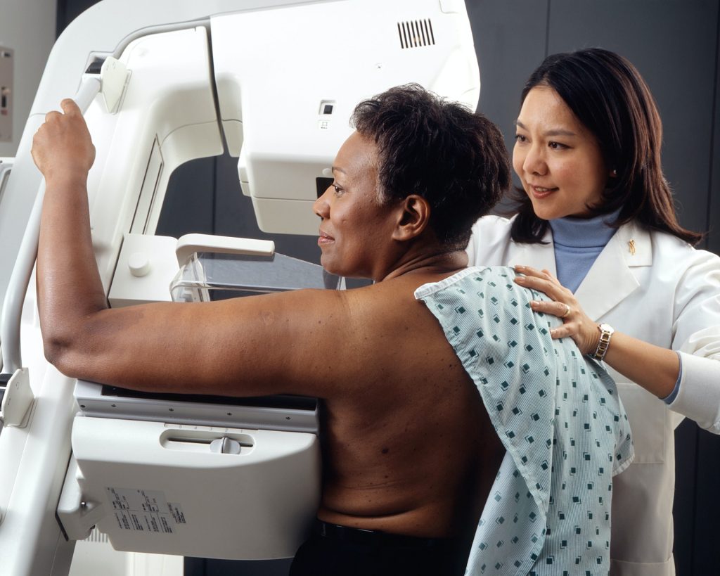
Photo by National Cancer Institute on Unsplash

Women who miss their first mammogram run a higher risk of being diagnosed with advanced breast cancer and dying from the disease. This is shown in a new study from Karolinska Institutet published in The BMJ.

Since the early 1990s, women in Sweden have been offered regular mammograms, which has contributed to a decrease in breast cancer mortality. Despite this, a significant proportion choose not to attend their first examination. The researchers behind the new study wanted to investigate the long-term consequences of this. 

The study is based on data from the Swedish mammography screening program and national health registries, and covers almost 433 000 women in Stockholm between 1991 and 2020, with follow-up for up to 25 years. 

The results show that 32% of all women who were invited to their first screening declined. These women were also less likely to participate in future examinations, which often led to a later diagnosis and poorer prognosis.

“Skipping the first mammogram is a strong indicator of who is at risk of late detection and higher mortality. Our results show that missing the first mammogram is not just a one-time choice, but often marks the beginning of a long-term pattern of not attending check-ups,” says the study’s first author, Ziyan Ma, a doctoral student at the Department of Medical Epidemiology and Biostatistics, Karolinska Institutet.

Were detected at a more advanced stage

When women who skipped their first screening were later diagnosed with breast cancer, the disease was more often detected at a more advanced stage. The risk of developing stage III cancer was approximately 1.5 times higher, and for stage IV, the risk was as much as 3.6 times higher compared to those who participated in the first mammogram. Over a 25-year follow-up period, almost 1 percent of those who did not participate had died of breast cancer, compared with 0.7 percent among the participants – a difference that corresponds to a 40 percent higher risk of dying from the disease. 

However, the total proportion of women who developed breast cancer was almost the same in both groups, approximately 7.7%. According to the researchers, this shows that the increased mortality is mainly due to delayed detection rather than more cases of the disease.

“Family history is a well-known, unchangeable risk factor for breast cancer. Our study shows that missing the very first screening examination carries a similar mortality risk – but unlike family history, this is a behaviour that we can change. Since over 30 percent of women skip their first screening, increased participation could save many lives. Since this group can be identified early, decades before deaths occur, healthcare providers have a chance to intervene with reminders or support to encourage participation, says the study’s last author, Kamila Czene, professor at the Department of Medical Epidemiology and Biostatistics, Karolinska Institutet

Source: Karolinska Institutet
At-home Melanoma Testing with Skin Patch Test

A microneedle patch captures cancer biomarkers in the top-most layer of skin to detect melanoma in animal tissue samples

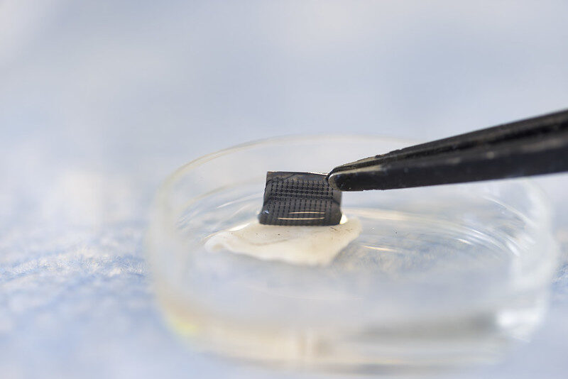
The newly designed ExoPatch being removed from a sample of mouse skin successfully distinguished melanoma from healthy skin in mice. A gel coating the microneedles picks up cancer indicators from the top-most layer of the skin. Dissolving the gel releases exosomes into a solution, which is then used on a two-lined test strip, similar to an at-home COVID-19 test. Image credit: Jeremy Little, Michigan Engineering.

Melanoma testing could one day be done at home with a skin patch and test strip with two lines, similar to COVID-19 home tests, according to University of Michigan researchers. Developed with funding from the National Institutes of Health, the new silicone patch with star-shaped microneedles, called the ExoPatch, distinguished melanoma from healthy skin in mice.

The patch and test move toward rapid at-home melanoma testing, helping patients catch the most aggressive form of skin cancer early without a biopsy or blood draw.

“The star-shaped needles make puncture easier and less painful, but they are so small that they only go through the top-most layer of the skin, the epidermis, and do not draw blood,” said Sunitha Nagrath, the Dwight F. Benton Professor of Chemical Engineering at U-M and co-corresponding author of the study published in Biosensors and Bioelectronics.

The ExoPatch microneedles, at just 0.6mm long with a width of less than 100 nm (0.0001 mm) at the tip, are coated with a gel that picks up exosomes, tiny packages released by cells, from the interstitial fluid that fills the spaces between cells in the epidermis.

Once thought to be trash ejected from cells for cleanup, exosomes actually contain DNA and RNA fragments that cells use to communicate with each other. Cancer cell exosomes can help tumours spread by preparing tissues to accept tumour cells before arrival, and detecting them can catch cancer earlier than past methods.

The gel that coats the ExoPatch contains a protein called Annexin V that attracts and sticks exosomes to the microneedles’ surface. Once removed from the skin, placing the patch in an acid dissolves the gel, which releases the exosomes into a solution. After dipping a test strip into the solution, two lines form if the sample contains melanoma exosomes, and one line forms for a negative test – the same way an at-home COVID-19 test strip works.

“A fair-skinned person with moles must go to the doctor about every six months to send off a biopsy to see if they’re malignant or benign. With this test, they could instead test at home, get the results right away and follow up with a dermatologist for a positive result,” Nagrath said.

As a first step in the proof-of-concept study, the researchers tested the ExoPatch on a tissue sample of pig skin, which closely resembles human skin in thickness and composition. Using a microscope, they found the microneedles penetrated about 350 to 600nm into the skin. For scale, the epidermis on the human forearm is about 18 300nm thick.

To test whether the ExoPatch could capture melanoma exosomes from skin tissue, the research team tested tissue samples of mouse skin, half from healthy mice and half from mice injected with a fragment of a human melanoma tumour. After a 15-minute application, the ExoPatch was placed under a powerful microscope.

“When looking at microscopy images, I was happy to see how nicely the exosomes adhered to the microneedles and were within the 30 to 150 nanometre size range we expect,” said Scott Smith, U-M doctoral student of chemical engineering and co-lead author of the study.

After confirming the exosomes stuck to the ExoPatch, the researchers dissolved the gel and ran the sample through the test strips. The test successfully distinguished between melanoma and healthy tissues with a 3.5-fold darker line in melanoma samples.

The ExoPatch isolated 11.5 times more exosomal protein from melanoma tissue samples compared to healthy tissue, showing it can specifically target cancerous exosomes.

A pilot study in humans followed by a series of clinical trials will be the next steps to move the technology toward use. Beyond melanoma, the ExoPatch gel coating could be modified to detect exosomes released by other cancers with a solid tumour, including lung, breast, colon, prostate and brain cancer.

“This is the first patch designed to capture disease-specific exosomes from fluid under the skin. The potential applications are huge,” said Nagrath.

Source: University of Michigan

Researchers Pioneer New Breathing Test to Detect Lung Disease Earlier

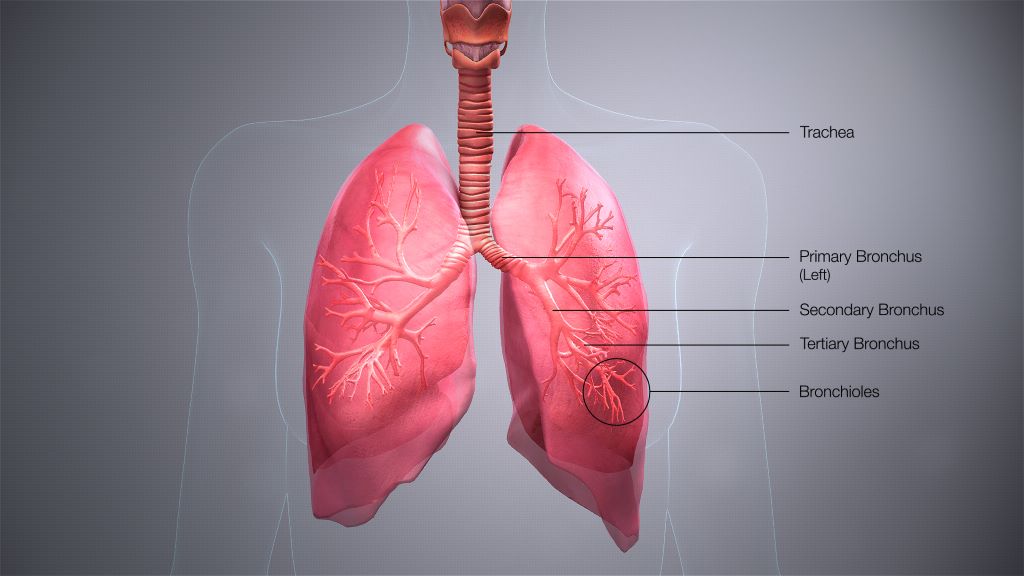
Credit: Scientific Animations CC4.0

A new project led by Oxford University aims to develop a novel breathing test that could detect asthma and COPD earlier, more accurately, and closer to home – reducing pressure on the NHS and improving outcomes for patients.

This work is included in a portfolio of research funded by the Engineering and Physical Sciences Research Council (EPSRC), part of UK Research and Innovation, to make diagnostic testing more accessible by designing simple, affordable tools that can be used in everyday settings. The project, called ACCESS (A Community-based diagnostiC for early airwayS disease), focuses on airway diseases, and will receive £1.3 million over three years.

Chronic respiratory diseases such as asthma and chronic obstructive pulmonary disease (COPD) affect more than half a billion people worldwide and cause over four million deaths every year. In the UK, these conditions hit disadvantaged communities hardest, with people in areas of socioeconomic deprivation facing particular challenges in accessing hospital-based diagnostic care.

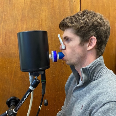
A volunteer undertaking a computed cardiopulmonography based breath test. (The volunteer is Dr Nick Smith, a member of the research team developing the CCP technology). Credit: Department of Chemistry, University of Oxford.

Currently, asthma and COPD diagnosis relies on a test called spirometry. This measures how much air a person can forcefully breathe out, but it is difficult for many patients to perform and often fails to detect disease in its earliest stages. By the time spirometry picks up abnormalities, damage to the lungs is usually irreversible, resulting in late diagnoses, missed chances for early treatment, and poorer outcomes.

The project is enabled by the strong ethos for collaborative and innovative research in this University and will be delivered by a multi-disciplinary team, working across university departments and the NHS.

Project lead Professor Grant Ritchie

To address these issues, Oxford researchers have developed a new test, known as computed cardiopulmonography (CCP), that could transform the way lung diseases are diagnosed. CCP is performed while a patient breathes normally for 12 minutes through a mouthpiece linked to a highly accurate gas analyser. Using cutting-edge laser technology and advanced mathematical modelling, the test captures a detailed picture of how evenly air flows through the lungs. This “fingerprint” of lung function changes early in the course of disease, making CCP a powerful tool for spotting subtle problems that spirometry can miss.

Early studies in people with asthma, COPD, and even in otherwise healthy smokers suggest CCP is a highly sensitive marker of small airways damage. But while the results are promising, the test currently takes too long and requires specialist gas supplies, restricting its use to hospitals and research labs.

The goal of the ACCESS project is to adapt CCP for community use, for instance in GP surgeries, pharmacies, and community diagnostic hubs. Specifically, the research team will work to reduce the time duration and volume of gas needed for each test, and speed up the data analysis so that results can be shared with the patient during the same appointment. Towards the end of the project, CCP will be trialled in a community diagnostic centre to gather feedback from patients and healthcare professionals.

The long-term goal is to support earlier diagnosis and treatment, helping reduce hospital visits and tackle health inequalities. By enabling earlier intervention and delivering care closer to home, CCP has the potential to improve outcomes for millions of people living with lung disease.

Source: Oxford University

Simple Blood Test Can Predict Risk of Severe Liver Disease

Photo by National Cancer Institute on Unsplash

A new study from Karolinska Institutet, published in The BMJ, shows how a simple blood analysis can predict the risk of developing severe liver disease. The method may already start to be applied in primary care to enable the earlier detection of cirrhosis and cancer of the liver.

“These are diseases that are growing increasingly common and that have a poor prognosis if detected late,” says Rickard Strandberg, affiliated researcher at Karolinska Institutet’s Department of Medicine, Huddinge, who has developed the test with his departmental colleague Hannes Hagström. “Our method can predict the risk of severe liver disease within 10 years and is based on three simple routine blood tests.” 

For the study, the researchers at Karolinska Institutet and their colleagues in Finland evaluated how well the method can estimate the risk of severe liver disease. The model, which is called CORE, was produced with advanced statistical methods and is based on five factors: age, sex and levels of three common liver enzymes (AST, ALT and GGT), which are commonly measured during regular health checks. 

A web-based calculator 

Their aim has been to produce a tool that is easy to use in primary care, where most patients first seek medical attention. A web-based calculator is already available for doctors and nurses at www.core-model.com. 

“This is an important step towards being able to offer early screening for liver disease in primary care,” says principal investigator Hannes Hagström, adjunct professor at the Department of Medicine, Huddinge, and senior consultant at Karolinska University Hospital. “Drug treatment is now available, soon hopefully also in Sweden, for treating people at a high risk of developing liver diseases such as cirrhosis or liver cancer.” 

The study is based on data from over 480 000 people in Stockholm who underwent health checks between 1985 and 1996. On following the participants for up to 30 years, the researchers could see that some 1.5% developed severe liver disease, such as liver cirrhosis and liver cancer, or required a liver transplant. 

Highly accurate risk prediction 

The CORE model proved highly accurate and was able to differentiate between people who either did or did not develop the disease in 88 per cent of cases, which is an improvement on the currently recommended FIB-4 method. 

“Primary care hasn’t had the tools to detect the risk of severe liver disease in time,” says Professor Hagström. “FIB-4 is not suited for the general population and is less effective at predicting the future risk of severe liver disease.” 

The model was also tested on two other population groups in Finland and the UK, where it again demonstrated a high accuracy in predicting this risk. The researchers make the point, however, that it needs to be further tested on groups at especially high risk, such as people with type 2 diabetes or obesity. They also recognise a need to integrate the model into medical records systems to facilitate its clinical use. 

Source: Karolinska Institutet

Pocket Ultrasound Reduces Hospital Stays for Patients with Shortness of Breath

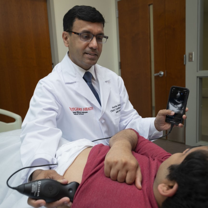
Credit: Rutgers Health/RWJBarnabas Health

When hospitalised patients struggle to breathe, doctors typically reach for their stethoscopes, but results from a clinical study in JAMA Network Open suggest they should diagnose the problem with portable ultrasounds instead.

The study, by Rutgers Universitty and RWJBarnabas Health, found initial exams with portable ultrasounds led to better diagnoses, shorter hospital stays and big cost savings. However, the findings revealed a need for additional training and workflow integration to help clinicians transition from traditional tools to this promising new technology.

“The study clearly shows that ultrasound is the superior diagnostic technology, even for long-time stethoscope users who get a few hours of ultrasound training,” said senior study author Partho Sengupta, Henry Rutgers professor and chief of cardiology at Rutgers Robert Wood Johnson Medical School (RWJMS), chief of cardiology at Robert Wood Johnson University Hospital (RWJUH) and member of the RWJBarnabas Health Medical Group.

The explanation here is simple. Ultrasound gives you more information, and more concrete information, about what’s going on

Partho Sengupta, Henry Rutgers professor

The study enrolled 208 patients admitted with shortness of breath to Robert Wood Johnson University Hospital in New Brunswick. About half of them underwent diagnosis via point-of-care ultrasound devices that attach to smartphones. The rest underwent diagnosis via existing standards of care. 

Initial diagnosis with ultrasound trimmed a patient’s average length of hospital stay from 11.9 days to 8.3 days. In all, initial diagnosis with ultrasound saved 246 bed-days and about $751 000 in direct costs across the cohort, while 30-day readmissions were similar between groups.

“The explanation here is simple. Ultrasound gives you more information, and more concrete information, about what’s going on,” Sengupta said.  “When clinicians can see fluid in the lungs, a failing heart or a stiff inferior vena cava in minutes, they can target therapy sooner or rule out a cardiopulmonary cause and look elsewhere.”

To keep things simple and encourage buy-in, scans focused on a handful of cardiac views and a six-zone lung sweep. The exam was designed to be quick and binary: signs of congestion or not, systolic function reduced or not.

The study provided several hours of ultrasound training to participating hospitalists (doctors who oversee and coordinate hospital patient care) so that each could perform and interpret the ultrasound exam in 10 to 15 minutes. 

Nevertheless, most hospitalists who underwent the training let sonographers perform the exams and cardiologists interpret them. Only 20% of patients received an ultrasound diagnosis from one of the hospitalists.

Sengupta said time pressure on rounds and a lack of incentives make it hard to add a 10-minute procedure, even if it changes care.

“This is consistent with what we observe in day-to-day clinical practice,” Sengupta said. “Although the ultrasound probe fits in your pocket and attaches to the back of a smartphone, its use in clinical settings remains inconsistent. This study overcame those barriers by leveraging a multidisciplinary framework.”

Kameswari Maganti, professor of cardiology at RWJMS and section chief for non-invasive cardiology at RWJUH, led the image interpretation with the sonography team that worked closely with the RWJUH hospitalist team, led by RWJMS faculty Catherine Chen and Payal Parikh, as well as the engineering and data science team, headed by Naveena Yanamala. 

“This coordinated team effort was key to developing and delivering a streamlined protocol that significantly reduced hospital stays and healthcare costs,” Sengupta said.

The researchers reported that ultrasound findings altered medical decisions in roughly a third of cases, including new diagnoses and changes in therapy. They also noted that longer-stay patients appeared to benefit the most, a hint that ultrasound-guided triage and treatment may pay particular dividends when cases are complex.

As with most single-center implementations, caveats apply. The model relied on trained sonographers and cardiology reads, which may not be available everywhere. Broader studies across multiple hospitals will be needed to confirm the cost and length-of-stay benefits, and to test strategies that make adoption stick.

Still, the argument for seeing more and guessing less is gaining ground. A bedside view of the heart and lungs, delivered early in admission, may help the right treatments arrive sooner.

Source: Rutgers University

From Symptoms to Solutions: Professional Testing Can Reveal Hidden Allergens

As spring arrives in South Africa, many people experience their most challenging allergy season. However, while pollen-filled air triggers obvious seasonal symptoms, allergies extend far beyond springtime discomfort, affecting millions year-round through food sensitivities, skin reactions, and environmental triggers.

“Spring allergies are usually just the tip of the iceberg,” says Tyron Hansen, Business Development Manager at BioSmart Lab. “You wake to spring sunshine and nature in full bloom, but instead of enjoying it, you’re shut inside with tissues and antihistamines. Meanwhile, your stomach acts up after breakfast, and that mysterious rash on your arms is back. Sound familiar?”

Hansen explains that many people assume that their symptoms stem from a single source, like pollen or food preservatives. However, BioSmart Lab’s test results often reveal they’ve actually been living with multiple triggers they never connected to their symptoms.

Why It’s Worth Looking Deeper

Professional allergen tests measure specific antibodies in your blood to identify how your body reacts to different substances. This matters because what looks like “seasonal” hay fever might actually be a mix of environmental, food, and even skin-related triggers.

“Our immune systems are like complicated alarm systems,” explains Hansen. “They go off loudly, but without proper testing, you only hear the siren – you don’t see what’s actually setting it off.”

A major source of confusion is the difference between allergies and intolerances. Both of which can make you feel unwell, but function very differently:

  • Allergies: They set off the immune system through immunoglobulin E (IgE) antibodies, causing anything from a runny nose or itchy eyes to potentially life-threatening anaphylaxis, often within minutes of exposure.
  • Intolerances: These don’t set off your immune system, but they can still cause digestive discomfort like bloating or nausea, and while not dangerous, they can nevertheless affect daily life.

“Knowing which is which can be the key to finally feeling better,” he adds.

Everyday Triggers You Might Be Missing

Allergies aren’t always obvious or seasonal. Many people live with daily discomfort without realising what’s behind it. Hansen provides some of the most common sources of ongoing allergic reactions.

  • Environmental triggers: Dust mites, mould spores, pet dander, and certain plants can cause year-round congestion, itchy eyes, or skin flare-ups.
  • Food sensitivities: That heavy, bloated feeling after meals could point to, for example, wheat, rice and corn sensitivity, or even certain food combinations.
  • Skin reactions: Chronic eczema or rashes are sometimes triggered by hidden allergens, not simply “sensitive skin.”
  • Allergic clues in children: Kids often can’t articulate their symptoms. What might appear to be frequent colds, skin rashes, or trouble concentrating could be subtle signs of allergies.

“Trying to figure out if your symptoms are caused by allergens through elimination diets, behavioural adjustments, or symptom tracking can take months and still leave you without answers, notes Hansen. “Blood-based testing provides a faster alternative by measuring IgE antibodies – the proteins your immune system releases when it detects threats.”

Taking Back Control

BioSmart Lab’s advanced blood panels analyse these IgE proteins across multiple allergens simultaneously. These tests can be purchased online and provide accurate, quantitative results without the risk of sparking reactions or being influenced by medication use.

Some people discover that they need to remove wheat to manage food allergies. Others learn that their “pet allergy” is actually a dust mite sensitivity. Parents often find their child’s mid-afternoon meltdowns are directly linked to specific food triggers at lunch.

“The goal isn’t just to manage symptoms – it’s to restore people’s quality of life,” emphasises Hansen. “Once you understand what your body is reacting to, you can move from frustrating guesswork to making informed choices, giving you control of your health back,” he concludes.

For more information about BioSmart’s allergy testing options, visit https://biosmart.com/allergy-tests/.

Test Detects Brain Cancers in Cerebrospinal Fluid with High Accuracy

Photo by Cottonbro on Pexels

A novel, multi-analyte test developed by researchers at Johns Hopkins Medicine can accurately identify brain cancers using small samples of cerebrospinal fluid (CSF), offering a promising new tool to guide clinical decision-making.

The findings, supported by funding from the National Institutes of Health, were published in Cancer Discovery and demonstrate that combining multiple biological markers, including tumour-derived DNA and immune cell signatures, is more effective for diagnosing central nervous system cancers than using any one marker alone.

“This study highlights how much more information we can gain when we evaluate several analytes together,” says senior study author Chetan Bettegowda, MD, PhD, Professor and Director of the Department of Neurosurgery at the Johns Hopkins University School of Medicine. “The ability to detect cancers with high specificity and also gain insight into the immune environment of the brain could be an important advance in the care of patients with brain tumours.”

To evaluate the potential of a multi-analyte approach, investigators analysed 206 CSF samples, including samples from patients with high-grade gliomas, medulloblastomas, metastases and central nervous system lymphomas. Their test, called CSF-BAM (cerebrospinal fluid–B/T cell receptor, aneuploidy and mutation), measured chromosomal abnormalities, tumour-specific mutations, and T and B cell receptor sequences. In combination, these markers identified brain cancers with more than 80% sensitivity (ability to detect cancer) and 100% specificity (correctly identified those who were cancer-free) in the validation cohort. The 100% specificity means no false positives were recorded among individuals with noncancerous conditions.

The study also showed that the assay could distinguish between the immune cell populations present in cancer and noncancer cases, offering additional biological context that could be helpful in more-challenging clinical scenarios. Investigators say this ability to categorize T and B cell populations in the CSF provides insights into both disease presence and immune response.

“Many patients with brain lesions face invasive diagnostic procedures to confirm a cancer diagnosis,” says Christopher Douville, MD, assistant professor of oncology and a senior study author. “A tool like this could help us make better-informed decisions about who really needs a biopsy and who doesn’t.”

Researchers say the test could be particularly useful for cases in which conventional imaging or cytology is inconclusive, or in situations when obtaining tissue for diagnosis is risky or not possible. The multi-analyte approach, they say, enables clinicians to better detect cancer and better understand the disease status, supporting a more tailored approach to patient care.

Source: Johns Hopkins Medicine

SAHPRA Approves Mpox Test Using African Medicines Regulatory Harmonisation (AMRH) Continental EUL Procedure

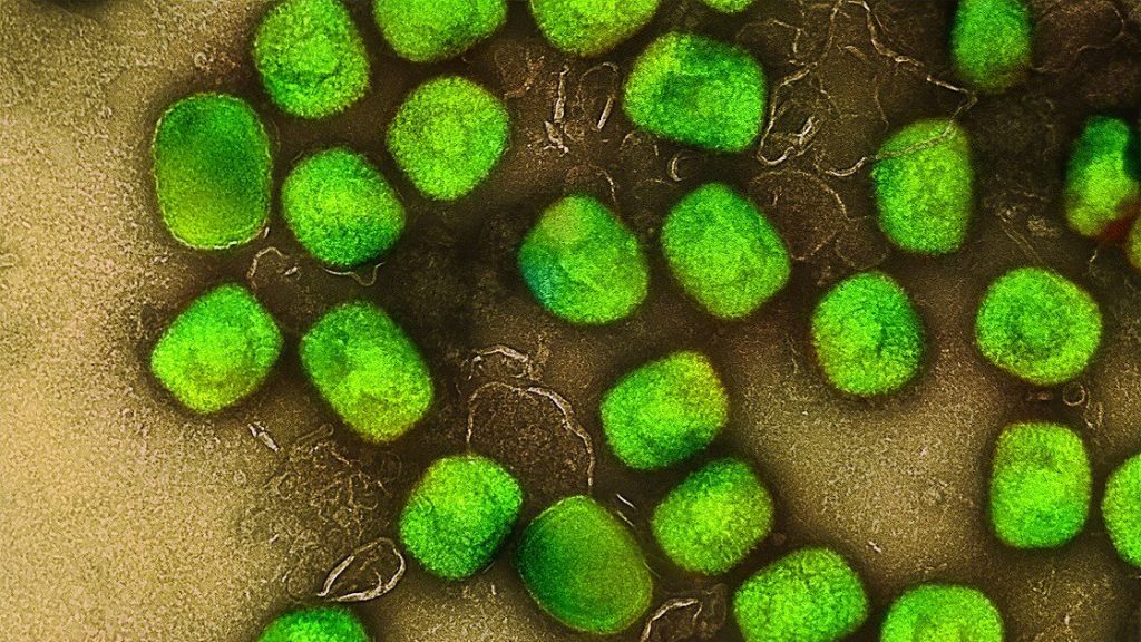
Mpox (monkeypox) virus. Source: NIH

Pretoria, 27 August 2025 – The South African Health Products Regulatory Authority (SAHPRA) has approved a molecular test kit for Mpox , Cobas MPXV, for use on the Cobas 6800/8800 Systems, within nine working days through a collaborative and harmonised review process under the African Medicines Regulatory Harmonisation (AMRH). Rapid and accurate testing is essential for early detection and to enable timely treatment, and effective containment of the virus.

SAHPRA’s Medical Device Unit, utilising both the World Health Organization (WHO) Emergency Use Listing (EUL) report and the African Medicines Regulatory Harmonisation (AMRH) Continental EUL Procedure, has successfully leveraged reliance mechanisms to facilitate the review and approval of Mpox diagnostic tools. This approach has proven instrumental in accelerating access to critical health technologies, particularly in public health emergencies.

The emergency use approval of the Cobas MPXV for use on the Cobas 6800/8800 Systems, developed by Roche Molecular Systems, Inc. and licensed to Roche Diagnostics South Africa (Pty) Ltd, represents a critical advancement in expanding diagnostic capacity amid Mpox outbreaks. Rapid and accurate testing is essential for early detection, timely treatment, and effective containment of the virus.

 “As part of our efforts to promote regulatory efficiency and responsiveness, SAHPRA has successfully leveraged reliance mechanisms to facilitate the review and approval of Mpox diagnostic tools. This marks a significant milestone in supporting regional and continental harmonisation of the regulatory processes for medical devices, including In-Vitro Diagnostics (IVDs),” says Dr Boitumelo Semete-Makokotlela, SAHPRA’s Chief Executive Officer.

For more information on the AMRH continental EUL listing publication, see here: https://www.nepad.org/news/public-notice-amrh-steering-committee-approves-emergency-use-listing-of-two-mpox

Source: SAHPRA

Researchers Discover a Significant Problem in Brain Imaging, Identify Fix

Photo by Mart Production on Pexels

In a new study, investigators from McLean Hospital (a member of Mass General Brigham), Harvard Medical School, and the National Institute on Drug Abuse Intramural Research Program (NIDA-IRP) discovered that the tendency of people’s arousal to wane over the course of brain scans has been distorting the brain connection maps produced by functional magnetic resonance imaging (fMRI).

The study is out now in Nature Human Behaviour.

The team found that as people’s arousal levels dwindle during an fMRI, such as if they become more relaxed and sleepy, changes in breathing and heart rates alter blood oxygen levels in the brain—which are then falsely detected on the scan as neuronal activity.

“You’re laying down in a snug scanner for quite some time, often with only a low-engagement button press task to attend to or nothing to do at all, as the scanner monotonously hums and vibrates around you,” said first author Cole Korponay, PhD, MPA, a research fellow at the McLean Hospital Imaging Center. “These arousal-dampening conditions create the illusion that people’s brain connection strengths continuously inflate throughout the scan.”

fMRI scans are commonly used to non-invasively map brain connectivity, but the technique relies on changes in brain blood oxygen to indirectly measure neuronal activity. It is therefore vulnerable to “noise” from other processes that can affect blood oxygen – such as changes in breathing and heart rates.

And because breathing and heart rate patterns are closely tied to arousal levels, changes in arousal can introduce significant noise into fMRI data. Problematically, the conditions of an fMRI scan tend to progressively lull people into lower arousal states.

In the present study, the research team identified a specific blood flow signal that seemed to track both the decline in subject arousal levels and the illusory inflation of functional brain connection strengths.

This non-neuronal, physiological noise signal, termed the “systemic low frequency oscillation” (sLFO) signal, grew over time during scanning, in a spatial and temporal pattern that closely followed the pattern of the connection strength increases.

The researchers then demonstrated that a method called RIPTiDe, developed by co-senior author Blaise Frederick, PhD, an associate biophysicist at the McLean Imaging Center, to remove the sLFO signal from fMRI data, was able to eliminate the illusory connection strength increases.

“By adopting this sLFO denoising procedure, future studies can mitigate the distortive effects of arousal changes during brain scans and enhance the validity and reliability of fMRI findings,” said Korponay.

This research was supported by the National Institute on Drug Abuse, the National Institute of Mental Health, and the National Institute on Aging, all part of the National Institutes of Health.

Source: Mass General Brigham McLean Hospital

New AI–based Test Detects Early Signs of Osteoporosis from X-ray Images

Photo by Cottonbro on Pexels

Investigators have developed an artificial intelligence-assisted diagnostic system that can estimate bone mineral density in both the lumbar spine and the femur of the upper leg, based on X-ray images. The advance is described in a study published in the Journal of Orthopaedic Research.

A total of 1454 X-ray images were analysed using the scientists’ system. Performance rates for the lumbar and femur of patients with bone density loss, or osteopenia, were 86.4% and 84.1%, respectively, in terms of sensitivity. The respective specificities were 80.4% and 76.3%. (Sensitivity reflected the ability of the test to correctly identify people with osteopenia, whereas specificity reflected its ability to correctly identify those without osteopenia). The test also had high sensitivity and specificity for categorising patients with and without osteoporosis.

“Bone mineral density measurement is essential for screening and diagnosing osteoporosis, but limited access to diagnostic equipment means that millions of people worldwide may remain undiagnosed,” said corresponding author Toru Moro, MD, PhD, of the University of Tokyo. “This AI system has the potential to transform routine clinical X-rays into a powerful tool for opportunistic screening, enabling earlier, broader, and more efficient detection of osteoporosis.”

Source: Wiley