Tag: 26/6/26

Faster Aging in Younger Generations Linked to Rise in Early-onset Cancer

Immune system aging linked to earlier lung cancer; adipose tissue aging linked to earlier colorectal cancer

Photo by Malvestida on Unsplash

Cancer is often considered a disease of aging. Older adults are at higher risk because they have had more time to accumulate cellular damage that can trigger tumour formation. But as cancer rates in younger adults rise, with each successive generation facing higher risks than the one before it, researchers are asking whether cellular damage is accumulating faster in recent generations, accelerating their body’s biological aging.

A new study led by researchers at Washington University School of Medicine in St. Louis provides evidence that younger generations are indeed aging faster biologically than their older counterparts. The causes remain under investigation around the world, including global efforts led by research members of Siteman Cancer Center, based at Barnes-Jewish Hospital and WashU Medicine, and Cancer Grand Challenges, a global initiative co-founded by the National Cancer Institute and Cancer Research U.K.; but importantly, the new research links this accelerated aging to an increased risk of early-onset cancers in younger generations. In general, early-onset cancers are those diagnosed at age 55 or younger.

The larger the gap between biological age — that is, how old our bodies appear to be — and chronological age — which is how many years we have actually lived — the higher the cancer risk, according to the researchers. They found that people in more recent birth cohorts had larger age gaps than those in older birth cohorts, which may help explain the rise in early-onset cancer in recent generations.

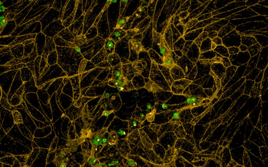
Their study also identified links between faster aging in particular organ systems and increased risks for certain cancers. For instance, an immune system that appears older than its actual age was associated with early-onset lung cancer. Similarly, fat tissue that appears older than its chronological age was associated with early-onset colorectal cancer.

The study, published June 22 in the journal Nature Medicine, suggests that measures of accelerated aging could help identify individuals at higher risk of early-onset cancer and guide new strategies for cancer prevention and early detection.

“Our ultimate goal is to decode how modern environments become biologically embedded to drive cancer risk, transforming prevention from broad recommendations to personalised interventions,” said Yin Cao, ScD, a molecular epidemiologist and an associate professor of surgery and of medicine at WashU Medicine. “This brings us closer to identifying risk earlier and developing prevention strategies that are tailored to an individual’s biology.”

Exploring biological aging

Cao’s team has been at the forefront of identifying individual factors that influence cancer risk across the life course, such as obesity, metabolic dysregulation, alcohol consumption, sedentary behaviour, poor diet quality and caesarean delivery. Although these discoveries have revealed important clues to the origins of cancer at younger ages, the contribution of any single factor is modest.

With that in mind, Cao, also a research member of Siteman, and her colleagues have sought ways to capture the influence of multiple risk factors operating together to spur cancer development. With support from Cancer Grand Challenges, Cao, as co-lead of Team PROSPECT, has been able to go after this problem.

For the current study, Cao’s team analysed data from more than 154,000 young adults in the UK Biobank, a large biomedical dataset containing biological, health and lifestyle data, and from more than 10,000 individuals in the U.S. participating in the National Institutes of Health’s (NIH) All of Us Research Program, an effort to build a comprehensive health dataset on more than 1 million people living in the U.S.

To estimate the level of biological aging — or age gap — the researchers, including first author Ruiyi Tian, a doctoral student in the Cao lab, examined aging at two levels: across the body as a whole, known as systemic aging, and within individual organs, known as organ-specific aging. For systemic aging, the researchers used established measures, including clinical biomarker-based measures such as PhenoAge and the Klemera-Doubal Method, as well as a metabolomic age score, which provides a measure of individual metabolism.

PhenoAge, for example, measures nine blood biochemistry markers such as albumin, made by the liver, and creatinine, a waste product removed by the kidneys. For organ-specific aging, the researchers used blood proteomic data, which measure levels of multiple proteins linked to specific organ systems, to estimate biological aging in individual organs.

The researchers calculated the average age gap for each birth cohort and used standard deviation to describe how much each group differed from the study average. Standard deviation is a measure of how spread out data points are around the average.

The researchers found that individuals in the UK born between 1965 and 1974 had systemic aging that was 23% of one standard deviation higher compared with those born between 1950 and 1954, after accounting for chronological age. In other words, people in the younger birth cohort showed a modest shift toward older biological profiles than people in the older birth cohort when at the same chronological age.

The researchers observed a similar pattern in the U.S cohort. Participants born between 1990 and 1999 had systemic aging that was 92% of one standard deviation higher compared with those born between 1965 and 1969.

This increased systemic aging in the younger group was associated with an 8% increased risk of early-onset solid cancers, especially lung, gastrointestinal and uterine cancers. When participants were divided into three groups based on their level of systemic aging, those with the most advanced systemic aging had a 15% increased risk of early-onset solid cancer compared with those with the least advanced systemic aging. According to the analysis, the increased risk persisted even after controlling for inherited genetic risks of cancer and genetic susceptibility to accelerated aging.

By zooming into organ-specific aging, the researchers found that advanced immune system aging was associated with increased risk of early-onset lung cancer, and advanced adipose (fat) tissue aging was associated with increased risk of early-onset colorectal cancer.

“If we can identify younger people with the highest cancer risk when they are still healthy, we can focus on prevention and early-detection strategies for the individuals who will benefit most from early interventions,” Cao said.

By Julia Evangelou Strait

Source: WashU Medicine

Finding the Lock and Key for the Cryptosporidium Parasite and Its Host

Cryptosporidium parasites. Credit: Cryptosporidiosis Laboratory. 

The Cryptosporidium parasite lives within cells that line the human intestine, but how does this pathogen find, recognise and then successfully invade these cells without triggering immunity? An international collaboration aims to find out. 

Parasites have lived on and within humans throughout history, co-evolving along with us to adjust to new climates and challenges. In some cases, this relationship has left significant genetic imprints. For example, the prevalence of sickle-cell disease in humans has been driven by selective pressure from the malaria parasite.

Cell biologist Adam Sateriale and his team at the Crick study a close relative of the malaria parasite, known as Cryptosporidium, which infects the intestinal tract of a wide range of animals, including humans. Most human infections are mild and asymptomatic, but Cryptosporidium can be deadly within immunocompromised people or very young children.  

“Certain species of the Cryptosporidium parasite are adapted to specifically invade and then live within the human intestinal lining, or epithelium,” Adam explains. “We’re interested to find out how the parasite recognises, and then successfully invades, these cells within the epithelium.”

Cryptosporidium and the malaria parasite, known as Plasmodium, diverged from one another nearly 500 million years ago, yet many of their features are conserved. Two organelles (or cellular compartments) can be found within both parasites: micronemes, which contain proteins that help parasites move, and rhoptries, which contain proteins that help parasites invade cells.

Micronemes of the malaria parasite are important vaccination targets, as proteins from these organelles are displayed on the surface of the parasite and are critical for infection. While Cryptosporidium has these invasion organelles, very little is known about their function and how they diverge from those of the malaria parasite.  

A recently awarded Wellcome Discovery grant has allowed Adam’s team to join forces with Gavin Wright’s team at the University of York and Amandine Guérin’s team at the University of Geneva to tackle these questions. Together, they will examine the function of micronemes and rhoptries and explore how Cryptosporidium specifically recognises and invades intestinal epithelial cells. To do that, they will harness the power of genetic editing.

“We’re going to use CRISPR gene editing to switch off, one by one, all the genes that code for the proteins in Cryptosporidium’s micronemes and rhoptries,” says Adam. “If the parasites can’t complete their normal functions without a particular protein, we’ll know that it’s a critical part of Cryptosporidium’s offensive strategy.”

Working together over the next five years, this multidisciplinary team are each bringing unique expertise: Adam’s team have developed CRISPR screening technology for the parasite, Amandine’s team work on Cryptosporidium invasion in detail and Gavin’s team specialise in identifying and studying cell surface protein interactions.

“Although there are many species of the Cryptosporidium parasite, some can only infect very specific hosts,” says Adam. “For instance, there are human species that can only invade and replicate within humans, suggesting a specific lock and key mechanism between the parasite and host. If we can find the essential parasite proteins that engage and unlock human cells for infection, we can then study these interactions and learn how to block them.”

He’s also excited about the impact this work could have, adding, “Ultimately this will lay the foundation for new treatments and preventions for children living in endemic areas.”

Source: Crick Institute

South Africa’s Tuberculosis Research Changes Global Medical Practice

Tuberculosis bacteria. Credit: CDC

A South African clinical study that began in a research unit in Gqeberha (PE), Eastern Cape, has transformed global treatment of drug-resistant tuberculosis. Furthermore, the study’s findings were published this week in the New England Journal of Medicine (NEJM), the highest-ranked medical journal in the world.

The publication recognises that this research study has set the global standard for TB care.

The BEAT Tuberculosis clinical study, conducted at the Clinical Health Research Unit (CHRU) Isango Lethemba TB Research Unit in the Eastern Cape and King Dinizulu Hospital Complex in KwaZulu-Natal, enrolled more than 400 participants over two years during the Covid-19 pandemic.

The study was executed by the University of the Witwatersrand in collaboration with the National Department of Health and funded by the United States Agency for International Development (USAID).

“This project has gone full circle,” says Dr Francesca Conradie, principal investigator of BEAT Tuberculosis and a researcher at the Clinical Health Research Unit (CHRU), University of the Witwatersrand. “The results from this trial have changed international guidelines. Being published in the New England Journal of Medicine is proof that South Africa produces world-class research that improves the lives of patients globally.”

Treatment for the whole family

The primary aim of BEAT Tuberculosis was to evaluate the safety and effectiveness of a novel, shortened treatment regimen for DR-TB compared with the established standard of care. The standard treatment at the time required a seven-drug regimen administered over a minimum of nine months. BEAT Tuberculosis tested a streamlined regimen of four to five medications, including the newer agents bedaquiline and delamanid, administered over six months.

The BEAT Tuberculosis trial enrolled children, pregnant women and breastfeeding mothers alongside adults. These former groups are usually excluded from clinical research. The result is a treatment regimen that can be used across the entire family.

“This is a one-size-fits-all treatment regimen,” explains Conradie. “Adherence is much easier when the three-year-old, the teenager, the mother and the father are all receiving treatment of similar duration and composition. That simplicity saves lives.”

The study enrolled 10 pregnant women. All 10 women gave birth to healthy babies, and nine of them were successfully treated. BEAT Tuberculosis has since been cited internationally as a model for inclusive clinical research methodology, and the findings have influenced World Health Organization policy on the treatment of DR-TB globally, including for pregnant women and children.

South Africa’s National Clinical Advisory Committee already reviews and approves the regimen for pregnant women presenting with Drug-Resistant TB, while other provinces are adopting the treatment, particularly when treating children.

During 2024, South Africa had 249,000 people who were infected with active tuberculosis, and 54 000 died from the disease,” says Professor Norbert Ndjeka, Chief Director: TB Control and Management, National Department of Health. “Not only did BEAT TB produce world-class research, but it is also being implemented progressively across South Africa and globally and is internationally recognised. South Africa has accomplished something exceptional.”

Source: Wits University

Does Iron Accumulation in the Brain Contribute to Neurodegeneration?

Salk Institute scientists discover chronoferroptosis, a chronic stress pathway in cells that causes neurons to become less resilient over time and more vulnerable to neurodegeneration

Representative neuronal cells are shown after acute iron exposure of six to eight hours (left) and after chronic iron exposure of nine days (right). The brain cell looks entirely different after chronic exposure, with dysregulated processes characteristic of the newly discovered cell stress pathway chronoferroptosis.
Click here for a high-resolution image.
Credit: Salk Institute

Neurodegenerative diseases affect tens of millions of people worldwide. Among these, Alzheimer’s and Parkinson’s diseases are the most common; in the United States alone, the Alzheimer’s Disease Association and Parkinson’s Foundation report roughly 7 million people with Alzheimer’s and another million with Parkinson’s. An intriguing clue lies in the tangled mystery of neurodegeneration that scientists are working to solve: iron accumulation.

Scientists have noticed that iron can slowly build up inside neurons. Early in life, this iron accumulation appears to have little effect on neuronal function. However, later in life, it can contribute to a slow neuronal demise. Salk Institute researchers studied nerve cells to figure out if and how this iron accumulation relates to neurodegenerative diseases. They found that the excess iron stuck in neurons lowers the cells’ defences, making them more vulnerable to stressors and other cellular insults through a process they named chronoferroptosis.

The study, published in Cell Death Discovery on June 18, 2026, points to iron accumulation as a key target in the effort to predict, prevent, and treat neurodegenerative diseases.

“Resilience has become a huge topic of discussion when it comes to Alzheimer’s disease and other neurodegenerative disorders, trying to make the brain more resilient in the face of stressors that contribute to neurodegeneration,” says senior and co-corresponding author Pam Maher, PhD, a research professor at Salk. “Our study reveals that cells lose resilience when iron hits a certain level, making neurons more susceptible to stressors that damage or even kill them.”

What do we already know about how the body uses iron, and is it linked to neurodegeneration?

Iron is an essential mineral for a healthy body. Found in dark leafy greens, starchy cereals, lean meats, seafood, and other common foods, iron helps red blood cells develop, carries oxygen around the body, makes hormones, and so much more, with a hand in everything from the immune system to energy production.

“It’s one of the most important minerals in the body,” says co-corresponding author Nawab John Dar, PhD, a postdoctoral researcher in Maher’s lab. “So, it isn’t the iron itself that is a problem with age. It is this accumulation of iron over time that is the problem.”

While the jury is still out on the exact mechanisms that initiate iron accumulation in neurons, the Salk team suspects the buildup is caused by a failure in the cells’ iron export machinery – iron enters neurons as usual but fails to get removed after use. But this failure doesn’t impact neurons for quite some time. The question is, why?

“People have been doing these experiments looking at iron exposure’s influence on cells over short 24- to 48-hour spans,” explains Dar. “But if neurodegenerative disorders are progressive, shouldn’t we have a cellular model that is progressive, too?”

Is iron accumulation making neurons less resilient?

Using a human-derived nerve cell line, the Salk team created the first progressive model of iron accumulation in neuronal cells. They compared the effects of both acute (between six and eight hours) and chronic (nine days) exposure to iron. What they discovered was an entirely new pathway, which they dubbed chronoferroptosis.

Maher has been studying ferroptosis for decades. Until now, ferroptosis was considered an iron-dependent cell death pathway, with cell death dependent on a process called lipid peroxidation. “It is like the cellular equivalent of when a cooking oil or nut goes bad. The fats in that oil or nut have undergone peroxidation,” explains Maher.

Chronoferroptosis adds the dimension of time to ferroptosis. To the researchers’ surprise, the pathway does not necessarily end in cell death. Instead, the findings reveal that ferroptosis can act as a cellular stress pathway.

In acutely exposed neurons, there was very little biochemical difference pre- and post-exposure to iron. However, in chronically exposed neurons, there were lots of changes: upregulation of some processes and downregulation of others; accumulation of harmful chemicals and depletion of helpful ones; and elevated lipid peroxidation. And when each exposure group was exposed to further stress, acutely exposed neurons could handle the stress, while chronically exposed neurons could not.

“We think these coordinated alterations in iron-handling and antioxidant defence proteins make chronically exposed neurons vulnerable to neurodegenerative pathology,” says Dar. “Entering this state of chronoferroptosis may set neurons up for age-related failure.”

How might chronoferroptosis inform neurodegeneration care?

By creating the first progressive model of iron accumulation in neuronal cells, the researchers were able to reveal surprising new clues in the case to crack neurodegeneration. “It’s not the amount of iron that seals the fate of these cells,” says Dar, “it’s the amount of time they spend under stress.”

Perhaps scientists will one day be able to detect when the brain begins entering this vulnerable state, when iron accumulation starts stressing neurons. They could then develop new interventions to address iron imbalances and keep neurons more resilient for longer.

“It’s not something we worked on in this paper, but our lab has developed several compounds to inhibit this pathway,” says Maher. “This could really be a promising therapeutic route for boosting neuron resilience and staving off neurodegeneration as we grow older.”

Source: Salk Institute

Bariatric Surgery Increases the Risk of Alcohol Problems

Sleeve gastrectomy. Credit: Scientific Animations CC4.0

The body absorbs alcohol much more rapidly after bariatric surgery, researchers from Norway have found. Patients need to know this when they choose the kind of surgery they will have.

“Bariatric surgery can come with a price. Patients have a significantly higher risk of developing alcohol problems than if they did not undergo surgery,” said Magnus Strømmen, a researcher at the Centre for Obesity Research at St. Olavs Hospital and a PhD research fellow at the Norwegian University of Science and Technology (NTNU).

Between one and two per cent of the population in Norway has undergone bariatric surgery. The most commonly used surgical methods are gastric bypass and gastric sleeve.

Both methods make patients eat less and feel full faster. This is partly due to reduced volume on the stomach, and partly due to hormonal changes. But a person’s changed anatomy also has consequences for what happens when they drink alcohol.

“In a normal stomach, a significant part of the alcohol will be broken down and thus not pass into the bloodstream. This is due to an enzyme that is secreted in the lining of the stomach. It is this protective mechanism that we deprive the patient of when we operate on the stomach. In addition, what you drink passes much faster into the intestine,” says Strømmen.

The small intestine’s big job is absorption. Since the stomach’s ability to break down alcohol more or less stops, significantly more alcohol passes directly into the bloodstream. That’s true even if the patient drinks the same as before the operation.

You get drunk faster

In a new study, Strømmen and his colleagues have had 33 adult patients undergo stress tests with alcohol. The participants consumed measured amounts of vodka mixed with orange juice both before bariatric surgery, and 3, 12 and 36 months after the operation, after which they had their blood alcohol levels measured after they had consumed the alcohol.

“Our findings show that alcohol uptake almost doubles, both after gastric bypass and gastric sleeve. Perhaps an even more dangerous finding, from a substance-abuse perspective, is that patients reach the maximum blood concentration in only half the time. These effects are lasting, probably lifelong,” Strømmen said.

In other words: The patients were intoxicated faster, and to a much greater extent, by the same amount of alcohol, and it took longer to get sober.

“The effects were more pronounced for people who had gastric bypass surgery. But that does not mean that the sleeve operation is harmless in terms of subsequent alcohol abuse,” Strømmen said.

Gastric bypass (left) and gastric sleeve are the most common forms of bariatric surgery today. In gastric bypass, a corner of the stomach is connected directly to the small intestine, so that both the stomach and one meter of the small intestine are disconnected. In gastric sleeve surgery, part of the stomach is removed so that the volume is reduced, without reconnecting the intestines. Illustration: Kari C Toverud, CMI

Bypass had a 69 per cent higher risk than sleeve

In another study, the researchers compared the risk of getting an alcohol abuse diagnosis after the two bariatric surgeries. The researchers analysed data from the Norwegian Patient Registry linked to the Norwegian Prescription Database for 17,800 patients operated on in the period from 2008 to 2018.

They found that patients who had gastric bypass surgery had a 69 per cent higher risk of being diagnosed with an alcohol-related problem than patients with a gastric sleeve. Bariatric patients who were given an alcohol-related diagnosis also had a higher mortality rate and used specialist health services more than patients who underwent bariatric surgery and who did not receive an alcohol diagnosis.

“It is important that patients, their relatives and health personnel, especially in general medicine, substance abuse and gastro medicine, share this knowledge. These Norwegian studies, based on different data sources and different methodologies, indicate that some of our patients struggle with alcohol problems as a complication from the surgery. Alcohol problems can cost the patient, their relatives, and society a great deal,” Strømmen said.

He likes to tell patients that they need to practice saying no.

“They will suddenly be in social contexts where friends and surroundings expect you to drink as much as before the operation. But your physiology has changed. That means you need to be more careful than before,” he said.

Risk factors for bariatric surgery must be investigated

“We can’t just tell people what to do or not to do,” says Associate Professor Magnus Strømmen. “I believe more that patients need to be educated about the mechanisms behind it. Knowledge can motivate people to be more careful with alcohol. They need to know how altered alcohol absorption can affect their actions while under the influence and that all alcohol intake doubles the stress on their organs. Photo: Aleksander Mjøen

He himself has been involved in building up the obesity outpatient clinic at St. Olavs Hospital in Trondheim. Now he wants to be sure that clinics incorporate this new information into their daily practice. Patients must be assessed individually in relation to the risk of alcohol problems.

“We find that many patients have a clear idea of what type of surgery they want when they are referred. And for a long time, this was given very great importance in the decisionmaking. But obesity is not just a single phenotype. Despite having a large body, patients are very different, also in terms of health,” he said.

Some patients may have type 2 diabetes, others struggle with heartburn. Some may have more extensive obesity and thus desire greater weight loss.

“For a patient like this, gastric bypass may be best. Other conditions may make you want to recommend gastric sleeve. Now we know that the operations result in different risks of alcohol problems. This means we must also investigate the patient’s risk factors for substance abuse before we decide which operation the patient should have. Where patients have several risk factors, gastric sleeve may be a better alternative, but we must also ask whether high-risk patients should be operated on at all,” Strømmen said.

Patients need to know

He wants patients to receive more specific information before the operation.

“It is important that patients make their decision to undergo surgery on a genuinely informed basis. It is not enough to say that their alcohol uptake will change. Patients should be educated about the mechanisms behind increased risk after surgery, and not least how to react differently to alcohol intoxication after surgery as a result of the sudden increase. This information can at best prevent patients from developing alcohol-related problems,” he said.

The patient’s risk factors for substance abuse should be considered before the type of surgery is chosen.

“I think most obesity clinics can get much better at their alcohol history, ie, a thorough conversation about the patient’s alcohol habits and any risk factors. We need to get better at asking the right questions, and make a more precise assessment. But this also requires transparency from the patients. I believe that good patient education can make patients understand why it is important to be honest about these things,” says Strømmen.

Drugs can replace bariatric surgery

In recent years, more effective drugs have been developed against obesity. Strømmen believes these should be considered for everyone before surgery, but especially for those at increased risk of alcohol abuse.

“The current guidelines state that we should not operate on patients with an active substance abuse problem, and that high-risk patients should abstain from alcohol after surgery. However, the guidelines do not provide any guidelines for how patients should be screened.  The lack of specification is a problem because this is information that many clinicians are reluctant to ask for, and which patients may be afraid to share,” he said.

By Ingebjørg Hestvik – Published 25.06.2026

References:

Strømmen, M., Dale, O., Klöckner, C. et al. Ethanol pharmacokinetics before and after sleeve gastrectomy and Roux-en-Y gastric bypass: a 3-year prospective study (the BAR-TRIAL)Int J Obes (2026).https://doi.org/10.1038/s41366-026-02113-3

Strømmen, M., Bakken, I.J., Sandvik, J. et al. Alcohol use disorders and related morbidity and mortality after sleeve gastrectomy and Roux-en-Y gastric bypass: a nation-wide registry study (the BAR-REGISTER). Int J Obes (2026). https://doi.org/10.1038/s41366-026-02123-1

Source: Norwegian SciTech News