Tag: melanoma

Huge Genetic Study of ‘Moliness’ Helps Unravel Mysteries of Melanoma

Photo by Bermix Studio on Unsplash

QIMR Berghofer scientists have uncovered hundreds of genes that play a role in the growth of both moles and melanoma, in a discovery that could lead to new ways of preventing and treating the deadliest form of skin cancer.

The world’s largest genetics study of ‘moliness’, published in Nature Communications, is unravelling the complex causes of both moles and melanomas that are not related to well-known risks caused by sun exposure, skin colour, and pigmentation.

The team found risk genes linked to biological pathways that could lead to the development of a mole or melanoma. These include an immune response pathway that may be failing to control cell growth, and genes implicated in harmful cell proliferation in other types of cancer, such as breast cancer, prostate, and brain cancers.

Working out how to stop these risk pathways could lead to new melanoma drug targets and prevention strategies that go beyond sun protection.

Watch the video here

Associate Professor Matthew Law, Team Head of QIMR Berghofer’s Genetics and Skin Cancer Lab, said research has made massive inroads but Australia still has the world’s highest incidence of melanoma. Around 1400 Australians lose their lives to the complex disease each year.

“We know how to reduce sun exposure and risk through SunSmart behaviours, and new immunotherapies have greatly improved survival rates. But people still get melanoma and people still die from melanoma,” A/Prof Law said.

“Existing immunotherapies fail to work for half of all patients with late-stage melanoma, so we need to find other ways to target the disease. By studying moles, we’re learning more about the biology of melanoma so we can find new ways of controlling it.”

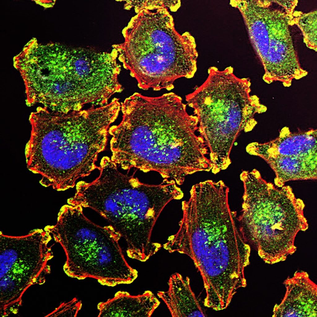
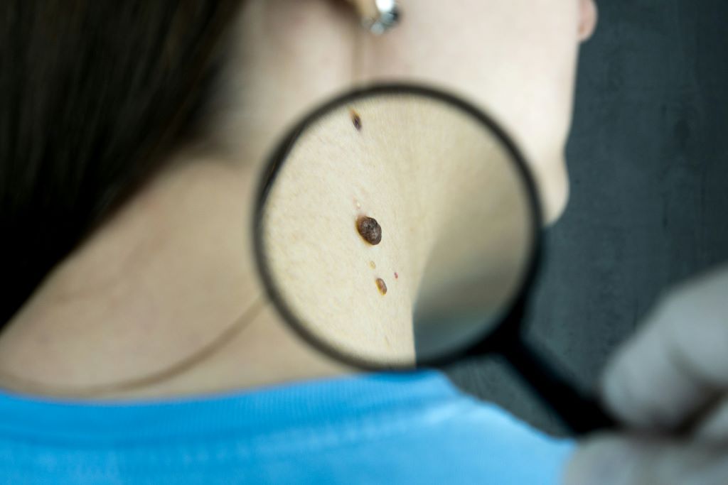
Moles and melanomas share the same cellular origin, forming from a pigment-producing cell called a melanocyte that gives skin its colour. In moles, the cell multiplies to form a cluster then stops growing, leaving a harmless spot. In melanoma, the cell growth continues aggressively.

Moliness is strongly influenced by your genes and having a high mole count is a major risk factor for melanoma. Around a third of melanomas develop from a mole. 

The QIMR Berghofer research analysed data from more than 85000 participants of European ancestry discovering 24 new genetic regions that determine the number of moles someone has. This is a five-fold increase on the five areas found in an earlier 2018 study also led by QIMR Berghofer researchers.

All but one of the genetic regions for mole count also play a role in melanoma. The team pinpointed more than 250 key genes in these regions to prioritise for further research.

One of the new genes, SIKE1, regulates immune responses to viral infections. The researchers think it could enable the development of melanomas by malfunctioning and affecting the immune system’s ability to detect and destroy melanocytes that are multiplying abnormally. This could be a promising target for a potential immunotherapy that could possibly prevent early stage melanoma growth.

Lead author Shanika Jayasinghe from QIMR Berghofer said the study builds on decades of world-leading skin cancer research at the Institute which has been involved in every major study of the genetics of moles and melanomas from twin studies to large-scale genome-wide research.

“I’m really proud to be continuing this long legacy of research. Our study increases understanding of why some people have a lot of moles and why some people develop melanoma so we can better treat and prevent this skin cancer,” Ms Jayasinghe said.

The researchers used the study insights to create a Polygenic Risk Score (PRS) for moliness to predict those who are genetically more likely to have a large number of moles, which could be integrated into melanoma screening tools in future to improve their accuracy in finding those at high risk so they can receive extra monitoring.

The next step is to analyse even larger data sets to find more genetic regions involved in moliness and melanoma. The researchers are also searching for existing drugs that could potentially target the newly identified biological pathways.

The scientists are grateful for the contribution of the many patients who participated in the 13 studies that were analysed for this project, including QIMR Berghofer’s QSkin Sun and Health Study and the Australian Genetics of Depression Study.

The study is available in Nature Communications with DOI 10.1038/s41467-026-70368-5.

Source: QIMR Berghofer

Vaccine and Immunotherapy Combo Halves Melanoma Recurrence

3D structure of a melanoma cell derived by ion abrasion scanning electron microscopy. Credit: Sriram Subramaniam/ National Cancer Institute

The combination of a vaccine and a drug, which both harness the immune system to attack cancer cells, has proven successful in cutting the risk of skin cancer recurrence by 49% , a new study shows. This reduction, which was calculated five years after patients had their tumours surgically removed, remains unchanged.

Led by researchers at NYU Langone Health and its Perlmutter Cancer Center, the study tested the vaccine, called intismeran, in combination with the mainstay immunotherapy pembrolizumab (Keytruda) in 107 patients who had been randomly chosen after melanoma surgery to determine whether the combination therapy prevented their cancer from recurring. Intismeran is a personalized immunotherapy strategy that is developed with information from a patient’s individual tumour. These results were compared with those from a randomly selected group of 50 melanoma patients who had only received pembrolizumab postoperatively, a current standard of care.

Results of the phase 2b trial, known formally as KEYNOTE-942, are being presented at the 2026 annual meeting of the American Society of Clinical Oncology on June 1 in Chicago and simultaneously published in the society’s Journal of Clinical Oncology.

After five years of follow-up, 68.8% of patients who took the combination therapy remained cancer-free, while 49.1% of the patients in the pembrolizumab-alone group had no signs of cancer. This means that adding intismeran to pembrolizumab reduced the risk for recurrence or death by 49%. The combination therapy also reduced the risk of distant metastasis by 59%. Overall survival, meaning no death from cancer or any other cause, was 92.2%for the vaccine with immunotherapy group, while for the immunotherapy-alone group it was 71.3%.

“Our study offers strong evidence to melanoma patients that intismeran vaccine therapy, when used in combination with immunotherapy, can demonstrably reduce their risk of having their cancer return and improve clinical outcomes,” said study senior investigator Janice Mehnert, MD, a professor in the Department of Medicine at NYU Grossman School of Medicine.

“Our findings also serve as encouragement to cancer researchers globally that mRNA vaccines like intismeran could work well in combination with immunotherapy for other cancers whose high rates of mutations have proven difficult to target,” said Dr Mehnert, who also serves as director of the melanoma medical oncology program and associate director of clinical research at Perlmutter Cancer Center.

The study results highlight the role of T cells, which are capable of attacking viruses as well as cancers. To spare normal cells, the immune system uses checkpoint molecules on T cell surfaces to “turn off” their attack against viruses when they clear the infection. The body may recognide tumours as abnormal, but cancer cells hijack checkpoints to turn off and evade immune responses. Immunotherapies like pembrolizumab seek to block checkpoints, specifically the PD-1 protein receptor, making cancer cells more “visible” and vulnerable again to immune cells.

Immunotherapies, including PD-1 inhibitors like pembrolizumab, have become the mainstay for treating melanoma, although they do not work for all patients, because melanoma cells, known for their ability to evade the immune system, can become resistant to immunotherapy. For this reason, researchers have looked at adding vaccines.

The vaccine intismeran is based on messenger RNA, a chemical cousin of DNA that provides cells with instructions for making proteins. Intismeran and other mRNA cancer vaccines are meant to teach the immune system to recognize cancer cells as different from normal cells. In designing a vaccine against melanoma, researchers attempted to trigger an immune response to specific abnormal proteins, called neoantigens, made by cancer cells.

Because the study volunteers all had their tumours removed, researchers were able to analyse their cells for 34 neoantigens that were specific to each melanoma and create a personalised vaccine for each patient. As a result, T cells specific to the neoantigen proteins encoded by the mRNA were produced. Those T cells could then attack any melanoma cells trying to grow or spread.

Dr. Mehnert said that a phase 3, multicenter trial is already underway to determine if intismeran helps as a first-line therapy in combination with pembrolizumab for melanoma. Already, the vaccine is being tested to see if it also works to prevent recurrence of lung and other cancers.

For the KEYNOTE-942 trial, patients were enrolled at cancer centers in Australia and the United States from 2019 to 2021. All were men and women who had had surgery to remove their melanoma tumors. Seven patients in each treatment group died during follow-up, most from cancer. Side effects were considered manageable and included fatigue, pain at injection sites, and chills.

Source: NYU Langone Health

Targeting Immune Suppression to Overcome Melanoma Resistance

Photo by Bermix Studio on Unsplash

For patients with advanced melanoma without BRAF mutation who no longer respond to immune checkpoint inhibitors, treatment options remain frustratingly limited. A new study from Vanderbilt researchers led by Professor Emerita of Pharmacology Ann Richmond outlines a promising therapeutic strategy that may re-sensitise these resistant tumours to immunotherapy.

Figure 5b from the paper shows the difference between tumours treated with a just an antibody and vehicle (solution) or with trametinib and rigosertib and a CD40 agonist. Image cropped and shared from the paper by Yan et al. published in Nature Communications in 2026 in accordance with a CC BY-NC-ND 4.0 license.

The research introduces a three-drug combination that enhances immune activity and suppresses tumour-promoting immune cells by leveraging a low dose of the MEK inhibitor trametinib and multi-kinase inhibitor rigosertib alongside a CD40 agonist to shift the tumour microenvironment toward immune activation. Notably, all three agents have been either approved by the U.S. Food and Drug Administration or are currently in clinical trials, which may speed their path to patient testing.

“While agonist CD40 therapy can be helpful for treatment of melanoma, this therapy also induces the CD11b+ B regulatory cells that suppress the T cell response to tumours,” Richmond said. “We showed that combining CD40 therapy with trametinib and rigosertib prevents the induction of these B regulatory cells.”

Immune checkpoint inhibitors have become a mainstay of melanoma treatment, working by releasing the molecular “brakes” that prevent T cells from attacking cancer. But resistance to ICI is common in metastatic melanoma, especially in tumours that evolve immune-suppressive microenvironments. While CD40 agonists can activate immune cells, this therapy also unexpectedly expands CD11b+ regulatory B cells.

By combining CD40 activation with MEK and PI3K inhibition, the researchers blocked the expansion of suppressive B cells while retaining the benefits of CD40 stimulation. In preclinical mouse models of melanoma, the triple combination not only suppressed tumour growth but also restored responsiveness to checkpoint blockade.

Key findings

  • B cells as a resistance mechanism: CD40 therapy alone induced regulatory B cells that dampen T cell–mediated tumor immunity.
  • Triple combination prevents immune suppression: Co-treatment with trametinib and rigosertib blocked the agonist CD40 induction of regulatory B cells, allowing immune responses to proceed.
  • ICIs regain effectiveness: The drug cocktail slowed tumor progression and re-sensitized resistant melanomas to anti-PD-1 therapy.

Translational promise

Because trametinib, rigosertib, and CD40 agonists are already in human trials or approved for other indications, this therapeutic strategy may advance more quickly than approaches requiring new drug development. Richmond’s team sees potential for testing the triple therapy in clinical trials for melanoma patients who have progressed on ICI.

“This approach provides a new route to enhance antitumor immunity in patients with tumors that no longer respond to immunotherapy,” Richmond said.

By Marissa Shapiro

Source: Vanderbilt University

Faecal Transplant Pills Show Promise in Clinical Trials for Multiple Types of Cancer

Two Canadian clinical trials show poop pills could help patients respond to immunotherapy while also reducing toxic side effects of cancer drugs

Faecal microbiota transplants (FMT), can dramatically improve cancer treatment, suggest two groundbreaking studies published in the prestigious Nature Medicine journal. The first study shows that the toxic side effects of drugs to treat kidney cancer could be eliminated with FMT. The second study suggests FMT is effective in improving the response to immunotherapy in patients with lung cancer and melanoma.

The findings represent a giant step forward in using FMT capsules – developed at Lawson Research Institute (Lawson) of St. Joseph’s Health Care London and used in clinical trials at London Health Sciences Centre Research Institute (LHSCRI) and Centre de recherche du Centre hospitalier de l’Université de Montréal (CRCHUM) – for safe and effective cancer treatment.

A Phase I clinical trial was conducted by scientists at LHSCRI and Lawson to determine if FMT is safe when combined with an immunotherapy drug to treat kidney cancer. The team found that customised FMT may help reduce toxic side effects from immunotherapy. The clinical trial involved 20 patients at the Verspeeten Family Cancer Centre at London Health Sciences Centre (LHSC).

“Standard treatment for advanced kidney cancer often includes an immunotherapy drug that helps the patient’s immune system tackle cancer cells,” says Saman Maleki, PhD, Scientist at LHSCRI. “But, unfortunately, the treatment frequently leads to colitis and diarrhoea, sometimes so severe that a patient must stop life-sustaining treatment early. If we can reduce toxic side effects and help patients complete their treatment, that will be a gamechanger.”

Separate Phase II lung and skin cancer studies were led by researchers at CRCHUM in collaboration with Lawson and LHSCRI. The studies found that 80 per cent of patients with lung cancer responded to immunotherapy after FMT, compared to only 39-45 per cent typically benefiting from immunotherapy alone. Similarly, 75 per cent of patients with melanoma who received FMT experienced a positive response to treatment, compared to only 50-58 per cent response in patients who receive immunotherapy alone. Twenty patients participated in the lung cancer clinical trial and 20 patients participated in the skin cancer clinical trial.

“Our clinical trial demonstrated that faecal microbiota transplantation could improve the efficacy of immunotherapy in patients with lung cancer and melanoma,” says Dr Arielle Elkrief, co-principal investigator and Physician Scientist, Université de Montréal-affiliated hospital research centre (CRCHUM). “The results also uncovered one possible mechanism of action of faecal transplantation – through the elimination of harmful bacteria following the transplant. Our results open up a novel avenue for personalised microbiome therapies, and faecal transplant is now being tested as part of the large pan-Canadian Canbiome2 randomised controlled trial.”

“Faecal microbiota transplantation in melanoma and lung cancer opens an entirely new therapeutic avenue, made possible by the exceptional commitment of our patients and the teamwork,” adds Dr. Rahima Jamal, Director of the Unit for Innovative Therapies (UIT) at CRCHUM. “At the Unit for Innovative Therapies (UIT) of the CRCHUM, we have had the privilege of translating laboratory discoveries into early phase clinical trials and witnessing their concrete impact on people living with cancer.”

Both studies use advanced, world-leading FMT capsules, also known as LND101, produced by Lawson in London, Ont. The research reinforces London’s place as a global leader in FMT innovation and treatment. The capsules are processed from healthy donor stools and ingested to help restore a patient’s healthy gut microbiome and treat different types of cancer.

“To use FMT to reduce drug toxicity and improve patients’ quality of life while possibly enhancing their clinical response to cancer treatment is tremendous, and it had never been done in treating kidney cancer before this,” says Dr Michael Silverman, Scientist at Lawson and Head of St. Joseph’s Infectious Diseases Program. “And none of this would be possible if not for this close collaboration: innovating the FMT capsules in Lawson labs and introducing them at LHSCRI and CHUM to advance vital research initiatives. Also, because LND101 comes from healthy donors, production can be scaled up to eventually help large numbers of cancer patients.”

The studies build on earlier London and CHUM-generated Phase I research showing FMT can safely augment treatment for people with melanoma. FMT is also being studied in people with pancreatic cancer and triple-negative breast cancer, and is already a well-established treatment for serious gut infections such as C. difficile, which can cause severe diarrhoea.

“Our hope is that our research will one day help people with cancer live longer while reducing the harmful side effects of treatment,” adds Dr Ricardo Fernandes, Scientist at LHSCRI and Medical Oncologist at LHSC. “We are world leaders in FMT research and we’re excited about its potential.”

Source: London Health Sciences Centre Research Institute

Lower Doses of Immunotherapy for Skin Cancer Give Better Results

Photo by Bermix Studio on Unsplash

According to a new study, lower doses of approved immunotherapy for malignant melanoma can give better results against tumours, while reducing side effects. This is reported by researchers at Karolinska Institutet in the Journal of the National Cancer Institute.

“The results are highly interesting in oncology, as we show that a lower dose of an immunotherapy drug, in addition to causing significantly fewer side effects, actually gives better results against tumours and longer survival,” says last author Hildur Helgadottir, a researcher at the Department of Oncology-Pathology at Karolinska Institutet, who led the study.

The traditional dose of nivolumab and ipilimumab is the one that is approved and established. Due to the extensive side effects, Sweden has increasingly begun to use a treatment regimen with a lower dose of ipilimumab, which is both gentler and cheaper. Ipilimumab is the most expensive part of this immunotherapy and causes the most side effects.

“In Sweden, we have greater freedom to choose doses for patients, while in many other countries, due to reimbursement policies, they are restricted by the doses approved by the drug authorities,” says Hildur Helgadottir.

Lower dose is more effective

The study included nearly 400 patients with advanced, inoperable malignant melanoma, the most serious form of skin cancer. The study shows that the regimen with the lower dose of ipilimumab is more effective, with a higher proportion of patients responding to treatment, 49%, compared to the traditional dose, 37%.

Progression-free survival, the time the patient lives without the disease worsening, was a median of nine months for the lower dose, compared to three months for the traditional dose. Overall survival was also longer, 42 months compared to 14 months.

Serious side effects were seen in 31% of patients in the low-dose group, compared to 51% in the traditional group.

“The new immunotherapies are very valuable and effective, but at the same time they can cause serious side effects that are sometimes life-threatening or chronic. Our results suggest that this lower dosage may enable more patients to continue the treatment for a longer time, which is likely to contribute to the improved results and longer survival,” says Hildur Helgadottir.

There were some differences between the two treatment groups, but even after adjusting for several factors such as age and tumour stage, the better outcome for the lower dose of ipilimumab remained. The study is a retrospective observational study and therefore it is not possible to definitively establish a causal relationship.

Source: Karolinska Institutet

Craters on Surface of Melanoma Cells Serve as Sites for Tumour Killing

Studying these craters could better assess immunotherapy’s success in treating solid tumours

3D structure of a melanoma cell derived by ion abrasion scanning electron microscopy. Credit: Sriram Subramaniam/ National Cancer Institute

Like the surface of the moon, new research published today in Cell finds the existence of craters on the surface of melanoma cells that serve as immune hubs, becoming major sites for tumour killing. These craters could serve as good markers for immunotherapy success.

This research provides insight into a key function of immune check-point blockade (ICB) cancer therapy that was previously unknown. ICB works by re-activating CD8+ T cells against tumours which shrinks and eventually kills the cancer cells. However, what facilitates local tumour killing by the infiltrating CD8+ T cells has remained a mystery.

Using a zebrafish model, researchers were able to monitor the infiltrating CD8+ T cells for up to 24 hours as they moved through the 3D architecture of endogenous melanoma tumours. Zebrafish provide the only tumour model where continuous live imaging over a 24 hour time period is feasible.  

“We found that rather than patrolling the entire tumour surface, CD8+ T cells aggregated in pockets on the melanoma border, forming prolonged interactions with melanoma cells,” says Leonard Zon, MD, Director of the Stem Cell Program at Boston Children’s Hospital and lead investigator of the study. “We termed these pockets Cancer Regions of Antigen presentation and T cell Engagement and Retention (CRATERs) and saw that, following immune stimulation, the CRATERs expanded and facilitated an effective immune response against the tumour.”

Zon, first author Aya Ludin, and the team also discovered CRATERs in human melanoma samples. Moreover, they saw similar structures in human lung cancer, indicating that CRATERs are likely not limited to melanoma and may form in other solid tumours.

To date, efficacy of therapeutic response to ICB therapy has been assessed mainly by estimating the degree of tumour necrosis and fibrosis. Indicators of CD8+ T cells infiltration has been associated with patient survival and treatment outcome, but direct evidence of effective immune cell-tumour cell interaction has been missing.

“Pending thorough clinical verification and taken together with other measurements, CRATERs may serve to more accurately assess the efficacy of an ongoing treatment and improve treatment outcomes,” said Zon.

The research team is now planning a prospective clinical trial to test if CRATERs are the best marker of ICB success.

Source: Boston Children’s Hospital via EurekAlert!

At-home Melanoma Testing with Skin Patch Test

A microneedle patch captures cancer biomarkers in the top-most layer of skin to detect melanoma in animal tissue samples

The newly designed ExoPatch being removed from a sample of mouse skin successfully distinguished melanoma from healthy skin in mice. A gel coating the microneedles picks up cancer indicators from the top-most layer of the skin. Dissolving the gel releases exosomes into a solution, which is then used on a two-lined test strip, similar to an at-home COVID-19 test. Image credit: Jeremy Little, Michigan Engineering.

Melanoma testing could one day be done at home with a skin patch and test strip with two lines, similar to COVID-19 home tests, according to University of Michigan researchers. Developed with funding from the National Institutes of Health, the new silicone patch with star-shaped microneedles, called the ExoPatch, distinguished melanoma from healthy skin in mice.

The patch and test move toward rapid at-home melanoma testing, helping patients catch the most aggressive form of skin cancer early without a biopsy or blood draw.

“The star-shaped needles make puncture easier and less painful, but they are so small that they only go through the top-most layer of the skin, the epidermis, and do not draw blood,” said Sunitha Nagrath, the Dwight F. Benton Professor of Chemical Engineering at U-M and co-corresponding author of the study published in Biosensors and Bioelectronics.

The ExoPatch microneedles, at just 0.6mm long with a width of less than 100 nm (0.0001 mm) at the tip, are coated with a gel that picks up exosomes, tiny packages released by cells, from the interstitial fluid that fills the spaces between cells in the epidermis.

Once thought to be trash ejected from cells for cleanup, exosomes actually contain DNA and RNA fragments that cells use to communicate with each other. Cancer cell exosomes can help tumours spread by preparing tissues to accept tumour cells before arrival, and detecting them can catch cancer earlier than past methods.

The gel that coats the ExoPatch contains a protein called Annexin V that attracts and sticks exosomes to the microneedles’ surface. Once removed from the skin, placing the patch in an acid dissolves the gel, which releases the exosomes into a solution. After dipping a test strip into the solution, two lines form if the sample contains melanoma exosomes, and one line forms for a negative test – the same way an at-home COVID-19 test strip works.

“A fair-skinned person with moles must go to the doctor about every six months to send off a biopsy to see if they’re malignant or benign. With this test, they could instead test at home, get the results right away and follow up with a dermatologist for a positive result,” Nagrath said.

As a first step in the proof-of-concept study, the researchers tested the ExoPatch on a tissue sample of pig skin, which closely resembles human skin in thickness and composition. Using a microscope, they found the microneedles penetrated about 350 to 600nm into the skin. For scale, the epidermis on the human forearm is about 18 300nm thick.

To test whether the ExoPatch could capture melanoma exosomes from skin tissue, the research team tested tissue samples of mouse skin, half from healthy mice and half from mice injected with a fragment of a human melanoma tumour. After a 15-minute application, the ExoPatch was placed under a powerful microscope.

“When looking at microscopy images, I was happy to see how nicely the exosomes adhered to the microneedles and were within the 30 to 150 nanometre size range we expect,” said Scott Smith, U-M doctoral student of chemical engineering and co-lead author of the study.

After confirming the exosomes stuck to the ExoPatch, the researchers dissolved the gel and ran the sample through the test strips. The test successfully distinguished between melanoma and healthy tissues with a 3.5-fold darker line in melanoma samples.

The ExoPatch isolated 11.5 times more exosomal protein from melanoma tissue samples compared to healthy tissue, showing it can specifically target cancerous exosomes.

A pilot study in humans followed by a series of clinical trials will be the next steps to move the technology toward use. Beyond melanoma, the ExoPatch gel coating could be modified to detect exosomes released by other cancers with a solid tumour, including lung, breast, colon, prostate and brain cancer.

“This is the first patch designed to capture disease-specific exosomes from fluid under the skin. The potential applications are huge,” said Nagrath.

Source: University of Michigan

Existing Drug Class May Help Patients with Immunotherapy-resistant Melanoma

3D structure of a melanoma cell derived by ion abrasion scanning electron microscopy. Credit: Sriram Subramaniam/ National Cancer Institute

Increased activity in a specific biological pathway may explain why many patients with a deadly form of skin cancer do not respond to the latest cancer treatments, a new study shows.

Publishing in the journal Cancer Research, the study featured data generated from experiments with human tissues and cells from patients with advanced melanoma that were implanted into mice. Results uncovered therapeutic targets that could limit melanoma growth in patients whose cancer failed to respond to initial treatment with immune checkpoint inhibitors.

Led by researchers at NYU Langone Health and its Perlmutter Cancer Center, the study focused on a subgroup of melanoma patients with mutations in the neurofibromin 1 (NF1) gene. NF1 mutations are just one type among several mutations, including those in the BRAF, NRAS, and PARP genes, that are linked to many cases of cancer, particularly melanoma. As many as 27% of melanoma patients are estimated to have NF1 mutations.

While immunotherapy, which stimulates the immune system to attack cancer cells as it would an invading virus, has proved to be a successful treatment, it does not work well for more than half of NF1-mutant melanoma patients.

“There is a pressing need for new drug therapies for melanoma patients with neurofibromin 1 mutations that do not respond to the latest immunotherapy, and for which there are no subsequent effective treatment options,” said study lead investigator Milad Ibrahim, PhD. Ibrahim is a postdoctoral fellow in the Dr Iman Osman Laboratory in the Ronald O. Perelman Department of Dermatology at the NYU Grossman School of Medicine.

To investigate why these patients were treatment resistant, investigators examined tumour cells from 30 melanoma patients who did not respond to immunotherapy. NF1 mutations were found in 40% of these melanoma samples. The samples came from NYU Langone’s extensive repository from more than 6000 melanoma patients.

Molecular testing showed that the signalling pathway built around a protein called epidermal growth factor receptor (EGFR) was more active in NF1 mutant melanoma cells than in cells with other melanoma-gene mutations. Increased EGFR activity has long been linked to abnormal cell growth in tumours and shorter survival with various cancers. The researchers also found that NF1 mutant melanoma cells depended on increased EGFR activity for survival, regardless of the presence of other mutations.

Because EGFR-inhibiting drugs are already used to treat some head and neck cancers, as well as colorectal and lung cancers, researchers then tested two drugs in the class, cetuximab and afatinib, in both NF1 mutant cell cultures and cancer cell lines without NF1 mutations. After transplanting both tumour cell types into mice and treating them with these drugs, results showed that both EGFR inhibitors were effective against cells and transplanted tumours with NF1 mutations, and they had no effect on melanomas without NF1 mutations.

“Our study results reveal a unique vulnerability in melanoma patients with neurofibromin 1 mutations, that an overexpression of the epidermal growth factor receptor pathway is essential for their survival and growth,” said the study’s senior investigator, Professor Iman Osman, MD.

“While further tests are needed, our results support a novel approach of deploying EGFR inhibitors either alone or in combination with other immunotherapies for treatment of melanoma patients whose tumours harbour NF1 mutation,” said the study’s co-senior investigator, Associate Professor Markus Schober, PhD.

However, Schober says this requires further testing in a clinical trial, which the research team plans to develop. He adds that if trial findings prove successful, the team’s research could provide a lifeline for many of these melanoma patients.

Source: NYU Langone Health

Study Identifies Mitochondrial Drivers of Skin Cancer Aggressiveness – and Possible Treatments

Mitochondrial pathways help melanoma cells become aggressive, and some currently available drugs target these pathways.

3D structure of a melanoma cell derived by ion abrasion scanning electron microscopy. Credit: Sriram Subramaniam/ National Cancer Institute

Researchers have discovered that the most aggressive melanomas, the deadliest form of skin cancer, overactivate two key processes in mitochondria. Blocking these pathways with currently available drugs effectively killed melanoma cells. The findings are published by Wiley online in CANCER, a peer-reviewed journal of the American Cancer Society.

By mapping the proteins expressed in 151 tumour and normal skin samples, investigators found that the most aggressive melanomas hyper-activate the machinery that builds mitochondrial proteins and the mitochondrial system that turns nutrients into energy.

Remarkably, blocking these pathways effectively halted or killed melanoma cells cultured in lab dishes. Two types of drugs accomplished this: antibiotics, originally designed to block bacterial protein synthesis machinery, which closely resembles the machinery found in mitochondria, and specialised energy-production inhibitors. Importantly, non-cancerous skin cells remained mostly unaffected, highlighting the safety and specificity of these treatment approaches.

“This discovery identifies melanoma’s excessive reliance on mitochondrial energy as its Achilles’ heel, revealing a therapeutic vulnerability that we can exploit with existing drugs,” said senior author Jeovanis Gil, PhD, of Lund University in Sweden. “By pairing mitochondrial blockers with today’s standards of care, we may cut off a major escape route that cancers use to resist therapy and come back.”

Dr Gil added that the mitochondrial-protein signature his team discovered can be measured in routine biopsy material and could serve as a biomarker to identify patients most likely to benefit from mitochondrial-targeted add-on therapies. By enabling clinicians to match treatments to each patient’s tumour biology, these findings mark a step forward for precision medicine in melanoma. Moreover, because mitochondrial rewiring fuels resistance across many cancers, success in melanoma could open the door to similar personalised combination strategies in other hard-to-treat cancers. 

Source: Wiley

How to Stop Melanoma’s Incredibly Swift Evasion of Treatment

Melanoma Cells. Credit: National Cancer Institute

Researchers have uncovered a stealth survival strategy that melanoma cells use to evade targeted therapy, offering a promising new approach to improving treatment outcomes.

The study, published in Cell Systems and conducted by researchers at the Institute for Systems Biology (ISB) and Massachusetts Institute of Technology (MIT) identifies a non-genetic, reversible adaptation mechanism that allows melanoma cells to survive treatment with BRAF inhibitors. By identifying and blocking this early response, researchers proposed a combination therapy that could delay resistance and enhance the effectiveness of existing treatments.

Cracking the Code of Melanoma’s Drug Escape

Melanoma, the deadliest form of skin cancer, is often driven by mutations in the BRAF gene, which fuels uncontrolled tumor growth. While BRAF inhibitors (such as vemurafenib) initially halt tumor growth, many tumors quickly adapt and survive treatment, leading to therapy failure.

Unlike traditional resistance driven by genetic mutations, this study uncovers an early, dynamic adaptation process that occurs within hours to days of drug treatment – long before genetic resistance takes hold. Surprisingly, this process does not rely on reactivating the BRAF-ERK pathway, which is the usual resistance mechanism.

Using cutting-edge mass spectrometry-based phosphoproteomics and deep transcriptomics analyses, researchers mapped the molecular shifts in melanoma cells over minutes, hours, and days of BRAF inhibitor treatment.

“We found that while the BRAF-ERK signaling pathway was quickly and durably suppressed, cancer cells did not rely on reactivating ERK to survive. Instead, they triggered an alternative SRC family kinase (SFK) signaling pathway, which promoted cell survival and eventual recovery,” said Chunmei Liu, PhD, a bioinformatics scientist at ISB and co-first author of the paper.

Turning a Weakness Into a Target

A key discovery in this study came when researchers linked SFK activation to reactive oxygen species (ROS), a cellular stress response that builds up under BRAF inhibition. As ROS levels surged, SFK activity spiked, helping melanoma cells withstand treatment. However, this adaptation was reversible – when treatment was removed, cells returned to their original state.

Recognizing this Achilles’ heel, the team tested a combination approach: pairing BRAF inhibitors with the SFK inhibitor dasatinib.

“By adding dasatinib, we blocked this adaptive escape mechanism, significantly reducing melanoma cell survival and stabilising tumours in animal models,” said ISB Associate Professor Wei Wei, PhD, co-corresponding author.

Importantly, SFK inhibition alone had little effect on melanoma cells, highlighting the need for a strategic combination therapy to suppress melanoma adaptation before resistance fully develops. 

“This approach has the potential to prolong the effectiveness of BRAF inhibitors and improve patient outcomes,” said ISB President and Professor Jim Heath, PhD, co-corresponding author.

Looking Ahead: A Path to the Clinic

Beyond uncovering a key mechanism of drug adaptation, this research underscores the importance of early intervention to prevent it from happening. It also highlights ROS accumulation and SFK activation as potential biomarkers for identifying patients who may benefit from this combination therapy.

Further preclinical studies and clinical trials will be necessary to validate this combination therapy strategy and determine its potential for broader clinical use.

Source: Institute for Systems Biology