Adaptive Spine Board Could Revolutionise ER Transport

ASB overlay is divided into five distinct sections—head and neck, upper trunk, buttocks and pelvis, thighs, and feet and heels

In combat zones and emergency rescues, rapid evacuation and treatment can mean the difference between life and death. But prolonged immobilisation during transport poses another life-threatening risk: pressure injuries.

A newly developed adaptive spine board (ASB) overlay aims to change that, offering an innovative solution to prevent pressure injuries and dramatically improve patient outcomes. Developed by researchers at The University of Texas at Arlington and UT Southwestern Medical School, the adaptive spine board sits atop a standard stretcher or spine board, using air-cell technology to redistribute pressure more effectively than traditional evacuation surfaces. The team’s newly published study in the Journal of Rehabilitation and Assistive Technologies Engineering shows the ASB outperforms other immobilisation options.

“The ability to dynamically adjust pressure so that no vulnerable body regions experience excessive weight is a breakthrough for medical evacuation,” said Muthu B.J. Wijesundara, principal research scientist at the University of Texas at Arlington Research Institute. “This innovation could set a new standard in casualty transport protocols.”

Also called bedsores or ulcers, pressure injuries result from prolonged pressure on the skin and underlying soft tissue, leading to cell death, tissue breakdown and open wounds. They are a constant risk for trauma patients during long-range transport, which sometimes lasts more than 16 hours. Research shows that more than 50% of casualties transported during the Iraq War developed pressure injuries before reaching a hospital.

While some existing technologies, such as vacuum spine boards, can help redistribute pressure, their effectiveness is limited. Many conventional supports fail to keep pressure below the thresholds recommended to prevent injury. Military stretchers and pads have shown to create high-pressure points on vulnerable areas of the body, including the back of the head, base of the spine, buttocks and heels.

“Beyond military use, the ASB overlay could prove valuable in civilian medical transport, particularly for spinal injury patients who are at high risk for pressure ulcers,” Dr Wijesundara said. “The research also highlights potential applications in other environments where prolonged immobilisation is necessary, such as disaster relief and space exploration.”

The ASB overlay features a multi-segmented air-cell design that target pressure-prone areas more effectively than previous solutions. It is divided into five distinct sections—head and neck, upper trunk, buttocks and pelvis, thighs, and feet and heels—each equipped with sensor-driven pressure modulation for responsive, localised support.

“One key innovation is the system’s ability to autonomously adjust the air-cell pressure to maintain optimal distribution for each patient,” Wijesundara said. “We developed an algorithm that compensates for environmental variables, such as temperature and barometric pressure changes, ensuring consistent performance across varying conditions. Testing showed that the ASB overlay outperformed typical equipment used in casualty transport.”

For critically injured patients, pressure injuries can significantly complicate treatment and recovery, leading to longer hospital stays, higher infection risks and additional surgeries. They’re also costly. The Agency for Healthcare Research and Quality (AHRQ) estimates that pressure injuries in the US can cost up to $151 700 per case, adding $11.6 billion in additional health care expenses annually. Alarmingly, the AHRQ also reports that approximately 60 000 patients die each year because of pressure injuries. The ASB overlay’s advanced pressure modulation could help mitigate these risks—especially for patients who cannot be repositioned during extended transport.

The research team is now planning additional studies to improve the device’s usability in real-world conditions. As the military increasingly relies on prolonged aeromedical evacuation, such advancements are critical for enhancing patient care in conflict zones.

Everything We Thought About Running Injury Development Was Wrong, Study Shows

Photo by Barbara Olsen on Pexels

A new study from Aarhus University turns our understanding of how running injuries occur upside down. The research project, published in The BMJ, is the largest of its kind ever conducted and involves over 5000 participants. It shows that running-related overuse injuries do not develop gradually over time, as previously assumed, but rather suddenly – often during a single training session.

“Our study marks a paradigm shift in understanding the causes of running-related overuse injuries. We previously believed that injuries develop gradually over time, but it turns out that many injuries occur because runners make training errors in a single training session,” explains Associate Professor Rasmus Ø. Nielsen from the Department of Public Health at Aarhus University, who is the lead author of the study.

The study followed 5205 runners from 87 countries over 18 months and shows that injury risk increases exponentially when runners increase their distance in a single training session compared to their longest run in the past 30 days. The longer the run becomes, the higher the injury risk.

Incorrect guidance for millions of runners

According to Rasmus Ø. Nielsen, the results cast critical light on how the tech industry has implemented so-called “evidence.” Millions of sports watches worldwide are equipped with software that guides runners about their training – both for training optimisation and injury prevention.

However, the algorithm used for injury prevention is built on very thin scientific grounds, according to Rasmus Ø. Nielsen.

“This concretely means that millions of runners receive incorrect guidance from their sports watches every day. They think they are following a scientific method to avoid injuries, but in reality they are using an algorithm that cannot predict injury risk at all,” he says.

Non-existing evidence behind guidance

The current algorithm, called “Acute:Chronic Workload Ratio” (ACWR), was introduced in 2016 and is now implemented in equipment from companies that produce sports watches, while organisations and clinicians, such as physiotherapists, also use the algorithm.

The ACWR algorithm calculates the ratio between acute load (last week’s training) and chronic load (average of the past 3 weeks). The algorithm recommends a maximum 20% increase in training load to minimise injury risk.

According to Rasmus Ø. Nielsen, the algorithm was originally developed for team sports and was based on a study with 28 participants. Due to the few participants in the study combined with data manipulation, the evidence base for using the algorithm to prevent running injuries is therefore “non-existent.”

Realtime guidance

The research team has therefore worked for the past eight years to develop a new algorithm that will be much better at preventing injuries for runners.

Rasmus Ø. Nielsen emphasises that he and the other researchers behind the study have no commercial interests in launching a new algorithm as a potential replacement for a method he himself criticises.

The algorithm will be made freely available to runners, companies, clinicians and organisations who can use it actively to guide training and injury prevention.

Rasmus Ø. Nielsen hopes that the new insights will be implemented in existing technology.

“I imagine, for example, that sports watches with our algorithm will be able to guide runners in real-time during a run and give an alarm if they run a distance where injury risk is high. Like a traffic light that gives green light if injury risk is low; yellow light if injury risk increases and red light when injury risk becomes high,” explains Rasmus Ø. Nielsen.

Source: Aarhus University

Student Designs a Prostate Checking Device to Replace the Digital Exam

Pro check, designed by Loughborough University student Devon Tyso.

A Loughborough University student has developed a new medical device that could transform how prostate health is assessed and monitored.

Devon Tyso, a Product Design and Technology student, has designed ‘PRO check’, an innovative tool designed to replace the traditional digital rectal examination (DRE), which involves a doctor manually assessing the prostate with a finger.

According to Devon, the current approach is heavily reliant on a clinician’s subjective judgement and experience, and many see the method as ‘intrusive’.

“As one in seven men will get prostate cancer, it’s vital to detect abnormalities early and track changes over time,” said Devon, “The current examination method involves a lot of guesswork.

“PRO check provides objective, measurable data and allows prostate health to be visualised – enabling more accurate diagnosis, and improved long-term monitoring.

“Having a device conduct the exam may also feel less invasive, which may encourage more men to get checked, potentially catching issues earlier.”

How the device works

Designed for use by GPs during routine prostate assessments, PRO check allows doctors to evaluate the size and texture of the prostate — two key indicators of potential health issues — in a more objective and consistent way than the traditional digital rectal examination.

The device is a handheld probe, and it is covered with a condom before being inserted into the body. Once in position, the condom inflates to different pressures, pressing against the surface of the prostate, causing it to compress. A laser grid is projected onto the inner surface of the condom so the shape of the underlying prostate can be captured.

Stereoscopic cameras capture images of the laser grid, tracking where the gridlines intersect and how these intersections shift as pressure changes. This information is then fed into mathematical equations to create 3D images — or ‘topographical representations’ — that reveal the prostate’s shape and surface structure under different pressures.

Studying the prostate’s surface details could help clinicians identify areas requiring further investigation. Healthy prostate tissue is typically soft and compressible, so regions that appear stiff or resist pressure could indicate potential abnormalities and warrant further investigation.

The device can also produce data on prostate volume – one of the measurements used to calculate prostate-specific antigen (PSA) density, which helps assess prostate cancer risk. Devon says currently volume estimates are often based on a clinician’s best judgement.

In addition, data from PRO check can be used to generate a compressibility-versus-pressure graph – a novel data type not currently available in clinical practice. This graph shows how the prostate compresses at different pressure levels, which Devon hopes could offer new insights into prostate health and complement existing diagnostic tools.

PRO check is designed to integrate with artificial intelligence, enabling automatic extraction of video data, real-time calculations, and the generation of 3D images for live display on a laptop or tablet during the examination.

The idea is that all examination data from PRO check would be stored on the patient’s records, helping to build a personalised prostate health profile that can be tracked and monitored over time.

Inspiration

Devon’s inspiration for PRO check came from a mix of personal experience – after his grandfather’s prostate cancer diagnosis – and unexpected technical research.

“It really hit home how common prostate issues are after my family member was found to have an enlarged prostate,” said the 22-year-old from Cardiff, “I realised nearly everyone I spoke to about it knew someone affected by it.

“When I started looking into prostate examinations, I kept thinking ‘how can a doctor remember what your prostate felt like four months ago?’ and how horrible it must be just be told whether you’re fine or not without seeing any data or anything visual.”

While researching non-invasive ways to assess tissue structure inside the body, Devon came across a technique used by NASA to map the surface of asteroids — projecting laser grids onto them, capturing images with satellite-mounted cameras, and analysing the gridline intersections to reveal the contours of the surface.

“I saw that NASA were mapping surface heights on a massive scale, and I thought – if they can do that in space, why can’t we use similar principles to examine something here on Earth?” said Devon, “I’ve basically used the exact same technique and scaled it down for PRO check.”

Prototypes

Devon designed PRO check as part of his final year project – which was exhibited at the School of Design and Creative Arts’ 2025 Degree Show – and has prototyped several of its key components.

He has built and tested two working prototypes. The first demonstrates how a laser grid and camera can be setup to map the surface of the prostate.

Devon designed a custom rig that enabled him to capture images of a laser grid projected onto different silicone prostate models — representing a healthy gland, a small tumour, a large tumour, and an enlarged prostate — from an optimal angle using a smartphone camera.

First prototype of pro check
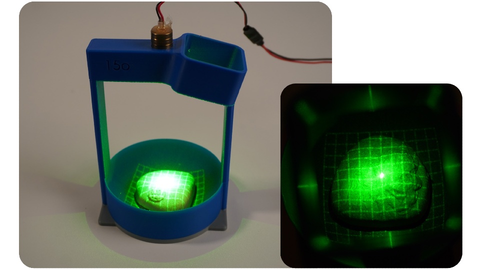
PRO check prototype one demonstrated how laser gridlines and a camera can be used to image the surface of the prostate.

The second prototype features electronics that inflate a small balloon at controlled pressures, regulated by a pressure-sensing chip. Devon consulted three healthcare professionals to measure the pressure typically applied during prostate exams and replicated those levels in his design.

Devon tested the prototype using the silicone prostate models but encased them in a sponge disc to simulate surrounding tissue.

Devon manually extracted data on the gridline intersections from the camera footage and applied mathematical equations to generate 3D images of the prostate surfaces and surrounding tissue under different pressures.

Next steps

Devon hopes to collaborate with medical professionals and product developers to turn PRO check into a fully realised medical device.

When speaking about his ultimate goal, Devon said: “I’d love to see this used in GP surgeries across the UK one day.

“With early detection being so critical, anything that helps men get checked sooner and more comfortably – and provides reliable data and visualisations – has huge potential. I really believe this could make a difference.”

Further information on PRO check can be found on the Degree Show website.

Source: Loughborough University

Hearing Devices Increase Social Connectedness and Reduce Mortality

Photo by Brett Sayles

Hearing loss doesn’t just affect how people hear the world — it can also change how they connect with it. New research from the University of Southern California, published in JAMA Otolaryngology – Head & Neck Surgery, is the first to link hearing aids and cochlear implants, surgically implanted devices that help those with profound hearing loss perceive sound, to improved social lives among adults with hearing loss. 

“We found that adults with hearing loss who used hearing aids or cochlear implants were more socially engaged and felt less isolated compared to those who didn’t use them,” said lead researcher Janet Choi, MD, MPH, an otolaryngologist with Keck Medicine at USC. “This suggests that hearing devices may help prevent the social disconnection and broader health consequences that can follow untreated hearing loss.” 

Hearing loss affects an estimated 40 million American adults, yet many go untreated. When left unaddressed, hearing loss can make communication difficult, leading people to withdraw from conversations and social activities, according to Choi.  

Previous research has shown that over time, social withdrawal can reduce mental stimulation and increase the risk of loneliness, anxiety, depression, cognitive decline and dementia. It has also linked chronic social isolation to biological and neurological changes, including increased brain inflammation and alterations in brain structure.  

“Understanding the link between hearing loss, hearing device use and social isolation is crucial,” said Choi. “Until this study, it has been unclear whether hearing devices could help reverse the isolation.”   

Choi and her fellow researchers conducted a comprehensive, systematic review and meta-analysis of 65 previously published studies, encompassing over five thousand participants, on how hearing aids and cochlear implants affect three key measures: social quality of life, perceived social handicap, which refers to the limitations and frustrations hearing loss can create in social situations, and loneliness.  

The researchers found that adults using hearing devices feel more socially connected and less limited in social situations. They are better able to engage in group conversations and feel more at ease in noisy or challenging listening environments. Participants also reported feeling less socially handicapped by their hearing loss, with fewer barriers and frustrations during interactions and an improved ability to stay engaged without feeling excluded. This increased confidence can help users connect more easily with family, friends and colleagues, leading to stronger feelings of belonging and reduced social anxiety. The study also suggested hearing devices may reduce loneliness, although further research is needed in this area, according to Choi. 

Those with cochlear implants reported the most improvement in their social quality of life. This is likely because cochlear implants offer greater hearing restoration than hearing aids, especially for individuals with more severe hearing loss. As a result, they may experience more noticeable improvements in social engagement once their hearing is restored. 

While it was outside the scope of the study to measure how better social lives relate to improved cognitive outcomes, Choi believes there may be a connection, as previous research has found managing hearing loss may be key to reducing the risk of cognitive decline and dementia. “While our study didn’t directly measure cognitive outcomes, the improvements we saw in communication and social engagement suggest that by restoring clearer communication, hearing devices may help preserve cognitive health by keeping the brain more actively involved and people more connected,” Choi said. 

This research follows a January 2024 study by Choi showing that adults with hearing loss who use hearing aids have an almost 25% lower risk of mortality, suggesting that treating hearing loss can improve lifespan as well as social quality of life.  

“These new findings add to a growing body of research showing that hearing health is deeply connected to overall well-being,” said Choi. “We hope this encourages more people to seek treatment and helps clinicians start conversations with patients about how hearing devices can improve their quality of life.”

Source: University of Southern California – Health Sciences

New Research Confirms that Neurons Form in the Adult Brain

A healthy neuron. Credit: NIH

A study in the journal Science presents compelling new evidence that neurons in the brain’s memory centre, the hippocampus, continue to form well into late adulthood. The research from Karolinska Institutet provides answers to a fundamental and long-debated question about the human brain’s adaptability.

The hippocampus is a brain region that is essential for learning and memory and involved in emotion regulation. Back in 2013, Jonas Frisén’s research group at Karolinska Institutet showed in a high-profile study that new neurons can form in the hippocampus of adult humans. The researchers then measured carbon-14 levels in DNA from brain tissue, which made it possible to determine when the cells were formed.

Identifying cells of origin

However, the extent and significance of this formation of new neurons (neurogenesis) are still debated. There has been no clear evidence that the cells that precede new neurons, known as neural progenitor cells, actually exist and divide in adult humans.

“We have now been able to identify these cells of origin, which confirms that there is an ongoing formation of neurons in the hippocampus of the adult brain,” says lead researcher Jonas Frisén, professor of stem cell research at the Department of Cell and Molecular Biology.

In the new study, the researchers combined several advanced methods to examine brain tissue from people aged 0 to 78 years from several international biobanks. They used a method called single-nucleus RNA sequencing, which analyses gene activity in individual cell nuclei, and flow cytometry to study cell properties. 

By combining this with machine learning, they were able to identify different stages of neuronal development, from stem cells to immature neurons, many of which were in the division phase.

To localise these cells, the researchers used two techniques that show where in the tissue different genes are active: RNAscope and Xenium. These methods confirmed that the newly formed cells were located in a specific area of the hippocampus called the dentate gyrus. This area is important for memory formation, learning and cognitive flexibility.

Hope for new treatments

The results show that the progenitors of adult neurons are similar to those of mice, pigs and monkeys, but that there are some differences in which genes are active. There were also large variations between individuals – some adult humans had many neural progenitor cells, others hardly any at all.

“This gives us an important piece of the puzzle in understanding how the human brain works and changes during life,” explains Jonas Frisén. “Our research may also have implications for the development of regenerative treatments that stimulate neurogenesis in neurodegenerative and psychiatric disorders.” 

Source: Karolinska Institutet

Muscle Metabolism in Men and Women Differs, with Implications for Diabetes

Photo by Ketut Subiyanto on Unsplash

The skeletal muscles of men and women process glucose and fats in different ways. A recently published study now provides the first comprehensive molecular analysis of these differences. The results possibly give an explanation why metabolic diseases such as diabetes manifest differently in women and men – and why they respond differently to physical activity.

Skeletal muscles are far more than just “movement driving motors.” They play a central role in glucose metabolism and therefore also in the development of type 2 diabetes. This is due to the fact that around 85% of insulin-dependent glucose uptake takes place in the muscles. This means that if muscle cells react less sensitively to insulin, for example in the case of insulin resistance, glucose is less easily absorbed from the blood. This process is specifically counteracted by physical activity. 

Women’s and Men’s Muscles Work Differently

The degree to which muscles work differently in women and men has long been underestimated. It is precisely this issue which has now been investigated by researchers at the Institute for Diabetes Research and Metabolic Diseases at Helmholtz Munich, the University Hospital of Tübingen and the German Center for Diabetes Research (DZD) e.V. The researchers, led by Dr Simon Dreher and Prof Cora Weigert, examined muscle biopsies from 25 healthy but overweight adults (16 women, 9 men) aged around 30 years. The test subjects had not taken part in regular sporting activities beforehand. Over a period of eight weeks, they completed one hour of endurance training three times a week, consisting of 30 minutes of cycling and 30 minutes of walking on the treadmill. 

Muscle samples were taken before they started, after they had the first training session and at the end of the program. Using state-of-the-art molecular biological methods, including epigenome, transcriptome and proteome analyses, the team investigated sex-specific differences at various levels. 

Men React with more Stress to Exercise 

The result: The first training session triggered a stronger stress response at the molecular level in men, which became manifest in the increased activation of stress genes and the increase in the muscle protein myoglobin in the blood. In addition, male muscles showed a distinct pattern of what are called fast-twitch fibers, which are designed for short-term, intensive exercise and preferably use glucose as an energy source. 

Women had significantly higher amounts of proteins that are responsible for the absorption and storage of fatty acids: an indication of more efficient fat utilisation. After eight weeks of regular endurance training, the muscles of both sexes matched and the muscle fibre-specific differences decreased. At the same time, women and men produced more proteins that promote the utilisation of glucose and fat in the mitochondria. 

“These adjustments indicate an overall improvement in metabolic performance, which can help to reduce the risk of type 2 diabetes,” says Weigert. “In future, our new findings might help to better predict individual diabetes risks and tailor recommendations for exercise therapies more specifically to women and men.” 

The scientists next want to investigate the role sex hormones play in these differences – and how hormonal changes in old age influence the risk of metabolic diseases. 

Original publication

Dreher et al., 2025: Sex differences in resting skeletal muscle and the acute and long-term response to endurance exercise in individuals with overweight and obesity. Molecular Metabolism. DOI: 10.1016/j.molmet.2025.102185

About the scientists

Prof. Cora Weigert is head of the research group “Molecular Diabetology and Exercise” at the Institute for Diabetes Research and Metabolic Diseases at Helmholtz Munich and Professor at the University of Tübingen.

Dr. Simon Dreher is scientist at the Institute for Diabetes Research and Metabolic Diseases at Helmholtz Munich and at the University of Tübingen.

Source: Deutsches Zentrum fuer Diabetesforschung DZD

Global Fund to Cut R1.4-Billion to SA for HIV, TB and Malaria

Photo by Reynaldo #brigworkz Brigantty

By Liezl Human

The Global Fund to Fight AIDS, TB and malaria (Global Fund) has notified Health Minister Aaron Motsoaledi that it will reduce funding to South Africa by R1.4-billion.

Global Fund said it would be reducing allocations for the seventh grant cycle from R8.5-billion to about R7.1-billion, a 16% reduction. Of this, 55% would be allocated to the National Department of Health and the rest to non-profit organisations such as the Networking HIV & AIDS Community of Southern Africa, Beyond Zero, and the AIDS Foundation of South Africa.

The fund informed recipient countries in May that it would be revising over 200 grants amidst funding shortfalls.

Global Fund was established in 2002 and provides funding for HIV, TB and malaria programmes in over 100 countries. According to its 2024 results report, 72% of its funding from 2021 to 2024 went to sub-Saharan Africa.

Other African countries also received notification of funding cuts. Mozambique’s allocation decreased by 12%, Malawi’s by 8% and Zimbabwe by 11%.

The shortfall in funding is due to Global Fund not having received money pledged by national governments. Over US$4 billion of the shortfall is due to the United States not fulfilling its pledge.

We reported last month how Mozambique’s health system has crumbled amidst USAID funding cuts.

In South Africa, funding cuts from PEPFAR earlier this year have led to clinics closing down, health staff getting retrenched, and people struggling to access HIV medication.

“As you know, the external financing landscape for global health programs is going through significant changes, with substantial impact on lifesaving services for the fight against the three diseases and health and community systems,” the Global Fund said in its letter to South African representatives.

The letter continued that while the Global Fund has “received some significant donor payments in recent days”, prospects to give the full grant cycle 7 (GC7) pledges “remain highly uncertain” and still face a risk of funding shortfalls.

“This is a difficult and unavoidable decision, which may require your country to reconsider how best to use the remaining GC7 grant amounts together with domestic resources and other sources of funds to keep saving lives,” the Global Fund said.

Foster Mohale, Department of Health spokesperson, said that the funding cut did not come as a surprise. Mohale said the department is “working with the provinces” to ensure that “service delivery” is not disrupted, and to apply measures to ensure “efficient use of limited resources”.

Republished from GroundUp under a Creative Commons Attribution-NoDerivatives 4.0 International License.

Read the original article.

Study Finds 1 in 6 Cancer Medications in Sub-Saharan Africa Are Defective

Source: Unsplash CC0

Serious quality defects were found in a significant number of cancer medications from sub-Saharan Africa, according to new research from the University of Notre Dame.

For the study published in The Lancet Global Health, researchers collected different cancer medications from Cameroon, Ethiopia, Kenya and Malawi and evaluated whether each drug met regulatory standards. Researchers considered a variety of factors, including appearance, packaging, labelling and, most importantly, the assay value.

The assay value is the quantity of active pharmaceutical ingredient (API) found in each drug. To meet safety standards, most products should be within a range of 90 to 110% of the right amount of API. Researchers measured the API content of each product and compared that number to what was designated on the medication packaging.

“It is important that cancer medications contain the right amount of the active ingredients so the patient gets the correct dose,” said Marya Lieberman, professor of chemistry and biochemistry at Notre Dame and lead author of the study. “If the patient’s dose is too small, the cancer can survive and spread to other locations. If the patient’s dose is too high, they can be harmed by toxic side effects from the medicine.”

One in six cancer medications tested was found to contain the incorrect quantity of API, with tested medications having APIs ranging from 28 to 120%. The study evaluated 251 samples of cancer medications collected from major hospitals and private markets in all four countries.

The study, funded by the National Cancer Institute of the National Institutes of Health, is among the first to evaluate cancer drug quality in sub-Saharan Africa. Currently, sub-Saharan Africa has no pharmaceutical regulatory laboratories carrying out chemical analyses for cancer drugs according to the standards required for regulatory purposes.

Yet, the need for cancer drugs is growing.

“We found bad-quality cancer medications in all of the countries, in all of the hospital pharmacies and in the private markets,” said Lieberman, an affiliate of Notre Dame’s Eck Institute for Global Health and Harper Cancer Research Institute. “We learned that visual inspection, which is the main method for detecting bad-quality cancer drugs in sub-Saharan Africa today, only found one in 10 of the bad products.”

In their study, the researchers explained how a combination of high demand for cancer medications, lack of regulatory capacity, and poor manufacturing, distribution and storage practices likely created a problematic environment throughout sub-Saharan Africa. They also argue that given these factors and the global supply chain for pharmaceuticals, substandard cancer medications are likely present in other low and middle-income countries as well.

Lieberman and her team identified several strategies that could help the global community address poor-quality cancer medications:

  • Provide inexpensive technologies at the point of care to screen for bad-quality cancer medicines and create policies for how to respond to products that fail screening tests.
  • Help regulatory agencies in low and middle-income countries get safety equipment and training so they can analyze the quality of cancer medicines in their markets, conduct root-cause investigations when products fail testing, take quick regulatory actions enabled by lab data and share data about bad-quality products.
  • Perform cost-benefit analyses of interventions that tackle common problems (such as medications being out of stock, unsafe shipping, storage or dispensing practices, and lack of availability or affordability of medications) to help policymakers and funders get the most impact on patient outcomes from their available resources.
  • Work with care providers to develop site-specific response policies and messaging for patients and engage regulators, donors and other resources.

Lieberman and her lab are developing a user-friendly technology called the chemoPAD for screening cancer medications. This low-cost paper device could potentially help hospitals, pharmacies and health care professionals in low and middle-income countries monitor drug quality without restricting a patient’s access to the medication.

“This is all part of a bigger project aimed at developing the ChemoPAD as a point-of-care testing device that we can use, something that’s more accurate in detecting poor-quality products than just visual inspection,” Lieberman said.

“There are lots of medicines where the regulators don’t have enough resources to verify the quality, and some manufacturers take advantage of that to cut corners. There are also problems with distribution systems, so even if a product is good quality when it leaves the manufacturer, it may be degraded during shipping or storage. These products flow into low and middle-income countries, and they get used on patients. I want to change that.”

Source: University of Notre Dame

Podcast: Paracetamol’s Newly Discovered Mechanism Could Unlock Powerful New Analgesics

Credit: Pixabay CC0

Despite being used for decades as a pain reliever, paracetamol’s mechanism of action was never fully known. Now, new research from the University of Jerusalem points to an unexpected effect, one which may usher in a whole new era of analgesics.

In QuickNews’ first podcast, you can listen to a discussion on how a newly discovered mechanism of action for paracetamol helps it achieve its analgesic effect, and how this could be applied to the development of novel, highly specific pain relievers.

Is It Really ADHD? Serious Flaws in Trials With Adult ADHD Patients

Photo by Alex Green on Pexels

Millions of adults around the world are diagnosed with ADHD every year, and there is a great need for research in the field. Yet much clinical research on adult ADHD suffers from serious methodological shortcomings that make it difficult to use the results in practice, researchers from the University of Copenhagen and the University of Sao Paulo show in a new study.

Originally developed for children, the diagnosis of ADHD is often difficult to make in adults. This is partly because the diagnostic criteria are based on behaviour in children. When diagnosing adults, however, these criteria are often based on adults’ subjective experiences, eg, of having difficulty concentrating or being very impulsive.

“The rising number of adults diagnosed with ADHD raises important questions about diagnostic validity – especially since many were never identified in childhood and are now seeking help, sometimes prompted by ADHD content on social media. That made us curious: how have randomised controlled trials on ADHD dealt with this diagnostic challenge?” explains Dr Igor Studart, who is first author of the study published in European Psychiatry.

Moreover, ADHD shares its symptoms with a number of other mental disorders such as depression, schizophrenia, and bipolar disorder, making it crucial to exclude these disorders when diagnosing ADHD. This requires a thorough diagnostic assessment by an experienced psychologist or psychiatrist.

But it is not always the case that such a thorough assessment is made. The study now shows that even psychiatric research into ADHD often neglects this fundamental work.

“We have examined how 292 of the most credible studies in evidence-based medicine – the so-called randomised controlled trials – diagnosed their adult subjects,” says Professor of Psychiatry and Consultant Psychiatrist Julie Nordgaard, who conducted the study together with Associate Professor and Senior Researcher Mads Gram Henriksen and Dr Igor Studart.

She continues:

“We conclude that half of the studies did not ensure a broad and thorough diagnostic assessment of the patients before the trial to rule out other disorders. This means that they can’t actually know, if their subjects have other mental disorders such as depression or schizophrenia. And that’s not all. More than half of the studies included subjects, who have also been diagnosed with other mental disorders, making the diagnosis even more difficult to allocate”, Julie Nordgaard explains.

According to the researchers, these methodological shortcomings are problematic, because they imply that it is impossible to know which disorders and symptoms the treatment investigated in these trials potentially had an effect on.

“This makes the research results from many of these clinical trials difficult to utilise. Yet, the results of randomised controlled trials are considered particularly trustworthy, and they may inform the guidelines we use to treat adult ADHD patients, even though the results from many of these trials should be assessed very carefully,” says Mads Gram Henriksen.

A need for consistent and robust diagnoses

According to the researchers, one of the problems with the diagnostic assessment in many of the clinical trials is that it seems to have been carried out by people who are not trained to do so. And often with methods that are not thorough enough.

“In 61% of the studies, they do not state who diagnosed the subjects. In only 35% of the studies, it is stated that a psychiatrist or psychologist made the diagnosis. But diagnostic assessment should always be performed by an experienced professional with the necessary training to ensure that the diagnosis is made correctly, and this should be stated in the studies’ method section,” explains Mads Gram Henriksen.

In some cases, the assessment and thus the diagnosis was made by the subject themselves, and in one particularly egregious case, it was done with the help of a computer, the researchers explain.

“In psychiatry, we really need that all diagnoses, not just ADHD, are made with the same uniform criteria and by trained professionals. Otherwise, we cannot rely on the results or compare them across studies,” says Julie Nordgaard and concludes:

“Especially in a situation where a diagnosis such as ADHD in adults is increasing, we need to be very thorough and have a solid foundation. Otherwise, we risk too many people getting a wrong diagnosis and not being able to give them the most effective treatment. Or they risk receiving unnecessary treatment that causes side-effects.”

Source: University of Copenhagen