Tag: neurons

Physical Pressure on the Brain Triggers Neurons’ Self-destruction Programming

Gliobastoma (astrocytoma) WHO grade IV – MRI sagittal view, post contrast. 15 year old boy. Credit: Christaras A.

The brain and spinal cord is made up of billions of neurons connected by synapses and managed and modified by glial cells. When neurons die, this communication network is disrupted and since this loss is irreversible, neuron death causes sensory loss, motor impairment and cognitive decline.

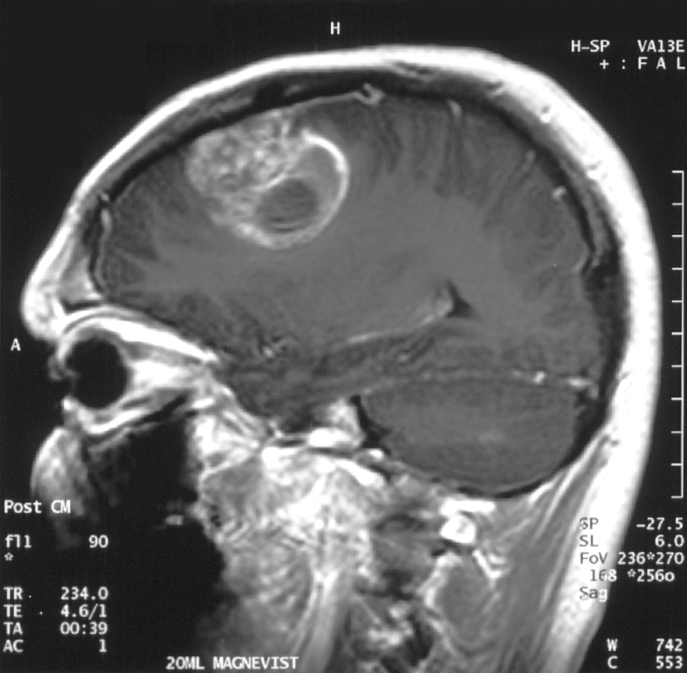
An interdisciplinary team of researchers from the University of Notre Dame is investigating the mechanisms of neuron death caused by chronic compression – such as the pressure exerted by a brain tumour – to better understand how to prevent neuron loss.

Published in the Proceedings of the National Academy of Sciences, their study found that chronic compression triggers neuron death by a variety of mechanisms, both directly and indirectly. The research is helping lay the groundwork for identifying therapies to prevent indirect neuron death.

“The impetus for this project was to figure out those underlying mechanisms. In cancer research, most researchers are focused on the tumour itself, but in the meantime, while the tumour is sitting there and growing, it’s damaging the organ that it’s living in,” said Meenal Datta, the Jane Scoelch DeFlorio Collegiate Professor of Aerospace and Mechanical Engineering at Notre Dame and co-lead author of the study. “We fully believe that these growth-induced mechanical forces of the tumor as it expands is part of the reason we see damage in the brain.”

As an engineer who leads the TIME Lab, Datta studies the mechanics of tumors and the microenvironment, specifically for glioblastoma, an incurable brain cancer. She had found in prior work that tumors damage the surrounding brain. But to understand the mechanisms by which tumors kill neurons from compression alone, Datta needed a “hardcore neuroscientist.”

Neurons captured on screen for research experiment.
Imaging of neurons from an experiment with the control group neurons on the left and the neurons impact by chronic compression on the right. (Provided by the Patzke lab.)

That neuroscientist is Christopher Patzke, the John M. and Mary Jo Boler Assistant Professor in the Department of Biological Sciences at Notre Dame and co-lead author of the study. Patzke utilises induced pluripotent stem cells (iPSCs), which are either obtained from external sources or generated directly in his lab. These cells function like embryonic stem cells and can be differentiated or changed in the lab into any cell type in the body, including neurons.

For this study, iPSCs were used to create neural cells and develop a model system of neurons and glial cells that behave as a neuronal network would in the brain. Researchers grew the cells and then applied pressure to the system to mimic the chronic compression of a glioblastoma tumour.

After compressing the cells, graduate students Maksym Zarodniuk and Anna Wenninger, from Datta and Patzke’s labs respectively, compared how many neurons and glial cells died versus lived.

“For the neurons that are still alive, many of them have this programmed self-destruction signaling activated,” Patzke said. “We wanted to understand which molecular pathway was responsible for this; is there a way to save neurons from going down the drain to this cell death mechanism?”

By sequencing and analysing all messenger RNA from the living neuronal and glial cells, the researchers found an increase in HIF-1 molecules, signalling for stress adaptive genes to improve cell survival, which leads to inflammation in the brain. The compression also triggered AP-1 gene expression, a type of neuroinflammatory response.

Both neurological reactions are indicators that neuronal damage and death is underway.

An analysis of data from the Ivy Glioblastoma Atlas Project shows that glioblastoma patients also reflect these compressive stress patterns and gene expression changes as well as synaptic dysfunction in line with the experiment’s results. The researchers confirmed these results by mimicking force via a live compression system applied to preclinical models of brains.

Overall, the findings may help explain why glioblastoma patients experience cognitive impairments, motor deficits and elevated seizure risk. Additionally, the signalling pathways offer opportunities for researchers to explore as drug targets to reduce neuronal death.

“Our approach to this study was disease agnostic, so our research could potentially extend to other brain pathologies that affect mechanical forces in the brain such as traumatic brain injury,” Datta said. “I’m all in on mechanics. Whatever it is that you’re interested in when it comes to cancer, above your question of interest, mechanics is sitting there and many don’t even know they should be considering it.”

The mechanics of compression and its effect on neuron loss is key for future research.

“Understanding why neurons are so vulnerable and die upon compression is critical to prevent excessive sensory loss, motor impairment and cognitive decline,” Patzke said. “This is how we will help patients.”

Source: University of Notre Dame

New Neural Maps Challenge Traditional Descriptions of the Brain

AI image of neurons created by Gencraft

For more than a century, maps of the brain have been based on how brain tissue looks under the microscope. These anatomical maps divide the brain into regions according to structural variations in the tissue. But do these divisions really reflect how the brain works? A new study on mice from Karolinska Institutet, published in Nature Neuroscience, suggests that this is often not the case.

By describing the brain in terms of electrical activity of its neurons, the researchers have found a new way to understand the functional organisation of the prefrontal cortex, the brain region responsible for planning, decision-making, and other advanced cognitive functions. 

“Considering that deviations in prefrontal cortex function have been linked to virtually all psychiatric disorders, it is surprising how little is known about how this region actually works,” says Marie Carlén, Professor at the Department of Neuroscience at Karolinska Institutet.

Did not align with previous maps

Her research group recorded and analysed the activity of more than 24 000 neurons in awake mice and created the first activity-based maps of the prefrontal cortex. The maps of spontaneous and cognition-related neuron activity did not match the traditional, tissue-based maps.

“Our findings challenge the traditional way of defining brain regions and have major implications for understanding brain organisation overall,” says Marie Carlén.

The researchers found that the activity patterns of neurons reflected the hierarchy of information flow in the brain rather than the structure of the tissue. Neurons with slow, regular activity turned out to be characteristic of the prefrontal cortex, which sits at the top of this hierarchy. The same activity pattern also marked regions at the top of the prefrontal cortex’s own internal hierarchy. Slow, regular activity is thought to characterise the integration of information flows, a process that is central to cognitive functions such as planning and reasoning. 

Different neuronal activity patterns work together

Carlén and her colleagues discovered that neurons involved in decision-making were concentrated in regions high up in the prefrontal hierarchy. Surprisingly, these neurons were characterised by very fast activity patterns. 

“This suggests that cognitive processes rely on local collaboration between neurons whose activity patterns complement one another. Some neurons appear to specialise in integrating information streams, while others have high spontaneous activity that supports quick and flexible encoding of information, for instance, information needed to make a specific decision,” says Marie Carlén.”

Source: Karolinska Institutet

New Research Confirms that Neurons Form in the Adult Brain

A healthy neuron. Credit: NIH

A study in the journal Science presents compelling new evidence that neurons in the brain’s memory centre, the hippocampus, continue to form well into late adulthood. The research from Karolinska Institutet provides answers to a fundamental and long-debated question about the human brain’s adaptability.

The hippocampus is a brain region that is essential for learning and memory and involved in emotion regulation. Back in 2013, Jonas Frisén’s research group at Karolinska Institutet showed in a high-profile study that new neurons can form in the hippocampus of adult humans. The researchers then measured carbon-14 levels in DNA from brain tissue, which made it possible to determine when the cells were formed.

Identifying cells of origin

However, the extent and significance of this formation of new neurons (neurogenesis) are still debated. There has been no clear evidence that the cells that precede new neurons, known as neural progenitor cells, actually exist and divide in adult humans.

“We have now been able to identify these cells of origin, which confirms that there is an ongoing formation of neurons in the hippocampus of the adult brain,” says lead researcher Jonas Frisén, professor of stem cell research at the Department of Cell and Molecular Biology.

In the new study, the researchers combined several advanced methods to examine brain tissue from people aged 0 to 78 years from several international biobanks. They used a method called single-nucleus RNA sequencing, which analyses gene activity in individual cell nuclei, and flow cytometry to study cell properties. 

By combining this with machine learning, they were able to identify different stages of neuronal development, from stem cells to immature neurons, many of which were in the division phase.

To localise these cells, the researchers used two techniques that show where in the tissue different genes are active: RNAscope and Xenium. These methods confirmed that the newly formed cells were located in a specific area of the hippocampus called the dentate gyrus. This area is important for memory formation, learning and cognitive flexibility.

Hope for new treatments

The results show that the progenitors of adult neurons are similar to those of mice, pigs and monkeys, but that there are some differences in which genes are active. There were also large variations between individuals – some adult humans had many neural progenitor cells, others hardly any at all.

“This gives us an important piece of the puzzle in understanding how the human brain works and changes during life,” explains Jonas Frisén. “Our research may also have implications for the development of regenerative treatments that stimulate neurogenesis in neurodegenerative and psychiatric disorders.” 

Source: Karolinska Institutet

These Newly Discovered Brain Cells Enable us to Remember Objects

Discovery of ‘ovoid cells’ reshapes our understanding of how memory works, and could open the door to new treatments for Alzheimer’s disease, epilepsy and more.

Ovoid cells. Photo credit: Dr. Mark Cembrowski

Take a look around your home and you’ll find yourself surrounded by familiar comforts – photos of family and friends on the wall, well-worn tekkies by the door, a shelf adorned with travel mementos.

Objects like these are etched into our memory, shaping who we are and helping us navigate environments and daily life with ease. But how do these memories form? And what if we could stop them from slipping away under a devastating condition like Alzheimer’s disease?

Scientists at the University of British Columbia have just uncovered a crucial piece of the puzzle. In a study published in Nature Communications, the researchers have discovered a new type of brain cell that plays a central role in our ability to remember and recognise objects. 

Called ‘ovoid cells,’ these highly-specialised neurons activate each time we encounter something new, triggering a process that stores those objects in memory and allowing us to recognise them months, even years, later.

“Object recognition memory is central to our identity and how we interact with the world,” said Dr Mark Cembrowski, the study’s senior author, and an associate professor of cellular and physiological sciences at UBC and investigator at the Djavad Mowafaghian Centre for Brain Health. “Knowing if an object is familiar or new can determine everything from survival to day-to-day functioning, and has huge implications for memory-related diseases and disorders.”

Hiding in plain sight

Named for the distinct egg-like shape of their cell body, ovoid cells are present in relatively small numbers within the hippocampus of humans, mice and other animals.

Adrienne Kinman, a PhD student in Dr Cembrowski’s lab and the study’s lead author, discovered the cells’ unique properties while analysing a mouse brain sample, when she noticed a small cluster of neurons with highly distinctive gene expression.

“They were hiding right there in plain sight,” said Kinman. “And with further analysis, we saw that they are quite distinct from other neurons at a cellular and functional level, and in terms of their neural circuitry.”

To understand the role ovoid cells play, Kinman manipulated the cells in mice so they would glow when active inside the brain. The team then used a miniature single-photon microscope to observe the cells as the mice interacted with their environment.

The ovoid cells lit up when the mice encountered an unfamiliar object, but as they grew used to it, the cells stopped responding. In other words, the cells had done their jobs: the mice now remembered the objects.

“What’s remarkable is how vividly these cells react when exposed to something new. It’s rare to witness such a clear link between cell activity and behaviour,” said Kinman. “And in mice, the cells can remember a single encounter with an object for months, which is an extraordinary level of sustained memory for these animals.”

New insights for Alzheimer’s disease, epilepsy

The researchers are now investigating the role that ovoid cells play in a range of brain disorders. The team’s hypothesis is that when the cells become dysregulated, either too active or not active enough, they could be driving the symptoms of conditions like Alzheimer’s disease and epilepsy.

“Recognition memory is one of the hallmarks of Alzheimer’s disease – you forget what keys are, or that photo of a person you love. What if we could manipulate these cells to prevent or reverse that?” said Kinman. “And with epilepsy, we’re seeing that ovoid cells are hyperexcitable and could be playing a role in seizure initiation and propagation, making them a promising target for novel treatments.”

For Dr Cembrowski, discovering the highly specialised neuron upends decades of conventional thinking that the hippocampus contained only a single type of cell that controlled multiple aspects of memory.

“From a fundamental neuroscience perspective, it really transforms our understanding of how memory works,” he said. “It opens the door to the idea that there may be other undiscovered neuron types within the brain, each with specialised roles in learning, memory and cognition. That creates a world of possibilities that would completely reshape how we approach and treat brain health and disease.”

Source: University of British Columbia

Glioma Cells can Also Fire off Electrical Signals in the Brain

Source: Pixabay

Researchers at Baylor College of Medicine and the Jan and Dan Duncan Neurological Research Institute at Texas Children’s Hospital have uncovered a new cell type in human brain cancers. Their study, published in Cancer Cell, reveals that a third of the cells in glioma, fire electrical impulses. Interestingly, the impulses, also called action potentials, originate from tumour cells that are part neuron and part glia, supporting the groundbreaking idea that neurons are not the only cells that can generate electric signals in the brain.

The scientists also discovered that cells with hybrid neuron-glia characteristics are present in the non-tumour human brain. The findings highlight the importance of further studying the role of these newly identified cells in both glioma and normal brain function.

“Previous studies have shown that patient survival outcomes are associated with tumour proliferation and invasiveness, which are influenced by tumour intrinsic and extrinsic factors, including communication between tumour cells and neurons that reside in the brain,” said Dr Benjamin Deneen, professor in the Department of Neurosurgery at Baylor.

Researchers have previously described that glioma and surrounding healthy neurons connect with each other and that neurons communicate with tumours in ways that drive tumour growth and invasiveness. 

“We have known for some time now that tumour cells and neurons interact directly,” said first author Dr Rachel N. Curry, postdoctoral fellow in paediatrics – neuro oncology at Baylor, who was responsible for conceptualising the project. “But one question that always lingered in my mind was, ‘Are cancer cells electrically active?’ To answer this question correctly, we required human samples directly from the operating room. This ensured the biology of the cells as they would exist in the brain was preserved as much as possible.”

To study the ability of glioma cells to spike electrical signals and identify the cells that produce the signals, the team used Patch-sequencing, a combination of techniques that integrates whole-cell electrophysiological recordings to measure spiking signals with single-cell RNA-sequencing and analysis of the cellular structure to identify the type of cells.

The electrophysiology experiments were conducted by research associate and co-first author Dr Qianqian Ma in the lab of co-corresponding author associate professor of neuroscience Dr Xiaolong Jiang. This innovative approach has not been used before to study human brain tumour cells. “We were truly surprised to find these tumour cells had a unique combination of morphological and electrophysiological properties,” Ma said. “We had never seen anything like this in the mammalian brain before.”

“We conducted all these analyses on single cells. We analysed their individual electrophysiological activity. We extracted each cell’s content and sequenced the RNA to identify the genes that were active in the cell, which tells us what type of cell it is,” Deneen said. “We also stained each cell with dyes that would visualise its structural features.”

Integrating this vast amount of individual data required the researchers to develop a novel way to analyse it.

“To define the spiking cells and determine their identity, we developed a computational tool – Single Cell Rule Association Mining (SCRAM) – to annotate each cell individually,” said co-corresponding author, Dr Akdes Serin Harmanci, assistant professor of neurosurgery at Baylor.

“Finding that so many glioma cells are electrically active was a surprise because it goes against a strongly held concept in neuroscience that states that, of all the different types of cells in the brain, neurons are the only ones that fire electric impulses,” Curry said. “Others have proposed that some glia cells known as oligodendrocyte precursor cells (OPCs) may fire electrical impulses in the rodent brain, but confirming this in humans had proven a difficult task. Our findings show that human cells other than neurons can fire electrical impulses. Since there is an estimated 100 million of these OPCs in the adult brain, the electrical contributions of these cells should be further studied.”

“Moreover, the comprehensive data analyses revealed that the spiking hybrid cells in glioma tumours had properties of both neurons and OPC cells,” Harmanci said. “Interestingly, we found non-tumour cells that are neuron-glia hybrids, suggesting that this hybrid population not only plays a role in glioma growth but also contributes to healthy brain function.”

“The findings also suggest that the proportion of spiking hybrid cells in glioma may have a prognostic value,” said co-corresponding author Dr Ganesh Rao, professor and chair of neurosurgery at Baylor. “The data shows that the more of these spiking hybrid glioma cells a patient has, the better the survival outcome. This information is of great value to patients and their doctors.”

“This work is the result of extensive equal collaboration across multiple disciplines – neurosurgery, bioinformatics, neuroscience and cancer modelling – disciplines strongly supported by state-of-the-art groups at Baylor,” Deneen said. “The results offer an enhanced understanding of glioma tumours and normal brain function, a sophisticated bioinformatics pipeline to analyse complex cellular populations and potential prognostic implications for patients with this devastating disease.”

Source: Baylor College of Medicine