Memory cells in the nose slow the influenza virus as soon as it enters the body. They reduce viral levels and may help protect against more severe illness. A new study from the University of Gothenburg may help guide the development of better influenza vaccines.
Today’s influenza vaccines are given as injections in the arm and mainly stimulate immune responses in the blood. At the same time, researchers are working to develop influenza vaccines that can be administered through the nose – an effort this study helps inform. The goal is to strengthen the body’s defences where the virus first encounters the immune system.
Memory cells remain in the nose
The researchers identified a group of memory cells, known as CD4 memory T cells, that remain in nasal tissue after an influenza infection. When the body encounters the virus again, these cells can rapidly reactivate and help other parts of the immune system fight the infection. The study shows that these cells can reduce viral replication in the nose and thereby contribute to better protection against illness.
“We show that CD4 memory T cells can remain in nasal tissue after an influenza infection and rapidly reactivate when the virus returns. This means the immune system can respond directly at the site where the virus first enters the body,” says Nimitha R. Mathew, a researcher at the Sahlgrenska Academy, University of Gothenburg, and one of the study’s lead authors.
In studies in mice, the researchers showed that these immune cells help limit viral levels and reduce tissue damage in the nose during a subsequent infection.
Similar cells found in humans
The researchers also analysed cells from the nasal mucosa of healthy adults. There, they found the same type of influenza-specific memory cells, suggesting that a similar local immune defence may also exist in humans. The study is published in the Journal of Experimental Medicine.
“Many people likely already have these kinds of memory cells in their noses after previous infections, but they are not always enough to stop the virus completely. The important thing about our findings is that we now know which immune cells can limit the virus where infection begins. That knowledge can be used when developing future nasal vaccines,” says Davide Angeletti, professor at the Sahlgrenska Academy, University of Gothenburg, and also one of the study’s lead authors.
Article: Nasal CD4⁺ tissue resident memory T cells provide cross protective immunity to influenza; 10.1084/jem.20251793
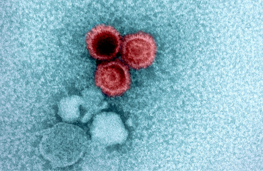
An electron micrograph showing three Epstein-Barr virions in red. Image: NIAID
Some viruses are cleared by the immune system within days, while others lurk in our bodies for a lifetime and reemerge later to cause new problems. How and why viral levels in the body change over time – and the health impacts of these changes – are only just starting to become clear.
A team led by scientists at Harvard Medical School, Massachusetts General Hospital, Brigham and Women’s Hospital, and the Broad Institute of MIT and Harvard recently reported the largest analysis to date of the human DNA virome – the collection of viruses in the body that have DNA as their genetic material.
The researchers tracked the viral load – the amount of viral DNA – for several common viruses in blood and saliva from over 900 000 individuals. They saw large variations in viral load from person to person depending on age, sex, lifestyle, and other factors, and discovered dozens of genetic factors strongly associated with viral load.
The team concluded that genetics plays a role in determining whether the effects of these viruses extend well beyond an initial infection.
“We’re getting to the point now where we can use human genetics to try to answer fundamental questions about pathology resulting from viruses,” including whether a virus is likely to play a role in causing cancer or other diseases later in life, said first author Nolan Kamitaki, research fellow in genetics in the Blavatnik Institute at HMS.
Study co-senior author Po-Ru Loh, HMS associate professor of medicine at Brigham and Women’s and an associate member at the Broad Institute, is broadly interested in the scientific information that can be mined from population-level datasets of DNA sequences that are readily available to researchers.
It turns out that these datasets capture information about the genomic material that people inherit, he said, as well as the makeup of their oral microbiomes, viruses hiding in their bodies, and acquired mutations in their DNA. Sometimes, scientists can link this information to downstream health consequences.
Recently, for example, Loh and Kamitaki analyzed whole-genome sequences from saliva samples collected from more than 12,500 individuals to investigate how genetics shapes the oral microbiome.
In their new study, the researchers analyzed whole-genome sequencing data from individuals in three biobanks: the UK Biobank, the National Institutes of Health’s All of Us Research Program, and Simmons Foundation Powering Autism Research for Knowledge. They tested blood and saliva samples for the viral loads of Epstein-Barr virus, two other human herpesviruses (HHV-6 and HHV-7), Merkel cell polyomavirus, as well as three common anelloviruses that are present in about 90% of people throughout life without causing disease.
They found that each virus had a markedly different trajectory over a lifetime. The viruses appeared most rapidly during the first several years of life, likely following primary infection. However, Epstein-Barr virus became more prevalent with age, while HHV-6 became less prevalent after childhood, possibly indicating more control by the immune system over time. The prevalence of HHV-7 similarly decreased sharply in middle age.
The team also found that Epstein-Barr viral load went up in the winter and down in the summer, while HHV-7 viral load showed the opposite pattern. Smoking was strongly associated with a higher Epstein-Barr viral load, nearly double in heavy smokers compared with nonsmokers, whereas smoking was associated with a lower HHV-7 viral load.
Notably, men consistently had a higher viral load in their blood and saliva than women across all seven viruses.
The role of genetics revealed
The researchers determined that many of the genetic factors most strongly linked to viral load were related to how the immune system responds to viruses and how infected cells dodge immune attacks.
Thus, the findings highlight the immune system’s role in controlling viral load in the body, showing how much immune responses can vary over time and between people.
“It’s amazing how much DNA can teach us about dynamic biological processes and the ways in which our habits, our genes, and our biology shape those processes,” said co-senior author Steven McCarroll, the Dorothy and Milton Flier Professor of Biomedical Science and Genetics at HMS and director of genomic neurobiology for the Stanley Center for Psychiatric Research at the Broad Institute.
The researchers identified the largest number of genetic associations for Epstein-Barr virus – which is thought to be a leading cause of multiple sclerosis and a risk factor for certain cancers – so they dug a little deeper. Their analysis revealed that the lifetime viral load for Epstein-Barr didn’t influence the risk of developing multiple sclerosis. Instead, the body’s immune response to Epstein-Barr is likely what increases the risk of the disease.
The researchers did find evidence that high Epstein-Barr viral load is a casual risk factor for Hodgkin lymphoma, a finding that needs further study in cell and animal models in the lab, they said.
“This finding is an example of why virus research in large genetic biobanks is important,” Kamitaki said.
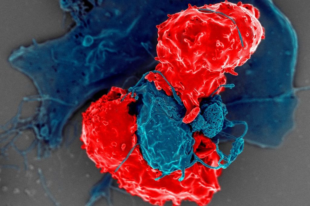
Scanning electron microscope image of T regulatory cells (red) interacting with antigen-presenting cells (blue). T regulatory cells can suppress responses by T cells to maintain homeostasis in the immune system. Credit: National Institute of Allergy and Infectious Diseases/NIH
Cedars-Sinai Health Sciences University investigators have identified for the first time a protein’s role as a “dimmer switch” that can calm an overactive immune system and restrain harmful inflammation. The protein, Butyrophilin 2A2 (BTN2A2), interacts with a key molecule that controls the strength of T-cell responses.
The findings, published inNature Communications, define a unique pathway that helps balance immune activity and could be harnessed to limit damage caused by a variety of autoimmune diseases.
In laboratory mice, loss of BTN2A2 led to exaggerated immune reactions and an increase in damaging kidney inflammation called glomerulonephritis. Treatment with BTN2A2 reduced disease severity by increasing immune-regulating T cells and lowering inflammation.
Supporting laboratory experiments in human T-cells demonstrated similar immune-calming effects.
“Glomerulonephritis remains a leading cause of chronic kidney disease and kidney failure worldwide, with limited treatment options,” said Ananth Karumanchi, MD, co-corresponding author of the study and director of the Renovascular Research Center at Cedars-Sinai. “Our findings provide a strong foundation for future studies aimed at modifying immune-driven kidney disease rather than simply managing its symptoms. The pathway could also be targeted in a range of autoimmune and inflammatory diseases including rheumatoid arthritis, multiple sclerosis, inflammatory bowel disease, and transplant rejections.”
Other Cedars-Sinai authors include Shafat Ali, Anders H. Berg, Michifumi Yamashita, Ambart E. Covarrubias, Jordan Mundell, Pranali N. Shah, Ruan Zhang, Vincent Dupont, Bong-Ha Shin, Shen Yang, Madhusudhanarao Katiki, Ramachandran Murali, Margareta D. Pisarska, Ravi Thadhani, Peter S. Heeger and Stanley C. Jordan
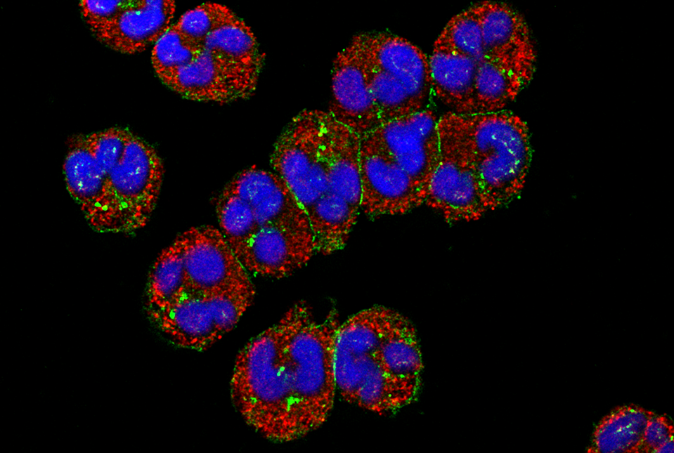
Human neutrophils visualised under a confocal microscope with cell membrane (red) and nucleus (blue). When faced with an infection during food scarcity, stress hormones trigger an immune response dependent on neutrophils, abundant cells that act as immediate, short-lived defenders. Credit: Thai Tran, National Institute of Arthritis and Musculoskeletal and Skin Diseases
When food is scarce, stress hormones direct the immune system to operate in “low power” mode to preserve immune function while conserving energy, according to researchers at Weill Cornell Medicine. This reconfiguration is crucial to combating infections amid food insecurity.
The answer could be important in helping those who are food insecure and face the risk of infectious diseases every day. “Mounting an immune response against infections requires a lot of energy. We have discovered a coordinated system that upholds immune function by shifting the composition and metabolism of immune cells,” Dr Collins said.
The results, published in Immunity, found that mice on a calorie-restricted diet fought off infection as well as mice that were fully fed, but did so while using very little glucose. This was possible thanks to glucocorticoids, stress hormones known for their role in regulating blood glucose. The researchers determined that glucocorticoids acted like master conductors, reorganizing immune cells and their energy usage to provide a survival advantage.
The research was co-led by Luisa Menezes-Silva, a visiting graduate student from the University of São Paulo, Brazil; Dr Mingeum Jeong, a postdoctoral associate; and Dr Seong-Ji Han, a research associate, all in the Collins lab at Weill Cornell.
Shifting Priorities
To understand the complex interactions involved in an immune response during scarcity, Dr Collins and his team put mice on a 50% restricted-calorie diet and then exposed the animals to bacteria that infect the gut. The mice that were fed a standard diet experienced a metabolic crash – their blood glucose levels and body weight plummeted.
The researchers had expected this would happen to all the animals because mounting an immune response can consume up to 30% of the entire body’s fuel reserves. But in the calorie-restricted mice, the immune system appeared to be functioning perfectly well without using much glucose.
To unravel this enigma, the researchers inventoried the immune cells of the infected animals and discovered that T cells, which normally target invading microbes, were depleted in the calorie-restricted mice. Instead, short-lived neutrophils, which serve as the body’s first responders to infection, were ramped up to twice the normal amount and had measurably enhanced pathogen-killing abilities. The cells seemed to be operating in energy-saving mode, consuming much less glucose than neutrophils from well-fed animals.
“So, this hormone rewires the immune system to eliminate the infection while keeping blood sugar from dropping, which rescues the calorie-restricted animals from malnutrition,” said Dr Collins.
Stress Hormones Lead the Charge
The researchers are breaking new ground by outlining how a sudden fall in food intake triggers glucocorticoid levels to rise, resulting in two major shifts. First, the body repositions certain immune cells – especially naïve T cells – into the bone marrow, which becomes a kind of “safe house” for when the cells are needed. Second, during an infection, glucocorticoids tilt the immune response away from energy‑intensive T cells toward neutrophils, abundant cells that act as immediate, short-lived defenders.
Beyond clearing a current infection, glucocorticoids prepare the immune system for repeat encounters with infectious agents. While the hormones direct killer T cells to stand down and neutrophils to step up, they also ensure memory T cells are preserved for future confrontations.
“Glucocorticoids reduce the immune cells that use up the most energy, while saving those that are critical for protection against future infections,” Dr Collins said. “So, these hormones are involved in every step of the infection-fighting process.”
“Since glucocorticoids are induced not only by nutrient restriction but also by any form of stress, our findings might have broader applicability,” said Dr Collins.
In the meantime, he and his team plan to explore what causes the system to fail when the degree and duration of calorie restriction are more severe. “We looked at reduced food intake over three weeks,” he said. “But when you cross the threshold into malnutrition, the whole system breaks down.” Understanding this collapse could inform better strategies to prevent infectious disease and infection-driven malnutrition in vulnerable populations.
Anxiety and insomnia have been shown to weaken the immune system and make us more prone to disease. Now, researchers found that this may be because experiencing symptoms of either can reduce the number of natural killer cells, our bodies’ machinery for defence. Their findings showed that in young women who experience insomnia symptoms, the number of total NK cells was lower. If they experienced anxiety symptoms, the number of NK cells that circulate through the body was lower. These findings could inform the development of novel strategies to raise awareness about the physiological consequences of anxiety and insomnia and help in the prevention of immune-related disorders and cancers, the team said.
Natural killer (NK) cells are the bodyguards of our immune system. As a first line of defense, they destroy invading pathogens, foreign bodies, and infected cells in early stages, thereby preventing them from spreading. NK cells can circulate within the blood stream (circulatory) or reside in tissue and organs. Having too few NK cells can lead to immune system dysfunction and increase susceptibility to disease.
Anxiety disorder and insomnia are two conditions that can disrupt the normal functioning of the immune system. Given these disorders are on the rise, researchers in Saudi Arabia have now examined the association between anxiety, insomnia, and NK cells in young, female students. They published their results in Frontiers in Immunology.
“We found that in students with insomnia symptoms, count and percentage of total NK cells and their sub-populations were declined,” said first author Dr Renad Alhamawi, an assistant professor of immunology and immunotherapy at Taibah University. “Students with general anxiety symptoms, on the other hand, had a lower percentage and number of circulatory NK cells and their sub-populations, compared to symptom-free students.”
Decimated defence
60 female students, aged between 17 and 23 years old, participated in the study. They filled out three questionnaires about sociodemographics as well as anxiety and insomnia symptoms. The symptoms of the latter two were self-reported. The surveys showed that around 53% of the participants reported sleeping disturbance suggestive of insomnia, and 75% reported anxiety symptoms, with around 17% and 13% reporting moderate and severe symptoms, respectively.
Participants also provided blood samples through which percentages of NK cells and their subtypes were determined. NK cells have two subtypes: CD16+CD56dim cells make up the majority of NK cells in the nervous system that connects the central nervous system to the rest of the body (peripheral NK cells). Cells belonging to this subtype also exhibit cytotoxicity, which means they can damage or kills cells that invade the body. The other subtype, CD16+CD56high cells, are less frequent and involved in the production of proteins that function as chemical messengers and in immunoregulation. Both subtypes are circulatory NK cells.
The results showed that students with anxiety symptoms had a lower percentage and number of circulatory NK cells and their sub-populations, compared to students who did not report symptoms. Severity of symptoms also played a role as students with moderate and severe anxiety symptoms had a significant lower percentage of circulatory NK cells compared to students without them. Among students with minimal or mild anxiety symptoms, only a statistically insignificant decline in NK cell percentage was observed. In students with insomnia symptoms, higher anxiety scores were negatively associated with the proportion of total peripheral NK cells.
A reduction of these cells can lead to the impairment of the immune system, which may result in diseases, cancers, and mental disease, including depression. “Understanding how these psychological stressors influence the distribution and activity of immune cells, especially peripheral NK cells, may provide valuable insights into the mechanisms underlying inflammation and tumorigenesis,” Alhamawi explained.
The study is limited in some respects, the team pointed out. It only included young females – the group amongst whom anxiety and sleeping disorders have been rising disproportionally, limiting the generalizability of the results. The researchers said that future studies that include different age groups, sexes, and people from different regions, are necessary to gain a better overall view of the hidden effects of anxiety and insomnia on the proportion and function of these immune cells.
Previous studies have suggested healthy lifestyles with regular physical activity, stress reduction, and a healthy and balanced diet can boost the number and function of NK cells. However, the impact of anxiety and insomnia can disrupt the normal functioning of various body systems, including the immune system, thereby contributing to the development of chronic and inflammatory diseases. “Such impacts ultimately compromise overall health and quality of life,” concluded Alhamawi.
Metabolic changes that “rewire” part of the immune system can intensify sepsis, the body’s dysregulated response to infection. This discovery may lead to new ways to block metabolic changes contributing to excessive and ineffective inflammation, reset the immune system, and bring sepsis under control, researchers at Vanderbilt Health reported January 15 in the journal Nature Immunology.
“Metabolism is potentially a means by which we could intervene in immune dysfunction in ICU (intensive care unit) patients including those with sepsis,” said the paper’s first and co-corresponding author, Matthew Stier, MD, PhD, assistant professor of Medicine in the Division of Allergy, Pulmonary and Critical Care Medicine at Vanderbilt Health.
“I think we can make great progress and great strides by aligning cutting-edge basic science tools with ICU patient samples to understand these mechanisms and prioritise therapies for future interventions,” he said.
Sepsis is characterised by the massive production and release of inflammatory molecules, including cytokines, that if unchecked, can lead to tissue damage, septic shock, organ failure, and death.
Despite decades of research focused on stopping this “cytokine storm” and hyperinflammation, “we have unfortunately not been able to identify successful drug therapies in sepsis,” said Stier, a physician-scientist who focuses on immunologic and metabolic dysfunction in critical illness. Targeting the inflammatory aspect of sepsis is likely important, but by itself may not be sufficient.
“We provide antibiotics and great supportive care to weather the cytokine storm,” he said, “but that doesn’t fully resolve the problem. It keeps people alive while we wait for their bodies to fix themselves — or not.”
In critical illness, including sepsis, the body’s normal metabolic processes become impaired. This includes immunometabolism, the energy-generating processes that fuel the immune system.
At the same time, the immune system’s protective functions become exhausted, resulting in an acquired immunosuppression, which leaves patients vulnerable to secondary infections, persistent organ dysfunction, repeated hospitalisations and death.
While prior research has defined the characteristics of metabolism and immune dysfunction, this study was among the first to explore the mechanisms of immunometabolic dysfunction in sepsis and their association with immunosuppression, often called “immunoparalysis.”
Stier and his colleagues used cutting-edge technologies, including single-cell sequencing and flow cytometry, to study immune cells collected from the blood of critically ill patients.
The blood samples were collected and stored through the Sepsis Clinical Resource and Biorepository (SCARAB), a unique, highly collaborative ICU biobank developed by Julie Bastarache, MD, and Lorraine Ware, MD, professors of Medicine in the Division of Allergy, Pulmonary and Critical Care Medicine.
Two of the most important elements of the body’s immune responses are CD4+ T “helper” cells, inflammatory “foot soldiers” of the immune system that are distinguished by the CD4 surface protein they express, and regulatory T (Treg or “suppressor”) cells, which guard against over-active immune responses.
To study the impact of critical illness and sepsis on these cells, the Vanderbilt Health team used SCENITH, a flow cytometry-based method developed by French researchers that enables researchers to functionally profile energy metabolism with single-cell resolution.
“This technique allowed us to do something prior studies hadn’t done … to look at the metabolism of every single cell set on its own and identify subset-specific metabolic adaptations,” Stier said.
The key finding: Treg cells undergo metabolic “reprogramming” in patients with critical illness and sepsis, leading to altered tryptophan metabolism and response to reactive oxygen species in a way that enhances their immunosuppressive capability, at the expense of CD4+ T “helper” cells.
“The metabolic turmoil of critical illness appears to give Treg cells a survival and functional advantage, contributing to the harmful immunoparalysis seen in sepsis,” he said.
“This is very much a preclinical paper,” Stier cautioned. Yet it demonstrates the feasibility of deeply dissecting immunometabolic mechanisms using ICU patient biospecimens and highlights the importance of such insights to prioritise future therapeutic targets in critical illness and sepsis, he said.
An international research group directed by UMC Utrecht have developed and characterised two first-in-class antibodies that specifically block the high-affinity IgG receptor FcγRI. Their findings open new perspectives for therapeutic modulation of FcγRI-driven inflammation in autoimmune diseases such as rheumatoid arthritis (RA), systemic lupus erythematosus (SLE) and immune thrombocytopenia (ITP).
FcγRI, also known as CD64, is a high-affinity receptor on myeloid cells that binds to the Fc region of immunoglobulin G (IgG) antibodies. It plays a key role in immune defence by triggering cellular functions such as phagocytosis and cytokine production. In a normal immune response, FcγRI is activated by immune complexes – clusters of antibodies bound to pathogens, which mark them for clearance. In autoimmune diseases, however, the immune system mistakenly targets the body’s own tissues (such as joint proteins in RA, nuclear antigens in SLE, or platelets in ITP), which results in the production of autoantibodies that form immune complexes. These misdirected complexes activate FcγRI unnecessarily, driving chronic inflammation and subsequent tissue damage.
The study, led by Prof Jeanette Leusen, PhD from the Antibody Therapy research group at the Center for Translational Immunology (UMC Utrecht) and carried out by PhD candidate Tosca Holtrop MSc, was a true team effort, in collaboration with experts from Kiel University (Germany), Leiden University Medical Center, Utrecht University, and Friedrich-Alexander University Erlangen-Nürnberg (Germany).
Discovery of first-in-class antibodies
For over 30 years, scientists have tried to generate antibodies against the IgG-binding domain of CD64, but the receptor’s extremely high affinity for IgG made this impossible with earlier technologies. Combining the UMAB unique immunisation method with novel phage display antibody libraries, the team bypassed this challenge by excluding the Fc region of the antibodies. This allowed them to discover two unique Fc-silent antibodies, C01 and C04, that bind purely via their Fab domains to FcγRI. Crystal structural analysis confirmed that C01 binds precisely within the IgG-binding site on EC2, making binding mutually exclusive.
High affinity for FcγRI
Quantitative binding studies revealed that both antibodies have higher affinity for FcγRI than human IgG, allowing them to efficiently displace IgG or pathogenic immune complexes up to 60 percent and block binding up to 90 percent. Importantly, neither antibody triggered FcγRI activation, a critical distinction from earlier anti-FcγRI antibodies, which could inadvertently trigger receptor clustering and cytokine release.
Subsequently. an in vitro model for immune thrombocytopenia was set-up, where C01 and C04 effectively inhibited opsonized platelets from binding to immune cells from ITP patients. In a preclinical in vivo ITP model, the antibodies significantly reduced IgG-dependent platelet depletion. The antibodies were also tested in an in vitro rheumatoid arthritis models, where they effectively inhibited patient-derived autoantibody–immune complex binding to monocytes, macrophages, and neutrophils from healthy donors.
Promising therapeutic candidate drugs
The study demonstrates that direct Fab-mediated inhibition of FcγRI is feasible and effective, opening a new avenue for treating autoimmune diseases characterised by IgG-autoantibody complexes. By preventing immune complex–driven activation without triggering the receptor, C01 and C04 represent a promising next step toward targeted, inflammation-sparing immunotherapy. “I think we found the needle in the haystack, after searching over a decade and thanks to a true team effort,” says Jeanette Leusen, principal investigator of the study. “Each research partner contributed a critical piece, from antibody discovery and structure determination to patient sample testing and preclinical models. Only together could we bring this to fruition. These antibodies not only provide a unique tool for studying FcγRI biology, but also hold promise as therapeutic candidates in autoimmune and infectious diseases.”
A woman with Systemic Lupus Erythematosus. Source: Wikimedia CC0
Epstein-Barr virus (EBV) one, of humanity’s most ubiquitous infectious pathogens, is to blame for systemic lupus erythematosus Stanford Medicine investigators and their colleagues have found.
The Epstein-Barr virus (EBV), which resides silently inside 95% of the world’s population, is directly responsible for making a minuscule number of immune cells go rogue and persuade far more of their fellow immune cells to launch a widespread assault on the body’s tissues, the scientists have shown.
“This is the single most impactful finding to emerge from my lab in my entire career,” said William Robinson, MD, PhD, a professor of immunology and rheumatology and the study’s senior author. “We think it applies to 100% of lupus cases.”
The study’s lead author is Shady Younis, PhD, professor and instructor in immunology and rheumatology.
About five million worldwide – 90% of them women – have lupus in which the immune system attacks the contents of cell nuclei. This results in damage to organs and tissues throughout the body, with symptoms varying widely among individuals.
Practically the only way to not get EBV is to live in a bubble.”
With appropriate diagnosis and medication, most lupus patients can live reasonably normal lives, but for about 5% of them the disorder can be life-threatening, said Robinson,. Existing treatments slow down disease progression but don’t cure it, he said.
The virus meets the B cell
By the time we’ve reached adulthood, the vast majority of us have been infected by EBV. Transmitted in saliva, EBV infection typically occurs in childhood, from sharing a spoon with or drinking from the same glass as a sibling or a friend, or maybe during our teen years, from exchanging a kiss. EBV can cause mononucleosis, “the kissing disease,” which begins with a fever that subsides but lapses into a profound fatigue that can persist for months.
“Practically the only way to not get EBV is to live in a bubble,” Robinson said. “If you’ve lived a normal life,” the odds are nearly 20 to 1 you’ve got it.
Once you’ve been infected by EBV you can’t get rid of it, Robinson said, even if you remain or become symptom-free. EBV belongs to a large family of viruses, including those responsible for chickenpox and herpes, that can deposit their genetic material into the nuclei of infected cells. There the virus slumbers in a latent form, hiding from the immune system’s surveillance agents. This may last as long as the cell it’s hiding in stays alive. Or, under certain conditions, the virus may reactivate and force the infected cell’s replicative machinery to produce myriad copies of themselves that break out to infect other cells and other people.
Among the cell types in which EBV takes up permanent residence are B cells, immune cells that do a couple of important things after they ingest bits of microbial pathogens. For one, they can produce antibodies: customised proteins that find and bind immune-system-arousing proteins or other molecules (immunologists call them “antigens”) on microbial pathogens that have infected an individual, or are trying to. For another, B cells are “professional antigen-presenting cells”: They can process antigens and display them on their surfaces to encourage other immune cells to raise the intensity of their hunt for the pathogen in question. That’s a substantial force multiplier for kick-starting an immune response.
Our bodies harbour hundreds of billions of B cells, which, through many cell divisions, develop an enormous diversity of antibodies. In the aggregate, these antibodies can bind an estimated 10 billion to 100 billion different antigenic shapes. This is why we’re able to mount a successful immune response to so many different pathogens.
Oddly, about 20% of the B cells in our bodies are autoreactive. They target antigens belonging to our own tissues – not by design, but due to the random way B-cell diversity comes about: through sloppy replication, apparently engineered by evolution to ensure diversification. Fortunately, these B cells are typically in a dopey state of inertia, and they pretty much leave our tissues alone.
But at times, somnolent autoreactive B cells become activated, take aim at our own tissues and instigate one of the disorders collectively called autoimmunity. Some awakened autoreactive B cells crank out antibodies that bind to proteins and DNA inside the nuclei of our cells. Such activated “antinuclear antibodies” — the hallmark of lupus — trigger damage to tissues randomly distributed throughout the body, because virtually all our body’s cells have nuclei.
The vast majority of EBV-infected people (most of us, that is) have no idea they’re still sheltering a virus and never get lupus. But essentially everyone with lupus is EBV-infected, studies have shown. An EBV-lupus connection has been long suspected but never nailed down until now.
The antinuclear B cell gets ornery
Although latent EBV is ubiquitous in the sense that almost everybody carries it, it resides in only a tiny fraction of any given person’s B cells. As a result, until the new study, it was virtually impossible for existing methods to identify infected B cells and distinguish them from uninfected ones. But Robinson and his colleagues developed an extremely high-precision sequencing system that enabled them to do this. They found that fewer than 1 in 10,000 of a typical EBV-infected but otherwise healthy individual’s B cells are hosting a dormant EBV viral genome.
Employing their new EBV-infected-B-cell-identifying technology along with bioinformatics and cell-culture experimentation, the researchers found out how such small numbers of infected cells can cause a powerful immune attack on one’s own tissues. In lupus patients, the fraction of EBV-infected B cells rises to about 1 in 400 — a 25-fold difference.
It’s known that the latent EBV, despite its near-total inactivity, nonetheless occasionally nudges the B cell it’s been snoozing in to produce a single viral protein, EBNA2. The researchers showed that this protein acts as a molecular switch – a “transcription factor” – activating a battery of genes in the B cell’s genome that had previously been at rest. At least two of the human genes switched on by EBNA2 are recipes for proteins that are, themselves, transcription factors that turn on a variety of other pro-inflammatory human genes.
The net effect of all these genetic fireworks is that the B cell becomes highly inflammatory: It dons its “professional antigen-presenting cell” uniform and starts stimulating other immune cells (called helper T cells) that happen to share a predilection for targeting cell-nuclear components. These helper T cells enlist multitudes of other antinuclear B cells as well as antinuclear killer T cells, vicious attack dogs of the immune system.
When that militia bulks up, it doesn’t matter whether any of the newly recruited antinuclear B cells are EBV-infected or not. (The vast majority of them aren’t.) If there are enough of them, the result is a bout of lupus.
What comes next?
Robinson said he suspects that this cascade of EBV-generated self-targeting B-cell activation might extend beyond lupus to other autoimmune diseases such as multiple sclerosis, rheumatoid arthritis and Crohn’s disease, where hints of EBV-initiated EBNA2 activity have been observed.
The million-dollar question: If about 95% of us are walking around with latent EBV in our B cells, why do some of us, but not all of us, get autoimmunity? Robinson speculates that perhaps only certain EBV strains spur the transformation of infected B cells into antigen-presenting “driver” cells that broadly activate huge numbers of antinuclear B cells.
Many companies are working on an EBV vaccine, and clinical trials of such a vaccine are underway. But that vaccine would have to be given soon after birth, Robinson noted, as such vaccines are unable to rid an already-infected person of the virus.
Stanford University’s Office of Technology Licensing has filed a provisional patent application on intellectual property associated with the study’s findings and technologies used to obtain them. Robinson, Younis and a third study co-author, Mahesh Pandit, PhD, a postdoctoral scholar in immunology and rheumatology, are named inventors on the application. They are co-founders and stockholders of a company, EBVio Inc., a company exploring an experimental lupus treatment, ultradeep B-cell depletion. This procedure involves total annihilation of all circulating B cells, which are replaced over the following few months by new, EBV-free B cells born continually in the bone marrow. Robinson is also a director of EBVio Inc. and a co-founder and shareholder of Flatiron Bio, LLC.
Sepsis is the No.1 cause of death in the intensive care unit of hospitals worldwide and a major concern for health scientists and medical professionals alike.
Dr Scott Widenmaier (PhD), an associate professor in the Department of Anatomy, Physiology and Pharmacology in USask’s College of Medicine, has zeroed in on a specific protein that might be key to helping the body fight back against the potentially life-threatening condition.
By manipulating this protein, researchers believe there is a new avenue to protect patients against sepsis. Widenmaier and his team have had their research recently published in Cellular and Molecular Gastroenterology and Hepatology.
“Sepsis is the largest cause of death in the intensive care unit globally,” Widenmaier said. “Sepsis can cause damage to organs like the heart, kidney, and lungs. It can also cause liver dysfunction, and when this occurs, the liver is not able to properly perform its functions that are useful in helping the body deal with an infection.”
Sepsis is caused by the body’s immune system response to infection causing damage to the body itself. As Widenmaier puts it, many people believe that bacteria or a virus they acquire are what causes people to get sick. However, it’s the body’s response to the infection that results in severe sickness and can escalate to sepsis – what Widenmaier identified as “a dysregulated immune response that leads to life-threatening complications.”
“The immune system releases cytokines and various factors that are trying to kill the bacteria or the virus, but the process of doing it actually dramatically changes our physiology and leads to us being really sick,” Widenmaier said.
While conventional methods for treating sepsis have been targeted at mitigating the infections that might lead to sepsis, Widenmaier said more recent studies have recognised that the body itself has built-in disease tolerance mechanism that could be harnessed to protect itself from the potential damage. In other words, when disease tolerance is working well, the process of killing the infection won’t cause the person to get nearly as sick and preserve healthy organ function.
Widenmaier and his team identified a “transcription factor” protein in the liver called NRF1, which acts as a “molecular switch” to help control the body’s own disease tolerance response. In experimental models infected with E. coli, over-expressing the NRF1 protein led to better overall responses to infection and protection against sepsis.
When over-expressed, the protein enables the liver to secrete more very low-density lipoprotein (VLDL) particles, which better protects organs against damage caused by sepsis. It’s this connection between the NRF1 “switch” and the liver’s production of VLDL that Widenmaier says may be a promising approach to improve the outcomes of patients with sepsis.
“Our lab is very interested in finding ways to either pharmacologically or genetically manipulate NRF1 to promote health,” he said.
Widenmaier credited his team – including colleagues, students and trainees – for their work in identifying this potential target for sepsis treatments and for the resulting research paper.
The next step for this research would be to see how feasible this pathway might be for treatment and whether it is still active in conditions when sepsis is very common – and while they aren’t at the stage of human trials yet, Widenmaier said he wants to delve deeply into this new area in the search for better sepsis care.
“We want to explore this quite intensively,” he said. “There’s a lot of clinical investigators across the country … I’m interested in continuing those connections and trying to strengthen them, and hopefully we can find a place where clinicians and our lab can benefit from the science.”
Study finds sensory cells that detect tissue damage, irritants also rein in harmful immune responses to protect the lungs
Clusters of mouse vagus nerve sensory cells reveal the presence of TRPV1, a molecular sensor that detects irritants, heat, and inflammation. A new HMS study reveals nerve cells with this sensor play a central role in taming inflammation and tissue damage in the lung during flu infection. Image: Chiu Lab.
A group of nerve cells known for their role in detecting chemical irritation, tissue damage, heat, and pressure now emerge as critical defenders against the worst ravages of the flu caused by an overactive immune response, according to new research by scientists at Harvard Medical School and the Harvard T.H. Chan School of Public Health.
The cells, called TRPV1 vagal nociceptors, live in the vagus nerve, which sends signals from internal organs – including the heart, lungs, and gut – to the brain to help regulate heart rate, breathing, digestion, and other functions. In the lungs, these cells trigger the protective cough reflex that forces the airways to expel foreign particles, mucus, and other irritants.
But the new research, published in Science Immunologyand conducted in mice, shows that in the setting of flu, these cells do much more – they rein in the immune system and avert the smoldering inflammation that often occurs in the aftermath of a viral infection and can injure healthy tissue.
Each year, the flu sickens millions and kills between 290 000 and 650 000 people worldwide, according to the World Health Organization. While the immune system helps fight off the virus, an excessive inflammatory response can inflict tissue damage and worsen illness. The findings are especially relevant in the wake of the COVID-19 pandemic, which revealed how an aberrant immune response following viral infection can sometimes lead to serious organ damage and even organ failure.
“Our research shows that the infected lung is a battleground where nerves and immune cells engage in a delicate dance to safeguard our health,” said co-senior study author Isaac Chiu, professor of immunology in the Blavatnik Institute at HMS. “Understanding this powerful neuro-immune signaling axis will be increasingly important as we design better ways to prevent and treat immune-mediated damage in viral infections, which can sometimes be worse than the direct damage caused by the virus itself.”
The findings, he added, raise the possibility that vagus nerve function may be one variable that explains why some people with the flu go on to develop long-lasting and devastating immune-driven damage in their lungs while others recover once the initial infection is resolved.
“Flu infections are highly variable in severity and there is a need to understand why certain groups of people, such as the elderly, experience worse disease,” said study first author Nicole Almanzar, a doctoral student in the Chiu Lab. “Our study demonstrates that the nervous system is actively involved in regulating the response of the lungs to infection, offering a new perspective on viral infections that could help explain why particular factors increase risk of severe infections.”
The new study illuminates the complex interaction between body and brain and adds to a growing body of research showing that the nervous and immune systems engage in highly orchestrated crosstalk during infection to modulate body defenses.
One of Chiu’s earlier studies found that during bacterial infections of the lung, the same set of vagal nerve neurons suppressed the immune defences. In the new study, however, the immune-taming function of these cells worked to shield the lung from excessive damage during viral infection.
“Context clearly matters,” Chiu said.
Disabling the neurons worsened flu damage in the lungs
In a set of experiments, researchers exposed a group of mice with genetically disabled or chemically silenced TRPV1 neurons to influenza A virus. Mice without these nerve cells fared notably worse than mice with functioning TRPV1 cells. Even though the overall amount of virus in the lungs was the same in both groups, mice lacking TRPV1 neurons suffered more severe lung pathology, higher levels of harmful inflammation, and lower survival rates. Interestingly, Almanzar noted, even though the overall viral load remained the same, the spread of the virus within the lobes of the lungs was more pronounced in mice without these protective neurons.
The researchers also found that the absence of TRPV1 neurons altered the lung’s immune landscape. The lungs of mice lacking these neurons had an overabundance of neutrophils and macrophages – two types of immune cells that, in excess, can worsen tissue damage. At the same time, interferon signalling – one of the body’s most important viral-defence pathways – was seriously impaired in these immune cells.
In another experiment, the researchers used an antibody to deplete inflammation-driving cells in flu-infected mice lacking TRPV1 neurons. These animals had notably better survival than untreated mice lacking these protective neurons. The observation further underscores how nerve cells help prevent harmful immune reactions that can sometimes be more dangerous than the virus itself.
The researchers noted that they do not yet know precisely how TRPV1 neurons restrain the march of inflammatory cells at the molecular level – a question they plan to explore in subsequent work.
“The vagus nerve is powerfully controlling inflammation, but how it does so remains a mystery to be solved,” Chiu said. “But we’re excited that it plays such a strong role in viral infections.”
Harnessing the immune brake for therapy
Moving forward, this insight opens the door to exciting new avenues for therapy. Instead of only targeting the flu virus or dampening immune activity, the research team said, future treatments could mimic the function of nerve cells to ensure the delicate balance between protective and damaging immune responses is not thrown off.
The idea is not that far-fetched, the researchers said, noting that the FDA recently approved a therapy for rheumatoid arthritis by vagus nerve stimulation.
“Imagine if you could harness this brake to control inflammation in the lungs and beyond,” Chiu said. “By stimulating related circuits where the vagus nerve shuts down immune cells, one could envision treating immune-mediated dysfunction of many kinds, including that caused by viral infections.”