Category: Cardiovascular Disease

Potential New Treatment to Reverse Inflammation and Atherosclerosis in Rheumatoid Arthritis

Image by Scientific Animations, CC4.0

Researchers from Queen Mary University of London have found that the molecule RvT4 enhances the body’s natural defences against atherosclerosis in patients with rheumatoid arthritis.

The mouse-based study, published in Nature Communications, shows that increasing levels of the RvT4 molecule in the body improves the ability of the body’s own defence mechanisms [macrophages] to reduce local inflammation and remove blockages in blood vessels.

This breakthrough in understanding the processes involved could lead to better treatments for people who have rheumatoid arthritis (RA), and who are at higher risk of developing cardiovascular disease.

Alongside the more widely-known symptoms of joint inflammation, people with the condition are also twice as likely as others to develop blood vessel disease.

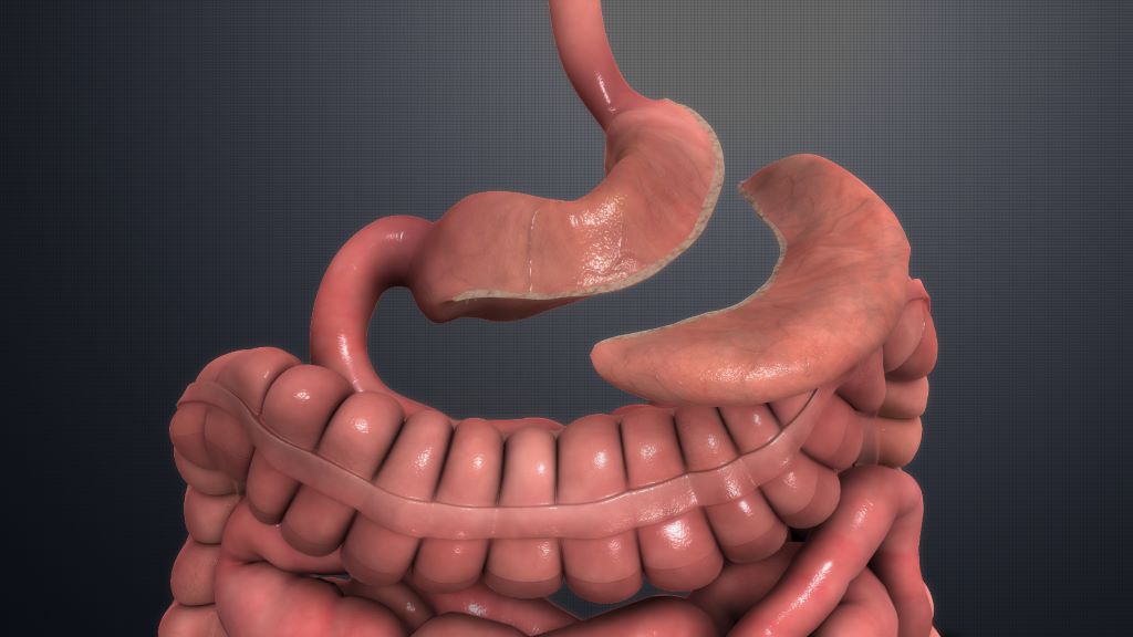
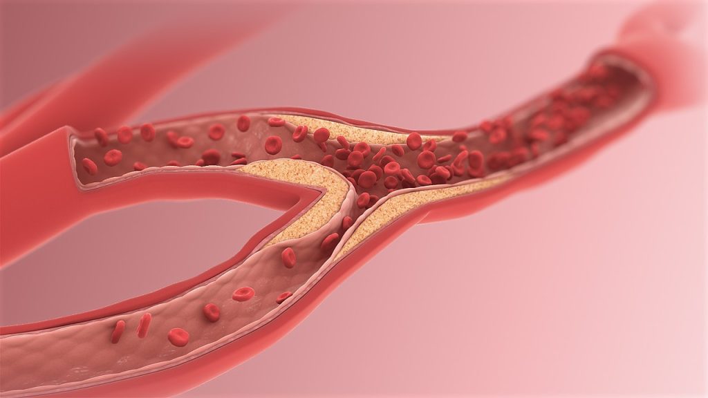
One type of blood vessel disease seen in people with RA is atherosclerosis, which is caused by a build-up of ‘plaque’ along the artery walls, which can break free and cause heart attacks and strokes.

Understanding the reasons why RA patients are at increased risk of these cardiovascular problems is critical in developing better treatments for this group and others.

To gain a better understanding of the causes of blood vessel disease in patients with RA, researchers explored the role of a group of molecules called 13-series resolvins (RvTs). In experimental arthritis the levels of one of these molecules, RvT4, are markedly reduced, a phenomenon that associates with a higher degree of blood vessel disease.

This study was designed to explore why this might be the case.

The findings

The study found that treating arthritic mice with RvT4 reduced blood vessel inflammation by re-programming macrophages, which accumulate in the diseased vessels, to release stored lipids.

Researchers observed that these lipids were preventing the macrophage from carrying out their usual work of clearing dead cells and reducing localised inflammation in blood vessels.

Once freed of their lipid burden, the macrophages were able to move and work much more effectively to reduce the causes of atherosclerosis.

The observation that RvT4 restores protective macrophage biological activities is an exciting finding.

RA patients also often present with metabolic dysfunction and this is thought to exacerbate vascular disease.

The study found that administration of RvT4 to mice engineered to develop characteristics of metabolic dysfunction, advanced atherosclerosis, and arthritis led to an overall decrease in lipoprotein-associated cholesterol in plasma and an increase in the ratio of HDL-associated cholesterol to total cholesterol.

Jesmond Dalli, Professor in Molecular Pharmacology and Lipid Mediator Unit Director at the William Harvey Institute, Queen Mary University of London, said: “The study is important because it identifies for the first time the loss of RvT4 production as a potential new cause of blood vessel inflammation in the context of arthritis, offering a mechanistic explanation on the cause of this important disease in RA patients. It also showed that RvT4 restores the biological activities of lipid loaded macrophages by promoting lipid breakdown and efflux from the cells, an observation that can guide the development of new treatments to limit the incidence and/or severity of cardiovascular disease in patients with RA.”

Source: Queen Mary University of London

Long-term Blood Pressure Control from Bariatric Surgery is Most Effective

Sleeve gastrectomy. Credit: Scientific Animations CC4.0

Compared to antihypertensives alone, bariatric surgery is more effective in controlling hypertension rates in people with obesity and uncontrolled hypertension, according to a study published in the Journal of the American College of Cardiology. People who underwent bariatric surgery had lower BMI and were on fewer medications after five years while maintaining normal blood pressure levels than those who only used antihypertensive medications.

“In clinical practice, obesity is an overlooked condition. As a consequence, there is a frequent failure in approaching obesity as a crucial step for mitigating the risk of important cardiovascular risk factors including hypertension,” said Carlos Aurelio Schiavon, MD, FACS, lead author of the study and a surgeon specialising in bariatric surgery at Heart Hospital (hcor) and BP Hospital in Sao Paulo.

Researchers in this study looked at the impact of treating obesity to lower hypertension. While new weight loss drugs exist, long-term adherence to medication can be challenging.

This study looks at bariatric surgery as a better long-term solution to control obesity and, as a result, hypertension.

The GATEWAY trial included 100 people (76% of whom were female) who had a body mass index (BMI) of around 36.9kg/m2. All participants had hypertension and were using at least two medications. People with previous cardiovascular events and poorly controlled Type 2 diabetes were excluded.

Subjects were assigned to either Roux-en-Y gastric bypass with medical therapy or medial therapy alone and the primary outcome was reduction of at least 30% antihypertensive medications while maintaining blood pressure levels less than 140/90mmHg at five years.

At five years, BMI was 28.01kg/mfor those who received bariatric surgery and 36.40kg/mfor those on medical therapy alone.

People who had bariatric surgery had an 80.7% reduction in the number of medications they were taking compared to a 13.7% reduction in those only using medical therapy.

Hypertension remission, defined as controlled blood pressure without medications, was 46.9% in those who underwent bariatric surgery compared to 2.4% in those on medical therapy alone.

“Our results underscore the importance of approaching obesity in reducing hypertension rates,” Schiavon said.

Limitations of the study include that it was a single-center, open-label study with a small sample size and there was loss of follow up in some patients.

In an accompanying editorial comment, Michael Hall, MD, MSc, professor and chair of the Department of Medicine at the University of Mississippi Medical Center, said the study provides important long-term data on the benefits of gastric bypass on weight loss and blood pressure control, but questions remain.

“Further studies assessing the threshold for bariatric surgery in people with obesity, optimal timing of bariatric surgery in obese people with cardiometabolic diseases, type of bariatric surgery and comparative studies of obesity pharmacotherapies and bariatric surgery are needed to clarify the optimal treatment pathways for this common and growing disease,” he said.

Source: American College of Cardiology

SGLT-2 Inhibitor Use Associated with Decreased Risk of Kidney Stones

Photo by Robina Weermeijer on Unsplash

Type 2 diabetes is associated with increased risk of kidney stones, but some forms of treatment for this condition may also have the benefit of lowering risk of kidney stones. In a study led by investigators from Mass General Brigham, researchers found that there was an association between the use of sodium-glucose cotransporter 2 (SGLT2) inhibitors and a lower risk of developing kidney stones. Their findings are reported in JAMA Internal Medicine.

Rates of kidney stones are on the rise in the United States and around the world. Type 2 diabetes is associated with increased risk of kidney stones, but some forms of treatment for this condition may also have the benefit of lowering risk of kidney stones.

The study included data from three nationwide databases of patients with type 2 diabetes who were seen in routine clinical practice.

The team analysed information from 716,406 adults with type 2 diabetes who had started taking an SGLT2 inhibitor or two other classes of diabetes medications known as GLP1 receptor agonists or dipeptidyl peptidase 4 (DPP4) inhibitors.

Patients who began taking SGLT2 inhibitors had a 30% lower risk of developing kidney stones than those taking GLP1 agonists and about a 25% lower risk than those taking DPP4 inhibitors.

The findings were consistent across sex, race/ethnicity, history of chronic kidney disease and obesity.

“Our findings could help inform clinical decision making for patients with diabetes who are at risk for developing kidney stones,” said corresponding author Julie Paik, MD, ScD, MPH, of the Division of Pharmacoepidemiology and Pharmacoeconomics and the Division of Renal (Kidney) Medicine at Brigham and Women’s Hospital.

Source: Mass General Brigham

Materials provided by Mass General BrighamNote: Content may be edited for style and length.


Journal Reference:

  1. Julie M. Paik, Helen Tesfaye, Gary C. Curhan, Heidi Zakoul, Deborah J. Wexler, Elisabetta Patorno. Sodium-Glucose Cotransporter 2 Inhibitors and Nephrolithiasis Risk in Patients With Type 2 DiabetesJAMA Internal Medicine, 2024; DOI: 10.1001/jamainternmed.2023.7660

Genetic Risks for ACE Inhibitor-induced Angioedema Identified

Credit: Pixabay CC0

Angioedema is a rare but potentially life-threatening adverse reaction to ACE inhibitors. In a joint analysis of eight European study collectives, researchers for the first time conducted a genome-wide association study (GWAS) with more than 1000 affected individuals, identifying a total of three risk loci in the genome. These included a new locus that had not previously been associated with the risk of ACE inhibitor-induced angioedema. The results of the study have now been published in the Journal of Allergy and Clinical Immunology.

Angiotensin-converting enzyme (ACE) inhibitors are effective antihypertensive drugs. They block the formation of the hormone angiotensin II, which plays a central role in the development of hypertension.

On the other hand, these drugs increase the concentration of the vasoactive signalling substance bradykinin. Among other things, this can lead to acute swelling of the skin or mucous membranes.

Such swellings are generally not life-threatening – but if they affect the tongue, throat or larynx, angioedema can be life-threatening for the patient due to the potential risk of suffocation.

Research to date suggests that susceptibility to such drug-induced angioedema is influenced by hereditary as well as lifestyle and environmental factors. This led researchers from the University Hospital Bonn (UKB), the University of Bonn and the Federal Institute for Drugs and Medical Devices (BfArM) to investigate potential genetic involvement.

“However, the understanding of the underlying biological processes, ie the pathophysiology, and thus the individual risk assessment is still limited. The identification of the responsible genes will provide completely new insights here,” says Prof Markus Nöthen at the University of Bonn.

Which biological processes play a role in ACE inhibitor-induced angioedema?

Based on data from eight European study collectives, the team from Bonn, together with cooperation partners, conducted the first GWAS with more than 1000 patients with ACE inhibitor-induced angioedema.

They identified a total of three loci in the genome that are associated with the risk of ACE inhibitor-induced angioedema.

“While two of the loci have already been described in previous studies, our study was the first to demonstrate a significant association for a new locus on chromosome 20,” explains corresponding author Prof.

Andreas Forstner from the Institute of Human Genetics at the UKB and the University of Bonn and at the Institute of Neuroscience and Medicine (INM-1) at the Research Center Jülich.

“Through further bioinformatic analyses, we were able to identify several candidate genes at the three risk loci indicating that genetic changes in the bradykinin, coagulation and fibrinolysis signalling play a role in the development of this type of angioedema,” adds first author Carina Mathey, doctoral student at the Institute of Human Genetics at the UKB and the University of Bonn.

Source: Universitatsklinikum Bonn

Radon Gas: Ubiquitous, Carcinogenic – and Possible Stroke Risk

Photo by Vladyslav Cherkasenko on Unsplash

A new study has found that exposure to radon, the second leading cause of lung cancer, is also linked to an increased risk of stroke. The study, which examined exposures in middle age to older female participants, found an increased risk of stroke among those exposed to high and even moderate concentrations of the gas compared to those exposed to the lowest concentrations. The study is published in Neurology®, the medical journal of the American Academy of Neurology.

Radon is a naturally occurring radioactive gas produced in certain rocks and soils which contain uranium or radium. In South Africa, some areas such as in the Western Cape have higher concentrations of radon due to underlying granite geology. It is also a concern near gold mine dumps, which have higher levels of uranium.

The gas can make its way into homes through cracks in basement walls and floors, construction joints and gaps around pipes.

“Radon is an indoor air pollutant that can only be detected through testing that measures concentrations of the gas in homes,” said study author Eric A. Whitsel, MD, MPH, of the University of North Carolina in Chapel Hill.

“Our research found an increased risk of stroke among participants exposed to radon above – and as many as two picocuries per litre (pCi/L) below – concentrations that usually trigger Environmental Protection Agency recommendations to install a home radon mitigation system.”

The study involved 158 910 female participants with an average age of 63 who did not have stroke at the start of the study.

They were followed for an average of 13 years. During the study, there were 6979 strokes among participants.

To determine radon exposures, researchers linked participants’ home addresses to radon concentration data from the U.S. Geological Survey and the U.S. Environmental Protection Agency (EPA).

The EPA recommends that average indoor radon concentrations do not exceed four picocuries per liter (pCi/L). For concentrations this high, the EPA recommends installing a radon mitigation system to lower radon levels in the home.

Participants were divided into three groups. The highest group had homes in areas where average radon concentrations were more than four pCi/L. The middle group lived in areas with average concentrations between two and four pCi/L. The lowest group lived in areas with average concentrations of less than two pCi/L.

In the group with the highest radon exposures, there were 349 strokes per 100 000 person-years compared to 343 strokes in the middle group and 333 strokes in group with the lowest exposure.

Person-years represent both the number of people in the study and the amount of time each person spends in the study.

After adjusting for factors such as smoking, diabetes and high blood pressure, researchers found participants in the highest group had a 14% increased risk of stroke compared to those in the lowest group.

Those in the middle group had a 6% increased risk.

“It’s important to note that we found an increased stroke risk among those exposed to radon concentrations as much as two pCi/L below the current lung cancer-based threshold for recommending radon mitigation,” said Whitsel.

“More studies are needed to confirm our findings. Confirmation would present an opportunity to improve public health by addressing an emerging risk factor for stroke.”

A limitation of the study was that it included only female participants who were middle age or older and primarily white, so the results may not be the same for other populations.

Source: American Academy of Neurology

Reduced Blood Lead Levels Tied to Lower Blood Pressure

Credit: Pixabay CC0

Researchers found that small declines in blood lead levels were associated with long-term cardiovascular health improvements in American Indian adults. Participants who had the greatest reductions in blood lead levels saw their systolic blood pressure fall by about 7mmHg, comparable to the effects of antihypertensives.

The findings as reported from researchers at Columbia University Mailman School of Public Health and NIEHS and NHLBI are published in the Journal of the American Heart Association.

“This is a huge win for public health,” said senior author Anne E. Nigra, PhD, assistant professor of environmental health sciences at Columbia Mailman School of Public Health.

“We saw that even small decreases in a person’s blood lead levels can have meaningful health outcomes.”

Nigra and her co- authors, including Wil Lieberman-Cribbin, MPH, also at Columbia Mailman School, credit these improvements in large part to public health and policy changes that have occurred over the last few decades.

In addition to seeing improvements in systolic blood pressure, the investigators found that reductions in blood lead levels were associated with reductions in a marker associated with hypertrophic cardiomyopathy and heart failure.

To conduct this research, investigators partnered with 285 American Indian adults through an extension of the Strong Heart Study, the largest study following cardiovascular health outcomes and risk factors among American Indian adults.

The researchers looked at blood lead levels and blood pressure readings over time in participants living in one of four tribal communities. Lead was first measured in blood collected during the 1997–1999 study visit and again in blood collected during a follow-up visit between 2006–2009.

During this time, participants’ blood pressure was taken and they participated in medical exams, including echocardiographs to assess their heart’s structure and function. Multiple factors were controlled for, including social variables, cardiovascular disease risks, and medical history.

At the start of the study, the average blood lead level was 2.04µg/dL. Throughout the study, the average blood lead level fell by 0.67µg/dL, or 33%.

The most significant changes, categorized by participants with average starting blood lead levels of 3.21 µg/dL and who experienced reductions of about 1.78 µg/dL, or 55%, were linked to a 7mmHg reduction in systolic blood pressure.

“This is a sign that whatever is happening in these communities to reduce blood lead levels is working,” said Mona Puggal, MPH, an epidemiologist in the Division of Cardiovascular Sciences at the National Heart, Lung, and Blood Institute (NHLBI). “The reductions in blood pressure are also comparable to improvements you would see with lifestyle changes, such as getting 30 minutes of daily exercise, reducing salt intake, or losing weight.”

The reductions in blood lead levels observed in the study are similar to those seen in the general US population following policies and efforts implemented within the past 50 years to reduce lead exposure through paint, gasoline, water, plumbing, and canned items.

Source: Columbia University’s Mailman School of Public Health

.

Researchers Urge Caution in Co-prescribing Potency Drugs and Nitrates

Pexels Photo by Freestocksorg

Co-prescribing potency drugs such as Viagra and organic nitrates for angina is associated with a 35–40% increased mortality risk and about 70% higher risk of heart attack and heart failure. This is according to a Swedish registry study published in the Journal of the American College of Cardiology. The Swedish researchers are now urging caution.

Drugs for erectile dysfunction or impotence containing phosphodiesterase inhibitors type 5 are contraindicated in the treatment of angina with organic nitrates. Because the two types of drugs enhance each other’s antihypertensive effect, they can cause serious side effects, including death, if taken together.

But many people who treat angina with organic nitrates use the medication as emergency relief for a sudden onset of angina. The medication is quickly absorbed by the body, exerts its effect, and then breaks down quickly again. It is not usually a permanent treatment, although maintenance treatment is possible. 

Does not necessarily indicate an increased risk

Potency drugs are also taken as needed, which theoretically makes it possible to separate the two treatments in time to avoid side effects. If patients are aware of these factors, co-prescribing does not necessarily mean an increased risk.

Previous studies have shown that an increasing number of men who treat their angina with organic nitrates are also prescribed potency drugs. However, there is no evidence that side effects have increased. 

The picture is not entirely clear, as it has also been shown that type 5 phosphodiesterase inhibitors for men with cardiovascular disease without angina reduce the risk of death and heart failure.

“There is an increasing demand for medication for erectile dysfunction from men with cardiovascular disease. And even if these drugs are beneficial for most men with cardiovascular disease, those who are also treated with nitrates need to consider the benefits of the drug against the cardiovascular risks,” says first author Ylva Trolle Lagerros, Associate Professor at the Department of Medicine at Karolinska Institutet.

To find out what the actual risk of concurrent prescribing is, the researchers used Swedish health registers between 2005 and 2013. They found nearly 61 500 men who had been prescribed organic nitrates, of which just over 5700 had also been prescribed one of the potency drugs in question. A clear majority of those who had a prescription for both medications used nitrates as an emergency treatment only.

Adjusted for differences

The men who received the drugs were on average nine years younger and significantly healthier than those who did not receive them. The researchers therefore had to adjust for these and other differences.

The adjusted results show that co-prescribing potency drugs with type 5 phosphodiesterase inhibitors and organic nitrates is associated with a 35–40% increased risk of death. In addition, the researchers show an approximately 70% increased risk of heart attack and heart failure. This suggests that the theoretical separation in time of the treatments does not seem to work fully.

“We want to point out the importance of careful and patient-centered consideration before prescribing this type of potency medication to men treated with nitrates,” says Ylva Trolle Lagerros.

Source: Karolinska Institutet

A New Pain-free Way to Treat Ventricular Arrhythmia

Source: CC0

A breakthrough study sets the foundation of a ground-breaking treatment regimen for treating ventricular arrhythmia. The study, published in Nature Communications, demonstrates the design and feasibility of a new hydrogel-based pacing modality.

The urgent need for an effective therapeutic regimen for ventricular arrhythmia inspired a team led by Dr. Mehdi Razavi at The Texas Heart Institute (THI), to collaborate The University of Texas at Austin (UT Austin) Cockrell School of Engineering led by Dr. Elizabeth Cosgriff-Hernandez, to co-develop an innovative strategy that addresses the pathophysiology of re-entrant arrhythmia.

Ventricular arrhythmia, which occurs in the lower chambers of the heart or ventricles, is the leading cause of sudden cardiac death in the United States.

When heart rhythm abnormality occurs in a self-sustained manner, it is called re-entrant arrhythmia, which is usually fatal.

“Re-entry occurs mainly from delayed conduction in scarred heart tissues, usually after coronary artery occlusion during a heart attack, which can be corrected by enabling pacing in these regions,” said Dr. Razavi, a practicing cardiologist and cardiac electrophysiologist.

“These hydrogels then can access the scarred tissue, thereby enabling direct pacing of the otherwise inaccessible regions of the heart.”

Given hydrogels’ biostability, biocompatibility, tunable properties, and the ease of incorporating electrical conductivity, the scientists are exploring them as potential electrodes that can be easily delivered inside coronary veins.

A clinical advantage of the unique system is that ischemia can be avoided by delivering the hydrogel using the veins.

The researchers successfully deployed the innovative hydrogel technology through minimally invasive catheter delivery in a pig model.

“The hydrogels have significant conductive properties that enable simultaneous pacing from multiple sites along the length of the hydrogel and create a conduction highway similar to those in Purkinje fibers,” according to Dr. Cosgriff-Hernandez.

Today, arrhythmia is treatable with medicines and procedures that control the irregular rhythms.

The current anti-arrhythmic drugs on the market are not always effective; although the drugs slow the conduction velocity, they facilitate re-entry arrhythmia.

Moreover, these drugs can be toxic and can lead to the destruction of tissues near the diseased regions of the heart.

Even with the widely used interventional ablation therapies, arrhythmia recurs in a significant proportion of patients. None of these procedures address the mechanism of re-entry.

Cardiac defibrillators implanted to compensate for the shortfalls in the current therapy options are painful when delivering electric shocks to restore heart rhythm and can severely deteriorate the patient’s quality of life.

If left untreated, arrhythmia can damage the heart, brain, or other organs, leading to stroke or cardiac arrest, during which the heart suddenly and unexpectedly stops beating.

“When injected into target vessels, the conductive hydrogel conforms to the patient’s vessel morphology. Adding a traditional pacemaker to this gel allows for pacing that resembles the native conduction in the heart — effectively mimicking the native electrical rhythm of the heart — and extinguishes the cause for arrhythmia, providing painless defibrillation,” added Dr. Cosgriff-Hernandez.

The work demonstrates for the first time the ability to confer direct electrical stimulation of the native and scarred mid-myocardium through injectable hydrogel electrodes as a pacing modality.

With minimally invasive catheter delivery and standard pacemaker technologies, this study indicates the feasibility of a novel pacing modality that resembles native conduction, potentially eliminating lethal re-entrant arrhythmia and providing painless defibrillation, which can be successfully adopted in a clinical workflow.

The scientific advance is significant considering pain management is highly relevant to overall wellness for patients with heart, lung, and blood diseases.

Such innovation in painless defibrillation and preventing arrhythmia could revolutionize cardiac rhythm management.

Source: Texas Heart Institute 

A Genetic Clue to Pulmonary Hypertension Risk

Photo by Sangharsh Lohakare on Unsplash

University of Pittsburgh Schools of Medicine researchers uncovered a fundamental mechanism that controls the body’s response to limited oxygen and regulates blood vessel disease of the lung.

By combing through genomes of more than 20 000 individuals in the US, France, England and Japan and combining the results with molecular studies in the lab, the team discovered a shared genetic trait that could predict a higher risk of pulmonary hypertension and its more severe form, pulmonary arterial hypertension, and influence the development of drug therapies that target the body’s response to limited oxygen. The findings were published in Science Translational Medicine.

“This new level of knowledge will help identify people who may be at a higher genetic risk of pulmonary hypertension and jump-start precision medicine practices to offer customised treatments,” said senior author Stephen Chan, MD, PhD.

Pulmonary hypertension encompasses a range of conditions of various causes that manifest in high blood pressure in the arteries of the lung and the right side of the heart.

The disease is accompanied by a decreased supply of oxygen to the lung tissue and the blood, is chronic and deadly, and its molecular origins and genetic background remain unsolved.

Using a combined approach of genomics and biochemistry, the Chan lab found a gene pair that had an important function in regulating blood vessel metabolism and disease.

This gene pair included a long non-coding RNA molecule – a messenger that facilitates the transformation of the body’s genetic code into protein products – and a protein binding partner, and their interaction was frequently active in cells exposed to low oxygen compared to normal cells.

Taking the findings a step further, the team discovered that a single DNA letter change directing expression of this RNA-protein pair under low oxygen conditions was associated with a higher genetic risk of pulmonary hypertension across diverse patient populations.

According to Chan, pulmonary hypertension is a borderline orphan disease, and the limited number of patients with pulmonary hypertension makes it challenging to find genetic variations that are rare but still impactful enough to eclipse individual differences.

With that in mind, Pitt scientists turned to collaborators around the globe and to public research datasets to ensure that the findings are relevant across a diverse global population.

Chan hopes that his findings will spur the development of targeted therapies relevant to oxygen sensitivity in blood vessel lining and that their pending patent application will contribute to the growth on an entirely new field of epigenetic and RNA drug therapeutics that work not by manipulating the genome but by changing how it is being read.

Source: University of Pittsburgh

High Cholesterol from Childhood Sedentary Time could be Reversed with Light Exercise

Photo by Victoria Akvarel on Pexels

Increased sedentary time in childhood can raise cholesterol levels by two thirds as an adult, but a new study has found light physical activity may completely reverse the risks and is far more effective than moderate-to-vigorous physical activity.

The study was published in The Journal of Clinical Endocrinology & MetabolismResearchers used data from the University of Bristol study Children of the 90s (also known as the Avon Longitudinal Study of Parents and Children), which included 792 children aged 11 years who were followed up until the age of 24.

Results from this study found that accumulated sedentary time from childhood can increase cholesterol levels by two thirds (67%) by the time someone reaches their mid-twenties. Elevated cholesterol and dyslipidaemia from childhood and adolescence have been associated with premature death in the mid-forties and heart problems such as subclinical atherosclerosis and cardiac damage in the mid-twenties.

Healthy lifestyles are considered important in the prevention of dyslipidaemia and one of the primary ways of lowering cholesterol, apart from diet, is movement behaviour. For the first time, this study objectively examined the long-term effects of sedentary time, light physical activity, and moderate-to-vigorous physical activity on childhood cholesterol levels.

The World Health Organization currently recommends children and adolescents should accumulate on average 60 minutes of moderate-to-vigorous physical activity a day and reduce sedentary time but have limited guidelines for light physical activity. Yet this new study and other recent studies has found light physical activity – which includes exercises such as long walks, house chores, or slow dancing, swimming, or cycling – is up to five times more effective than moderate-to-vigorous physical activity at promoting healthy hearts and lowering inflammation in the young population.

Dr Andrew Agbaje from the University of Exeter led the study and said: “These findings emphasise the incredible health importance of light physical activity and shows it could be the key to preventing elevated cholesterol and dyslipidaemia from early life. We have evidence that light physical activity is considerably more effective than moderate-to-vigorous physical activity in this regard, and therefore it’s perhaps time the World Health Organization updated their guidelines on childhood exercise — and public health experts, paediatricians, and health policymakers encouraged more participation in light physical activity from childhood.”

During the research, accelerometer measures of sedentary time, light physical activity, and moderate-to-vigorous physical activity were collected at ages 11, 15, and 24 years. High-density lipoprotein cholesterol, low-density lipoprotein cholesterol, triglyceride, and total cholesterol were repeatedly measured at ages 15, 17, and 24 years. These children also had repeated measurement of dual-energy X-ray absorptiometry assessment of total body fat mass and muscle mass, as well as fasting blood glucose, insulin, and high sensitivity C-reactive protein, with smoking status, socio-economic status, and family history of cardiovascular disease.

During the 13-year follow-up, sedentary time increased from approximately six hours a day to nine hours a day. Light physical activity decreased from six hours a day to three hours a day while moderate-to-vigorous physical activity was relatively stable at around 50 minutes a day from childhood until young adulthood. The average increase in total cholesterol was 0.69 mmol/L. It was observed without any influence from body fat.

An average of four-and-a-half hours a day of light physical activity from childhood through young adulthood causally decreased total cholesterol by (-0.53 mmol/L), however, body fat mass could reduce the effect of light physical activity on total cholesterol by up to 6%. Approximately 50 minutes a day of moderate-to-vigorous physical activity from childhood was also associated with slightly reduced total cholesterol (-0.05 mmol/L), but total body fat mass decreased the effect of moderate-to-vigorous physical activity on total cholesterol by up to 48%. Importantly, the increase in fat mass neutralised the small effect of moderate-to-vigorous physical activity on total cholesterol.

Source: University of Exeter