A New Oral Combo Drug for AML Eases Treatment Burden

The ASCERTAIN V clinical trial demonstrated that an all-oral drug combination for older patients with acute myeloid leukaemia (AML) is an effective alternative to the current standard, which requires repeated hospital or office visits for intravenous treatment. In the international phase 1/phase 2 trial, patients took a regimen of two pills, decitabine-cedazuridine and venetoclax, with strong response rates and survival outcomes. The study results were published in the New England Journal of Medicine.
Nearly half of patients (46.5%) achieved complete response, while 63% experienced either complete response or complete response with incomplete haematologic recovery, meaning cancer cells were undetectable, but the patient’s healthy blood cell counts had not yet returned to normal. The median overall survival reached 15.5 months – comparable to existing intravenous therapies.
The oral combination of decitabine-cedazuridine and venetoclax received U.S. Food and Drug Administration approval on May 13 for the treatment of AML in newly diagnosed adults 75 years or older and patients clinically unable to undergo traditional, intensive chemotherapy.
“Having received approval, we anticipate that this oral AML regimen will become the standard of care for patients who are older or more frail,” said lead author Dr Gail J. Roboz, professor of medicine and director of the Clinical and Translational Leukemia Program at Weill Cornell and a haematologist oncologist at NewYork-Presbyterian/Weill Cornell Medical Center. “We hope these results point to a future for AML patients where the treatment journey is less disruptive and less burdensome without sacrificing outcomes.”
Turning a Standard into an Oral Treatment
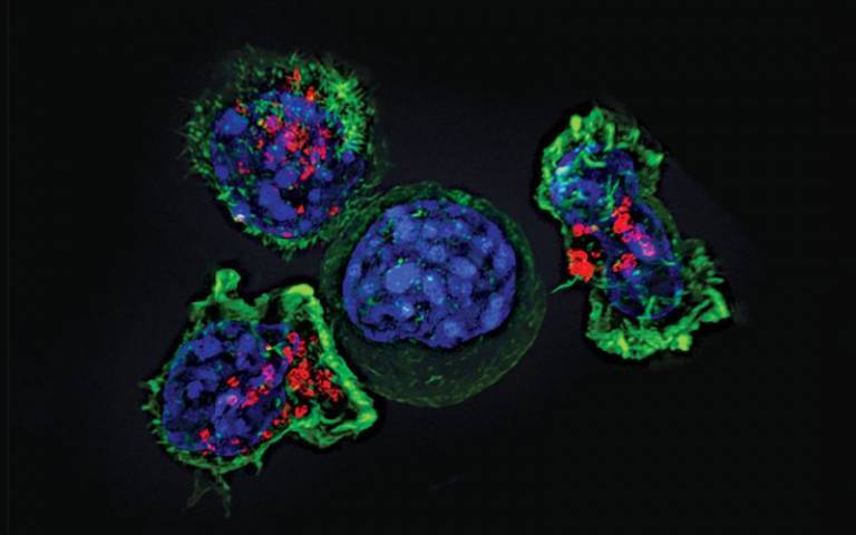
AML is an aggressive blood cancer that can be diagnosed at any age and is especially difficult to treat in older adults and patients with other serious health conditions. For these individuals, the current standard treatment combines venetoclax with a class of drugs known as hypomethylating agents, such as decitabine. Venetoclax inhibits Bcl-2, a protein that leukaemia cells overproduce to avoid cell death, while hypomethylating agents restore the activity of genes involved in cell growth and survival, helping slow leukaemia progression.
However, this regimen requires monthly treatment cycles that combine oral venetoclax with five to seven days of an injectable hypomethylating agent delivered in a clinic or hospital. These frequent visits create significant physical, logistical and emotional challenges for patients and families.
More recently, pharmacologists developed a pill version of decitabine by pairing it with another drug called cedazuridine that prevents decitabine from being broken down when ingested.
With the ASCERTAIN V trial, Dr Roboz and her colleagues tested whether decitabine’s oral version, combined with venetoclax, could match the efficacy of intravenous AML treatment.
The nonrandomised phase 1/phase 2 study enrolled 189 newly diagnosed AML patients at centres across the United States, Canada and Spain. The patients took a month of venetoclax, along with five days of decitabine-cedazuridine at the start of each treatment cycle.
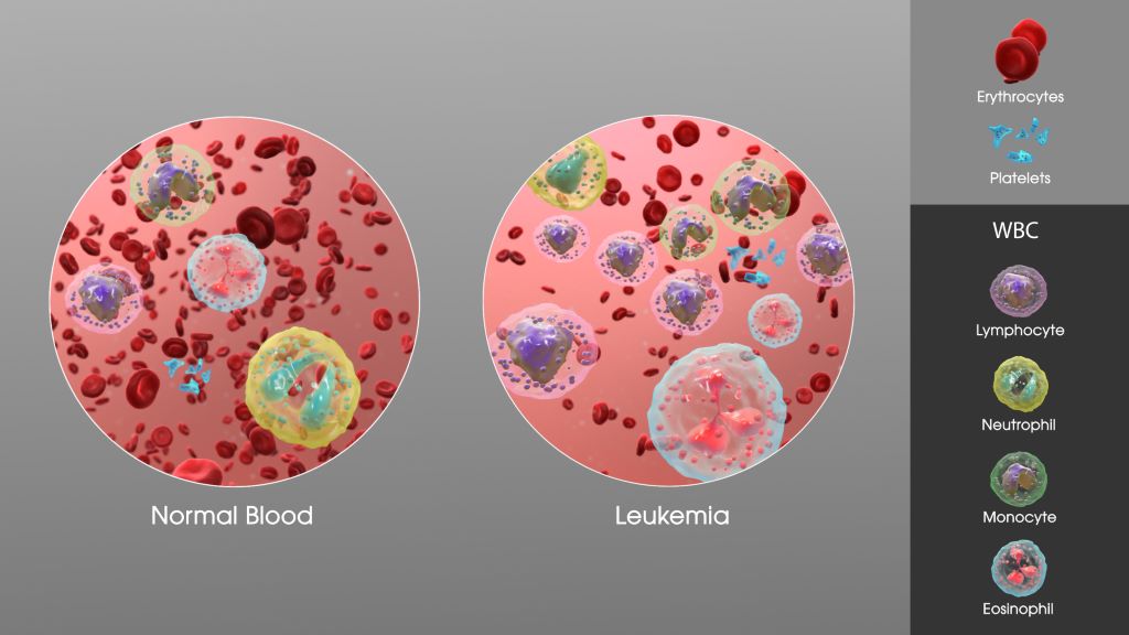
The oral regimen demonstrated a safety profile consistent with what doctors already expect from standard AML therapies, which commonly deplete healthy blood cells alongside leukaemia cells. The most common serious side effects included anaemia, neutropenia and fever associated with low white blood cell numbers.
Tailoring Treatment to Reduce Side Effects
During the trial, researchers also investigated how to fine-tune the treatment schedule to optimise leukaemia control, while minimising side effects related to low blood counts.
The paper offers recommendations for physicians, including careful monitoring of leukaemia cells until they reach a certain threshold and then strategically pausing venetoclax to allow the body to replenish normal white blood cells, red blood cells and platelets.
“The goal of the all-oral therapy is to keep people out of the hospital, especially once they have achieved remission,” said Dr Roboz, who is also a member of the Sandra and Edward Meyer Cancer Center at Weill Cornell. “Patients are thrilled not to have to deal with monthly chemotherapy injections or infusions.”
Looking Ahead
For now, most patients taking the oral regimen must continue treatment to maintain remission, much like a chronic condition. “AML patients taking ongoing cycles of treatment require close monitoring but can still have an excellent quality of life,” Dr Roboz said.
In the future, the researchers hope that increasingly sensitive blood monitoring tests may identify when patients can safely stop treatment.
Dr Roboz and AML researchers worldwide are also exploring “triplet therapies,” which add additional targeted drugs to the decitabine-cedazuridine and venetoclax combination.
“The goal is to get away from treatment cycles that go on indefinitely,” said Dr Roboz. “We want to drive the leukaemic cells to such low levels that patients can discontinue therapy and be cured.”
Source: Weill Cornell Medicine