Tag: rheumatoid arthritis

CAR T Cell Therapy for Rheumatoid Arthritis Highly Effective in First Patients

Patients own immune cells genetically modified in the laboratory, track down the disease driving B cells, even deep within tissues. Inflammatory foci such as those seen here around the knee joints of a study participant (magenta) are no longer detectable even several months after CD19-CAR T-cell therapy (right in the PET-MRI image). Swelling and pain have subsided, with improved mobility. © Charité | David Simon

Immunotherapies such as CAR T cell therapy are used primarily to treat cancer. In the future, these patient-specific therapies, manufactured from patients’ own immune cells, could also help cure autoimmune diseases. Six patients with particularly severe rheumatoid arthritis have now received this treatment at Charité – Universitätsmedizin Berlin. In Nature Medicine, the researchers report the results from the world’s first clinical trial of its kind: Disease activity decreased substantially in all participants. By the end of the observation period, three of the patients no longer required any medication for rheumatoid arthritis.

Rheumatoid arthritis is a chronic disease in which the immune system mistakenly attacks the body’s own joints. This causes recurrent inflammation and joint swelling and, as the disease progresses, can lead to joint damage. Currently available treatments can usually keep the inflammation under control, but do not cure the disease. Patients therefore require lifelong medication, including anti-inflammatory drugs and medications that suppress the immune system, with all the associated side effects.

In some patients, even several of the newer treatments fail to produce an adequate response. Doctors then call the disease treatment-refractory. For those affected, this means persistent pain, restricted mobility and a substantial impact on quality of life. “One reason could be disease driving B cells – memory cells of the adaptive immune system that may survive in the lymph nodes, bone marrow or joint tissue after an infection, where they produce harmful antibodies directed against the body’s own tissues and repeatedly reignite the inflammation,” explains Prof David Simon, who designed the trial for this patient group together with Prof Gerhard Krönke at Charité’s Department of Rheumatology and Clinical Immunology.

The researchers hope that CAR T cells, patients’ own immune cells genetically modified in the laboratory, could selectively track down the disease driving B cells, even deep within tissues, reset the pathological B-cell memory as fully as possible and thereby effectively give the B-cell system a fresh start.

Cell therapy resets the immune system

Originally developed for cancer treatment, CAR T cells are now being used more widely. In cancer treatment, patients’ immune cells are given a kind of “training” to specifically recognise and eliminate tumour cells; in autoimmune diseases, the aim is instead to direct immune cells toward disease driving memory cells. “The identifying marker on many B cells, both abnormal B cells in cancers of the blood or lymphatic system and disease driving B cells in rheumatoid arthritis, is the surface molecule CD19. You could think of it as a kind of ‘name tag’” explains David Simon. “To enable CAR T cells to detect and eliminate the disease-causing cells, we equip patients’ own immune cells with a receptor that acts like a search sensor for CD19.”

For this type of CD19 CAR T-cell therapy, T cells are first collected from the patient’s blood. These are immune cells that normally recognize and eliminate infected or abnormal cells. In the laboratory, the cells are genetically modified and equipped with the key recognition feature: a chimeric, or artificial, antigen receptor, known as a CAR, that binds specifically to the CD19 molecule. Before the modified immune cells can be returned to the patient in a single infusion, a short course of preparatory chemotherapy is required to create space in the immune system. This temporarily reduces the number of certain immune cells so that the CAR T cells can then multiply effectively and carry out their function. Once returned to the body, the modified immune cells begin searching for the CD19 marker and specifically attack cells that carry it. In doing so, they temporarily eliminate all CD19-positive B cells, allowing the immune system to reset and helping to eliminate even long-lived disease-driving B cells in the joints that would otherwise be difficult to reach.

Will the approach work in difficult-to-treat rheumatoid arthritis?

For the world’s first clinical trial to evaluate the safety and efficacy of a CD19 CAR T-cell therapy in rheumatoid arthritis, the research team at Charité initially enrolled six patients with particularly severe disease. The three women and three men, aged 31 to 69, had received up to eight targeted or biologic therapies over the previous ten years, none of which had been sufficiently effective.

The key question was whether CAR T cells can track down the disease-driving B cells in the joints and whether the treatment is not only effective but also safe. For the researchers, the results from the first part of the COMPARE trial are highly encouraging: “Disease activity decreased markedly in all six patients. During follow-up of up to one year, three patients were in sustained remission without any medication for rheumatoid arthritis,” reports Gerhard Krönke, who leads the joint Clinical Rheumatology research group at Charité and the German Rheumatology Research Center (DRFZ), a Leibniz Institute. “This is particularly remarkable given that none of the established treatments had previously been able to relieve their symptoms adequately.”

The cell therapy appears to have done more than temporarily curb inflammation in the joints: the modified immune cells also tracked down and eliminated disease-promoting B cells in deeper reservoirs such as the bone marrow, lymph nodes and joint tissue. At regular follow-up visits over the subsequent twelve months, the research team found that levels of the autoantibodies characteristic of rheumatoid arthritis had declined sharply.

David Simon adds: “When the B-cell system later recovered, predominantly naïve B cells that had not yet been shaped by the disease returned. In contrast, the B cells directed against the body’s own tissues that had been present before treatment were no longer detectable in almost all patients, an indication that the treatment may indeed be able to reset the pathological immune memory.” Protective antibodies from previous vaccinations, for example against chickenpox or tetanus, remained detectable. Despite the profound depletion of B cells, protective antibody memory appears to be largely preserved. However, any longer-term effects of the therapy on the immune system still need to be investigated.

Early successes, but still some way to go

The trial shows that, in some patients, a single CAR T-cell treatment can lead to a sustained symptom-free period without medication – a state of disease inactivity known as remission. For selected patients with rheumatoid arthritis who have not responded adequately to treatment, it may in the future be possible to reset the pathological immune memory in a targeted way and thereby stop the persistent inflammation, instead of having to suppress it continuously with drugs.

Nevertheless, CAR T-cell therapy for autoimmune diseases, and for rheumatoid arthritis in particular, remains experimental. There is not yet any long-term experience with this treatment. In addition, responses to the therapy varied among patients in the current trial: some did not achieve a complete response, and in one case the disease returned after an initial medication-free period of remission. In contrast, the researchers consider the safety data obtained thus far to be encouraging. “After the participants received the CD19 CAR T cells, we observed only a temporary, mild-to-moderate cytokine release syndrome (CRS) in all participants, which was readily manageable. There were no severe neurological complications or other serious adverse events, and infections were rare,” explains Dr Marie Luise Hütter-Krönke, Medical Director of the Hematology Early Clinical Trial Unit at Charité’s Department of Hematology, Oncology and Cancer Immunology.

In a second phase of the trial involving ten additional patients, the researchers will compare the new treatment approach with a drug already approved for rheumatoid arthritis that also targets B cells. In this way, they aim to determine whether CAR T cells have a stronger and longer-lasting effect and whether they can indeed reset immune memory. If the new concept is confirmed in this and other, larger trials, it could ultimately become an alternative for patients whose lives are severely affected and for whom no adequate treatment is currently available.
Source: Charité – Universitätsmedizin Berlin

Drug Reduces the Risk of Developing Rheumatoid Arthritis, Clinical Trial Finds

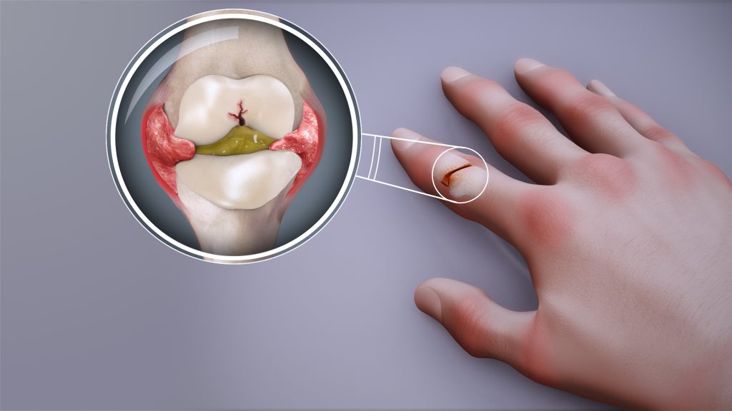
Rheumatoid arthritis. Credit: Scientific Animations CC4.0

A clinical trial led by researchers from Hospital Clínic de Barcelona, the August Pi i Sunyer Biomedical Research Institute (IDIBAPS) and The Institute for Advanced Chemistry of Catalonia (IQAC-CSIC) has compared the effectiveness of abatacept and hydroxychloroquine in preventing the development of rheumatoid arthritis in patients with palindromic rheumatism, an autoimmune disease that progresses to arthritis in approximately half of all patients. The trial was conducted over two years across 14 hospitals throughout Spain and involved 70 patients with palindromic rheumatism. The findings, published in Nature Medicine, indicate that abatacept is significantly more effective than hydroxychloroquine in preventing the onset of arthritis.

Palindromic rheumatism is characterised by intermittent episodes of joint inflammation, with acute flare-ups lasting a few days and resolving spontaneously. However, around half of patients eventually develop rheumatoid arthritis, a chronic disease that causes irreversible joint damage. This risk is particularly high in individuals with biomarkers such as rheumatoid factor and anti-citrullinated peptide antibodies. The presence of these two autoantibodies – proteins of the immune system that mistakenly attack the body’s own organs and tissues – is used in the diagnosis of the disease.

Until now, in the absence of clinical trial evidence, the standard approach has been to treat patients with palindromic rheumatism using hydroxychloroquine, a drug with anti-inflammatory and immunosuppressive properties aimed at improving disease symptoms. In this context, the team led by Raimon Sanmartí, head of the Inflammatory Arthropathies Research Group at IDIBAPS, conducted a two-year clinical trial involving 70 patients with palindromic rheumatism. The objective was to compare the effectiveness of hydroxychloroquine with abatacept, a lymphocyte inhibitor – a type of white blood cell that attacks the joints after mistakenly identifying them as a threat – in reducing progression from palindromic rheumatism to rheumatoid arthritis

The results show that treatment with abatacept significantly reduces progression to rheumatoid arthritis. Only 20% of patients receiving abatacept developed arthritis, compared with 50% of those treated with hydroxychloroquine. Furthermore, patients treated with abatacept not only avoided progression to rheumatoid arthritis in most cases, but also experienced a significant improvement in palindromic rheumatism symptoms. “The study shows that patients treated with abatacept are more likely to achieve complete remission of attacks associated with acute pain and joint swelling, and that their inflammatory episodes are less severe,” explains Isabel Haro, Head of the Peptide Synthesis and Biomedical Applications Unit at IQAC-CSIC. The research team also highlights that both drugs proved safe and well tolerated throughout the trial.

Early intervention

“The results of this study indicate that we can intervene at an early stage to modify the natural course of the disease and reduce the risk of patients developing more severe and irreversible conditions,” says Sanmartí. “This opens the door to a paradigm shift in the treatment of these patients.”

The study, which involved several national research centres, also analysed the evolution of a number of biomarkers (autoantibodies) developed by the CSIC research group during patient follow-up. “Although no significant differences were observed between abatacept and hydroxychloroquine in terms of autoantibody responses, this work demonstrates the value of immunomodulatory approaches in the early stages of disease, when it is still possible to prevent progression to more severe and chronic forms,” notes Haro.

Rheumatoid arthritis is a disease that significantly affects patients’ quality of life and places a considerable burden on healthcare systems. Preventing its development in a substantial proportion of cases represents an important advance in the management of rheumatic diseases.

Source: Institute for Advanced Chemistry of Catalonia

Early Treatment Can Delay Rheumatoid Arthritis for Years

Early treatment for people at high risk of developing rheumatoid arthritis can delay the disease for several years.

Photo by Towfiqu barbhuiya: https://www.pexels.com/photo/person-feeling-pain-in-the-knee-11349880/

Treating people who are at high risk of rheumatoid arthritis (RA) can delay the onset of the disease for several years, with benefits also continuing well after treatment has stopped. A new trial has shown that one year of treatment with the drug abatacept, a biologic therapy that targets immune cell activation, reduced progression to rheumatoid arthritis in people at high risk.

The new King’s College London study, published in The Lancet Rheumatology, builds on results from a trial reported by King’s researchers in 2024.

While the original trial followed 213 participants from the UK and the Netherlands for two years, the new study reports outcomes from an extended follow-up period of between four and eight years, making it one of the longest follow-up studies of its kind in people at risk of RA.

RA is a chronic autoimmune condition affecting around half a million people in the UK. It develops when the immune system mistakenly attacks the joints, causing pain, swelling, fatigue and long-term disability.

People at risk of developing the condition also often stop working before the disease starts, which creates employment instability and has economic consequences.

While effective treatments exist for people with established RA, there is currently no licensed therapy that can prevent the disease from developing in those at risk.

The researchers found that the benefits of just 12 months of abatacept therapy persisted well beyond the treatment period. People who received the drug took significantly longer to develop RA than those given placebo, with disease onset delayed by up to four years beyond the treatment period.

Although the drug did not permanently prevent RA, the findings show that early treatment can alter the course of the disease by postponing its development, potentially reducing the number of years people live with symptoms and complications.

Intervening early in people at high risk of RA can have lasting benefits. We have shown that this approach is safe and can prevent disease while patients are on treatment as well as substantially relieve symptoms. Importantly, it can also delay the onset of RA for several years, even after treatment has stopped. This could reduce how long people live with symptoms and complications, drastically improving their quality of life.

Professor Andrew Cope, Professor of Rheumatology in the Centre for Rheumatic Diseases at King’s College London

The study also showed that abatacept was most effective in individuals at highest risk of developing RA, identified through a blood test detecting specific autoantibodies. While these participants were at highest risk of progressing to RA they were also much more likely to benefit from early intervention.

During the at-risk phase, treatment with abatacept reduced symptoms such as joint pain and fatigue and improved overall wellbeing. But once treatment stopped, symptom levels became similar between the treatment and placebo groups, suggesting that continued immune modulation may be required to sustain symptom control.

The study found that abatacept was safe, with similar rates of serious adverse events in both the treatment and placebo groups, and no safety concerns linked to the drug.

The researchers suggest the latest findings provide evidence that early, targeted immune treatment can delay RA in people at highest risk, supporting further research into preventive approaches for autoimmune disease.

Source: King’s College London

Why Drugs Targeting Interleukin-17 Don’t Work in Rheumatoid Arthritis

Rheumatoid arthritis. Credit: Scientific Animations CC4.0

Cedars-Sinai investigators may have figured out why certain immunosuppressive treatments don’t work well in rheumatoid arthritis. In a study published in Science Immunology, scientists traced the problem to specific changes that occur in immune cells within the joints as the disease progresses.

The findings could lead to more effective therapies for the incurable autoimmune disease.

“Our discoveries point to the importance of the tissue environment in worsening rheumatoid arthritis and driving resistance to antirheumatic medications,” said Nunzio Bottini, MD, PhD, director of the Kao Autoimmunity Institute at Cedars-Sinai, professor of Medicine and corresponding author of the study.

Rheumatoid arthritis causes chronic inflammation in the joints. In other forms of autoimmune arthritis, inflammation can be relieved by targeting interleukin-17, one of several proteins that can contribute to joint inflammation.

In experiments involving human rheumatoid arthritis tissues and laboratory mice, investigators showed that, over time, the immune cells that produce interleukin-17 gradually stop making it. This finding helps explain why IL-17-targeted treatments do not work well against established rheumatoid arthritis.

“These immune cells can also change in ways that make them more aggressive and able to sustain inflammation even without interleukin-17,” Bottini said.

Changes to the immune cells appear to be driven by synoviocytes – nonimmune cells that produce the lubricating synovial fluid in the joints, according to the study.

Bottini said that the Department of Computational Biomedicine at Cedars-Sinai, particularly the laboratory of Kyoung Jae Won, PhD, played a key role in the study by contributing critical work in spatial biology, an emerging field that studies how cells function within their tissue environments.

The findings carry significant implications for treating rheumatoid arthritis, according to Joyce So, MD, PhD, chief genomics officer at Cedars-Sinai and medical director of the newly established Center for Genomic Medicine at Cedars-Sinai Guerin Children’s.

“This important new insight contributes to shifting the paradigm of how we understand rheumatoid arthritis progression and why IL-17 treatments haven’t worked as well as expected,” So said. “Only with a precise understanding of the biological mechanisms of disease can effective, precision therapies be developed. In the meantime, clinicians can help patients in early or presymptomatic stages make the most of treatments that may lose effectiveness over time.”

Source: Cedars–Sinai

Muscle Wasting Reversed in Patients with Rheumatoid Arthritis

Photo by Towfiqu barbhuiya

Patients with rheumatoid arthritis increased their leg muscle volume when treated with an anti-rheumatic drug, offering new hope for improved muscle health.

Publishing in the prestigious journal, The Lancet Rheumatology, a team from Newcastle University and The Newcastle upon Tyne Hospitals NHS Foundation Trust describe how 15 patients were given Tofacitinib, a Janus kinase (JAK) inhibitor used to treat RA, as part of an experimental medicine study. After six months, their leg muscles increased in size, particularly in the thigh.  

Sarcopenia is a progressive, age-related musculoskeletal disease characterized by the loss of muscle mass, strength, and physical performance, increasing the risk of falls, fractures, physical disability, and mortality. Currently there are no medicines approved to reverse this muscle-wasting disease. It is commonly seen in patients with rheumatoid arthritis where chronic inflammation contributes to the loss of muscle mass and strength.

The Rheumatoid Arthritis and MUScle (RAMUS) study

In this study, the leg muscles of 15 patients were measured by MRI scans at one month and at six months. After 6 months, participants’ muscle volume had increased by 4% for the whole leg, equating to an average increase of 242 cm3, and by 5% for the thigh.

Alongside the increase in muscle volume, the RAMUS study also reported changes in the gene expression in muscle tissue with treatment, which were opposite to changes seen in ageing muscle.

Dr Joshua Bennett, NIHR Clinical Lecturer at Newcastle University and Paediatric Rheumatology Registrar within the Newcastle Upon Tyne Hospitals NHS Trust and lead author of the paper said: “This small study is the first to show that JAK inhibitor drugs may be able to reverse sarcopenia in rheumatoid arthritis. We know that sarcopenia is also seen in many other diseases and in old age, but no drugs have been approved to reverse it. Larger studies are now needed to test our findings, but it is exciting to think of the potential for these drugs to treat muscle wasting.”

13 patients experienced adverse events, the majority of which were mild. The team say a larger,  follow up study is needed which would include a control group.

Supports growing evidence

Professor John Isaacs, Professor of Clinical Rheumatology, Newcastle University, Director of Research at Newcastle Hospitals and Deputy Director of the National Institute for Health and Care Research (NIHR) Newcastle Biomedical Research Centre (BRC) who led the study added: “Our findings support growing evidence that inflammation, as evidenced by pro-inflammatory cytokines, may contribute to muscle loss.

“Tofacitinib treatment led to increased muscle volume in patients and triggered changes in muscle gene activity that run counter to the patterns typically seen in age-related muscle decline, suggesting a distinct biological effect.

“While this research did not demonstrate improved muscle strength, patients with arthritis often struggle to perform strength tests due to joint pain and so this does not diminish the potential benefits of reversing sarcopenia as we know it increases the risk of falls, fractures, physical disability, and mortality.”

The primary study funding was by Pfizer, with The BMA Foundation, the JGW Patterson Foundation and the Newcastle Hospitals Charity also contributing. RAMUS was also supported by the NIHR Newcastle Biomedical Research Centre (BRC) and Clinical Research Facility.

Source: Newcastle University

Immune Ageing Found to Drive – Not Be Driven by – Rheumatoid Arthritis

Rheumatoid arthritis. Credit: Scientific Animations CC4.0

Features of immune system ageing can be detected in the earliest stages of rheumatoid arthritis (RA), even before clinical diagnosis, a new study has found which provides at-risk individuals with hope for early intervention.

The research led by academics at the University of Birmingham, delivered through the National Institute for Health and Care Research (NIHR) Birmingham Biomedical Research Centre, and published in the journal eBioMedicine shows that individuals with joint pain or undifferentiated arthritis already exhibit signs of a prematurely aged immune system, suggesting that immune ageing may play a direct role in the development of RA.

The study involved 224 participants across various stages of RA development and was funded by FOREUM and the European League Against Rheumatism (EULAR). It represents one of the most comprehensive analyses of immune ageing in RA to date.

Researchers found that patients with early immune ageing features were more likely to develop RA. These findings could lead to the development of predictive tools that identify at-risk individuals and enable timely treatment.

“We’ve discovered that immune ageing isn’t just a consequence of rheumatoid arthritis—it may be a driver of the disease itself,” said Dr Niharika Duggal, senior author of the study and Associate Professor in Immune Ageing at the University of Birmingham. “We found that people in the early stages of rheumatoid arthritis ie, before a clinical diagnosis show signs of faster immune system ageing.

“These findings suggest we might be able to intercept the disease development in at-risk individuals and prevent it from developing by using treatments that slow ageing, such as boosting the body’s natural process for clearing out damaged cells (autophagy).”

Key Findings

  • Hallmarks of immune ageing, including reduced naïve T cells and thymic output, were observed in patients with early joint symptoms.
  • An elevated IMM-AGE score revealed accelerated immune ageing in patients before RA diagnosis.
  • Elevated levels of inflammatory markers (such as IL-6, TNFα, and CRP) were found in preclinical stages.
  • Advanced ageing features, including senescent T cells and inflammatory Th17 cells, appeared only after RA was fully established.

The study suggests that targeting ageing pathways could offer new strategies to prevent RA. Future research should determine whether geroprotective drugs such as spermidine (autophagy booster), senolytics (clearance of senescent cells) and metformin (attenuates inflammation and boosts autophagy) may help slow or halt disease progression in high-risk individuals.

Source: University of Birmingham

Exercise Breakthrough Offers Relief for People with Rheumatoid Arthritis

The novel exercise involves applying a pneumatic cuff to restrict the flow of blood. Credit: University of South Australia

New research from the University of South Australia is offering fresh hope to people living with rheumatoid arthritis (RA).

Evaluating the effectiveness of a novel form of exercise – blood flow restricted resistance training – among people with RA, researchers found that this alternative workout method not only improved their strength and physical performance, but also reduced their pain.

Blood flow restricted resistance training involves placing a pneumatic cuff – much like a blood pressure cuff – around the top of the working limb. The cuff is then inflated so that it restricts blood flow out of the limb, creating a highly metabolic environment which forces the muscles to work harder, even when using lighter weights or less effort.

The Arthritis Australia-funded study is the first to trial blood flow restricted resistance training on both the upper and lower limbs in people with RA, using five exercises – leg press, machine hamstring curl, machine knee extension, cable tricep extension, and cable bicep curl – with gradually increasing weights.

All participants in the study reported that they “liked” the programme, and the group showed clear improvements in strength, movement and pain levels.

Lead researcher UniSA’s Dr Hunter Bennett says the training offers a practical and achievable option for people with RA.

“RA can cause a loss of muscle mass and strength, which affects day-to-day activities, independence, and increases the risk of falls and fractures,” he says.

“Resistance training is one of the best ways to rebuild that strength, but for people with RA, using heavy weights can be difficult or harmful due to pain, fatigue or injury risk. This is where blood flow restricted resistance training can help.”

Dr Bennett says this approach is ideal for people who need to do resistance exercises but find it hard to lift weights.

“Many people with health conditions are understandably deterred by exercise, yet it is often one of the best things they can do to improve their condition,” he says.

And while this exercise might look unusual, the research shows that it works.

“This kind of training could be a game-changer for people with rheumatoid arthritis.

“It offers a way to build strength and reduce pain without pushing through discomfort – and that’s incredibly empowering for people who’ve often been limited by their condition.”

While this was a small-scale trial, researchers say the results are promising and lay the foundations for a larger trial comparing blood flow restricted resistance exercise to more traditional exercise approaches.

Source: University of South Australia

Discovery of New Skeletal Tissue Holds Promise for Regenerative Medicine

“Lipocartilage” is a type of supportive skeletal tissue, that consists of densely packed, bubble-like cells containing fat. This image shows a scan of mouse ear lipocartilage stained with a green fluorescent dye. Charlie Dunlop School of Biological Sciences

An international research team led by the University of California, Irvine has discovered a new type of skeletal tissue that offers great potential for advancing regenerative medicine and tissue engineering.

Most cartilage relies on an external extracellular matrix for strength, but “lipocartilage,” which is found in the ears, nose and throat of mammals, is uniquely packed with fat-filled cells called “lipochondrocytes” that provide super-stable internal support, enabling the tissue to remain soft and springy – similar to bubbled packaging material.

The study, published in the journal Science, describes how lipocartilage cells create and maintain their own lipid reservoirs, remaining constant in size. Unlike ordinary adipocyte fat cells, lipochondrocytes never shrink or expand in response to food availability.

“Lipocartilage’s resilience and stability provide a compliant, elastic quality that’s perfect for flexible body parts such as earlobes or the tip of the nose, opening exciting possibilities in regenerative medicine and tissue engineering, particularly for facial defects or injuries,” said corresponding author Maksim Plikus, UC Irvine professor of developmental and cell biology. “Currently, cartilage reconstruction often requires harvesting tissue from the patient’s rib – a painful and invasive procedure. In the future, patient-specific lipochondrocytes could be derived from stem cells, purified and used to manufacture living cartilage tailored to individual needs. With the help of 3D printing, these engineered tissues could be shaped to fit precisely, offering new solutions for treating birth defects, trauma and various cartilage diseases.”

Dr Franz Leydig first recognised lipochondrocytes in 1854, when he noted the presence of fat droplets in the cartilage of rat ears, a finding that was largely forgotten until now. With modern biochemical tools and advanced imaging methods, UC Irvine researchers comprehensively characterised lipocartilage’s molecular biology, metabolism and structural role in skeletal tissues.

They also uncovered the genetic process that suppresses the activity of enzymes that break down fats and reduce the absorption of new fat molecules, effectively locking lipochondrocytes’s lipid reserves in place. When stripped of its lipids, the lipocartilage becomes stiff and brittle, highlighting the importance of its fat-filled cells in maintaining the tissue’s combination of durability and flexibility. In addition, the team noted that in some mammals, such as bats, lipochondrocytes assemble into intricate shapes, like parallel ridges in their oversized ears, which may enhance hearing acuity by modulating sound waves.

“The discovery of the unique lipid biology of lipocartilage challenges long-standing assumptions in biomechanics and opens doors to countless research opportunities,” said the study’s lead author, Raul Ramos, a postdoctoral researcher in the Plikus laboratory for developmental and regenerative biology. “Future directions include gaining an understanding of how lipochondrocytes maintain their stability over time and the molecular programs that govern their form and function, as well as insights into the mechanisms of cellular aging. Our findings underscore the versatility of lipids beyond metabolism and suggest new ways to harness their properties in tissue engineering and medicine.”

Source: University of California – Irvine

Study Identifies Hip Implant Materials with Lowest Risk of Revision

Photo by DanR on Flickr

Hip implants with a delta ceramic or oxidised zirconium head and highly crosslinked polyethylene liner or cup had the lowest risk of revision during the 15 years after surgery, a new University of Bristol-led study has found. The research could help hospitals, surgeons and patients to choose what hip implant to use for replacement surgery.

The aim of the study, which appears in PLOS Medicine, was to establish hip implant materials at risk of revision. This would help orthopaedic surgeons, and patients, and to improve shared decision making before surgery by identifying hip implants with the lowest risk of revision.

The researchers analysed the UK’s National Joint Registry (NJR) data from 1 026 481 hip replacement patients carried out in the NHS and private sectors in England and Wales for up to 15 years after initial hip replacement operations (between 2003 to 2019). 

After reviewing hip implants from the NJR data, the research team found the risk of revision following a hip replacement is influenced by the type of material used in the bearing surface. Bearing surfaces are the moving parts of an artificial hip joint that glide against each other during activity.

The data indicated that hip implants with a delta ceramic or oxidised zirconium head and highly crosslinked polyethylene liner or cup had the lowest risk of revision throughout the 15 years following hip replacement surgery.

These findings were confirmed when the research team investigated the specific reasons for revision hip replacements being performed.  The data also showed 20 869 (2%) of hip replacement patients had to undergo revision after the initial surgery.

Senior author Dr Erik Lenguerrand, Senior Lecturer in Medical Statistics and Quantitative Epidemiologist in the Bristol Medical School: Translational Health Sciences (THS), said: “Our research has found the risk of hip replacement revision depends on the hip implant materials used in the original surgery.  The lowest risk of revision are from implants with delta ceramic or oxidised zirconium head and a highly crosslinked polyethylene (HCLPE) liner or cup.

“Further research is needed to find out the association of implant materials with the risk of rehospitalisation, re-operation other than revision, mortality and the cost-effectiveness of these materials.”

Michael Whitehouse, Professor of Trauma and Orthopaedics at Bristol Medical School: THS, and senior clinical lead for the paper, explained: “Our study has used data from one of the largest registries in the world that includes all public and private health care sectors in England and Wales. This means that the data is more generally applicable than that available previously, which was limited by broad groupings of implant types or much smaller study size. It highlights the importance of considering the whole structure that is created when implants are put together to make up a hip replacement rather than focusing on individual components.

“Our findings will help hospitals, surgeons and patients to choose hip implants and combinations of them with the lowest risk of revision following an initial hip replacement operation.”

Tim Wilton, Medical Director of the National Joint Registry (NJR), added: “We are always delighted when the data from the NJR can be used by researchers to produce important research of this kind which gives meaningful analysis to guide surgeons and patients in their decisions. An important value of the NJR data is that it allows researchers a unique insight to assess the long-term performance of different hip implant materials.

“By tracking the combinations of materials used and subsequent revision rates, this research highlights the role of implant material choice in surgical outcomes. This ensures that the materials used can be optimised for longevity and patient health. Surgeons would be well advised to study these findings carefully in relation to the implant choices they make, and to use the information in pre-operative discussions with their patients. As the demand for joint replacements continues to rise, this insight can be invaluable in reducing revision surgery.”

The research was not a randomised controlled trial and therefore it was not possible to control all factors that can influence the risk of revision.

The categorisation of hip implants used as part of hip replacements is often broad in national joint replacement surgery registries and does not fully show differences in revision risks associated within the different types of implant materials grouped together.

Source: University of Bristol

Gut Health Signals could Transform Arthritis Treatment

Gut Microbiome. Credit Darryl Leja National Human Genome Research Institute National Institutes Of Health

Changes in the gut microbiome before rheumatoid arthritis is developed could provide a window of opportunity for preventative treatments, new research suggests.

Bacteria associated with inflammation is found in the gut in higher amounts roughly 10 months before patients develop clinical rheumatoid arthritis, according to a longitudinal study by researchers at the University of Leeds. 

This new research might give us a major opportunity to act sooner to prevent rheumatoid arthritis.

Dr Christopher Rooney, Leeds Institute of Medical Research

Previous research has linked rheumatoid arthritis to the gut microbiome, which is the ecosystem of microbes in your intestines. But this new study, published in the Annals of the Rheumatic Diseases, reveals a potential intervention point. 

Lead researcher Dr Christopher Rooney, NIHR Academic Clinical Lecturer at the University of Leeds and Leeds Teaching Hospitals NHS Trust, said: “Patients at risk of rheumatoid arthritis are already experiencing symptoms such as fatigue and joint pain, and they may know someone in their family who has developed the disease. As there is no known cure, at-risk patients often feel a sense of hopelessness, or even avoid getting tested.  

“This new research might give us a major opportunity to act sooner to prevent rheumatoid arthritis.” 

Major opportunity for treatment

Funded by Versus Arthritis, the longitudinal study was conducted on 19 patients at risk of rheumatoid arthritis, with samples taken five times during a 15-month period.  

Five of these patients progressed to clinical arthritis, and the research showed they had gut instability with higher amounts of bacteria including Prevotella, which is associated with rheumatoid arthritis, about ten months before progression. The remaining 14, whose disease didn’t progress, had largely stable amounts of bacteria in their gut. 

Potential treatments that the researchers want to test at the 10-month window include changes to diet like eating more fibre, taking prebiotics or probiotics, and improving dental hygiene to keep harmful bacteria from periodontal disease away from the gut. 

The exact relationship between gut inflammation and rheumatoid arthritis development remains unclear. In a small number of patients within the study, the gut changes occurred before there were any changes to the joints observed by a rheumatologist, but more research is needed to determine whether these influence each other. 

Although bacteria is associated with rheumatoid arthritis, the researchers want to make it clear that there is no evidence this is contagious. 

Lucy Donaldson, director for research and health intelligence at Versus Arthritis, said: “At Versus Arthritis, we welcome the findings of this study which could give the clinicians of the future a crucial window of opportunity to delay – or even prevent – the onset of rheumatoid arthritis. This success is testament to the dedication of UK researchers who are working to personalise treatment and prevent chronic conditions that have significant impacts on a person’s ability to work, raise families and live independently.” 

The study initially took data from 124 individuals who had high levels of CCP+, an antibody that attacks healthy cells in the blood, which indicates risk of developing rheumatoid arthritis. The researchers compared their samples to 22 healthy individuals and seven people who had a new rheumatoid arthritis diagnosis.  

The findings from this larger group showed that the gut microbiome was less diverse in the at-risk group, compared to the healthy control group. 

The longitudinal study, which took samples from 19 patients over 15 months, revealed the changes in bacteria at ten months before progression to rheumatoid arthritis. 

The Leeds research team will now carry out an analysis of treatments that have already been trialled, to inform future testing of treatments at this potential 10-month intervention point. 

Source: University of Leeds