Ischaemic and haemorrhagic stroke. Credit: Scientific Animations CC4.0
Early neurological deterioration (END) within the first 48 hours after acute ischaemic stroke (AIS) onset is relatively common, and is a predictor of poor outcomes. Treatment options are limited and unproven, but but a clinical trial has shown that the anticoagulant argatroban was safe and effective in improving outcomes. The results were published in JAMA Neurology.
Apart from straightforward causes, such as intracerebral haemorrhage and malignant oedema, the mechanism of END remains mostly unclear. Interventions for unexplained END can include plasma volume expansion, induced hypertension, and intensified antithrombotic therapy, but none has been formally proved so far.
The direct thrombin inhibitor argatroban is rapid acting, short acting, and has low bleeding rates, which could help prevent thrombus propagation and provide additional benefit after stroke/TIA. Argatroban has been associated with a reduction in ischaemic stroke damage but the safety and efficacy of argatroban is not well established for AIS treatment, and evidence is lacking for the effect of argatroban in patients with AIS and END.
Researchers conducted a randomised clinical trial that initially included 628 patients, average age 65 and 400 (63.7%) male. Eligible patients were adults with AIS who experienced END, which was defined as an increase of 2 or more points on the National Institutes of Health Stroke Scale within 48 hours from symptom onset.
Patients were randomly assigned to the argatroban group and control group within 48 hours of symptom onset. Both groups received standard therapy based on guidelines, including oral mono or dual antiplatelet therapy. The argatroban group received intravenous argatroban for 7 days (continuous infusion at a dose of 60mg per day for 2 days, followed by 20mg per day for 5 days) in addition to standard therapy.
The results showed that good neurological function at 90 days in those randomised to receive argatroban plus antiplatelet compared with antiplatelet alone was observed in 80.5% vs 73.7%)of participants, a statistically significant difference.
The authors concluded that the trial “shows that the combination of argatroban and antiplatelet therapy resulted in a significantly greater likelihood of good functional outcome at 90 days in patients with END after AIS, with no additional risk of major intracranial or extracranial haemorrhage.”
When 67-year-old Larry Christian suffered a sudden loss of balance, he was diagnosed with a haemorrhagic stroke, and referred to the University of Delaware’s Physical Therapy Clinic for rehabilitation.
“Initially, I had a lot of balance problems that we worked pretty intensely to correct,” Christian said.
He enrolled in a clinical trial at UD, led by co-investigator Darcy Reisman, professor and chair of the Department of Physical Therapy, that sought to explore whether high-intensity interval training (HIIT) aids in improved gait post-stroke. UD was one of three sites selected for the clinical trial led by primary investigator and associate professor Pierce Boyne of the University of Cincinnati. Sandra Billinger, professor and vice chair of stroke translation research at the University of Kansas Medical Center, is also a co-investigator and represents the third site involved in the clinical trial.
Now, seven years later, Christian is walking better.
“Participating in this study got me to a point where I could walk better and even take a walk outside,” Christian said. “I’ve been pretty healthy all my life, and while I can’t play volleyball anymore, walking again made me feel great.”
Christian is among the lucky ones. Among 7 million stroke survivors in the US, fewer than 10% have adequate walking speed and endurance to complete normal daily activities like grocery shopping.
Reisman said the results of the multi-million-dollar, five-year clinical trial showed HIIT helped more people than just Christian. The results, published in JAMA Neurology, show that chronic stroke survivors who engaged in high-intensity exercise with bursts of maximum-speed walking alternated with recovery periods saw a significant difference in their walking capacity over 12 weeks. The improvements were so dramatic Boyne and Reisman have secured a clinical trial grant renewal to triple the size of their study to 165 participants.
She added HIIT looks different for each stroke survivor, and the optimal exercise program for each person with stroke remains unknown.
“We want them to train at the fastest possible speed, which varies from person to person,” Reisman said. “But we don’t want them running.”
For those already walking at a reasonably fast pace, research associate Henry Wright in Reisman’s lab will add an incline or a weighted vest or wrap a bungee cord around their waist to create resistance.
“It’s self-reported data, but participants tell me they have more energy, or they’re able to do more around the house, or they’re not winded when they go shopping,” Wright said. “By the end of the training, I can see their walking is smoother, they’re getting farther on clinical testing, and it’s rewarding to see their gains.”
The results from the initial clinical trial showed Reisman and collaborators that HIIT was feasible and safe in a small group of stroke survivors, who saw sustained gains in walking capacity, more so than patients engaged in moderate-intensity exercise.
However, further study of the intervention in larger populations is crucial to change the standard of care.
“Many physical therapists were trained during a time when patients with neurologic conditions, particularly stroke, were treated with kid gloves, partly because they say stroke is the heart attack of the brain,” Reisman said. “It’s common they also have cardiovascular conditions, so people tend to be extra careful with those patients in terms of pushing them.
“But what we know now is at least moderate-intensity, and likely high-intensity interval training, is essential not only for stroke survivors’ cardiovascular system but also for their brain,” Reisman said. “The evidence shows that intensity is linked to the release of neurotrophins in the brain that help the brain remodel after a stroke.”
Kiersten McCartney, a physical therapist obtaining her doctorate in biomechanics and movement science, worked on the clinical trial with Reisman. She spent the 2022 Winter Session at Magee Rehabilitation Hospital in Philadelphia, helping them implement moderate-to-high-intensity exercise and saw the benefits first-hand.
“I’ll never be able to say there’s no risk of heart attack. Even the fittest people can have a heart attack when exercising,” McCartney said. “Still, the data points to the idea that you’re doing more harm than good by not engaging your patients with stroke in high-intensity exercise when we talk about those longer-term outcomes.”
The HIIT-Stroke Trial 2 will continue to examine dosing to confirm whether a full 12 weeks of vigorous exercise is needed to see significant improvements in walking. Reisman and collaborators will identify whether differences in sex and other factors played a role in rehabilitation. If the five-year study results are similar and show significant gains from high-intensity interval exercise in a larger population, investigators would next work with NIH Strokenet to launch a nationwide clinical trial in people with stroke.
“We’ve known about the value of moderate-intensity exercise for more than a decade, and it’s still not the standard of care,” Reisman said. “If we find that HIIT is the optimal intervention, the next phase would be the knowledge translation phase, where we’d systematically develop a methodology to get HIIT into clinics.”
For HIIT to work as an intervention, Reisman said therapists will need the proper tools. She’s been pushing for commercially available heart rate monitors, placed around the chest during exercise, to be the standard of care in clinics for years.
“They’re already a standard of care for people in the community,” Reisman said. “Getting them into clinics is imperative so PTs can monitor patients’ heart rate the entire time they exercise. That constant monitoring gives therapists data on how a person is responding beyond visible signs and symptoms, and in turn, more peace of mind.”
But beyond tools and training, Reisman said, it comes down to evidence and education.
“If we have hundreds and hundreds of stroke survivors who’ve gone through our high-intensity exercise intervention, and we’ve seen no major adverse events – that will help,” Reisman said. “The more data we have to show therapists, the better we can implement this intervention that will change lives.”
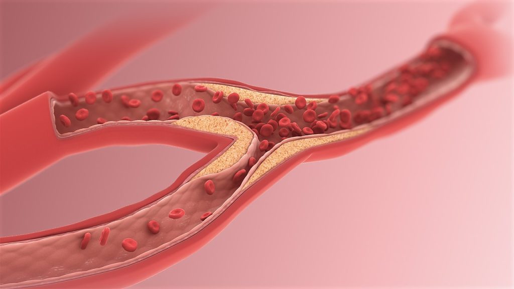
Atherosclerosis is almost twice as common in night owls compared to early birds, according to a study from the University of Gothenburg, Sweden. Circadian function appears to be particularly important during the early stages of cardiovascular disease.
Atherosclerosis involves fatty deposits gradually accumulating on the inside of the arteries, making it harder for blood to pass through. The disease is usually not noticed until it leads to blood clots causing angina, heart attack, or stroke.
Previous research has shown that people with late-night habits have an increased risk of cardiovascular disease, but this is the first study to show how circadian rhythms specifically affect calcification of the arteries.
Coronary artery calcification
The study, which has been published in the journal Sleep Medicine, involved 771 men and women aged between 50 and 64, all of whom are part of the larger population study SCAPIS.
The degree of artery calcification in the heart’s coronary arteries was examined using computer tomography.
Participants themselves indicated their so called chronotype on a five-point scale: extreme morning type, moderate morning type, intermediate type, moderate evening type, or extreme evening type.
Of the 771 participants, 144 identified as extreme morning types, and 128 as extreme evening types.
Among the group who were most alert in the morning, 22.2% had pronounced artery calcification — the lowest proportion of all five chronotypes.
The extreme evening type group had the highest prevalence of severe coronary artery calcification, at 40.6%.
The first author of the study is Mio Kobayashi Frisk, a doctoral student at Sahlgrenska Academy, University of Gothenburg:
“Our results indicate that extreme evening chronotype may be linked not only to poorer cardiovascular health in general, but also more specifically to calcification in the coronary arteries calcification and atherosclerosis,” Mio Kobayashi Frisk says.
Preventive treatment
The statistical analysis considered a range of other factors that can affect the risk of atherosclerosis, including blood pressure, blood lipids, weight, physical activity, stress level, sleep, and smoking.
The last author of the study is Ding Zou, a researcher at Sahlgrenska Academy, University of Gothenburg:
“As well as the previously known factors, the individual circadian rhythm also appears to be an important risk factor for atherosclerosis. We interpret our results as indicating that circadian rhythms are more significant early in the disease process. It should therefore particularly be considered in the preventive treatment of cardiovascular diseases,” says Ding Zou.
Self-reported chronotype
Those who had experienced a heart attack were excluded from the study, meaning that the study participants were healthier than the general population.
Another weakness identified by the researchers is that participants themselves provided their chronotype.
Each chronotype can be said to have an average time when half of the night’s sleep has passed.
In a previous study on the same population, though not necessarily the same individuals, this time occurred at 02:55 AM for the extreme morning type group and at 04:25 AM for the extreme evening type group.
With the remaining chronotype groups’ mid-sleep times were somewhere in between these extremes.
A study published in Nature Communications has revealed that the time at which we eat could influence our risk of developing cardiovascular disease. This study, suggests that eating a late first or last meal is associated with a higher risk of cardiovascular disease. It also appears that a longer night-time fasting duration is associated with a reduced risk of cerebrovascular disease such as stroke. The findings, suggest the importance of daily meal timing and rhythm in reducing cardiovascular disease risk.
The study was led by scientists from INRAE, the Barcelona Institute for Global Health, Inserm, and the Université Sorbonne Paris Nord.
Diet plays a major role in the development and progression of cardiovascular diseases. The modern lifestyle of Western societies has led to specific eating habits such as eating dinner late or skipping breakfast. In addition to light, the daily cycle of food intake (meals, snacks, etc) alternating with periods of fasting synchronizes the peripheral clocks, or circadian rhythms, of the body’s various organs, thus influencing cardiometabolic functions such as blood pressure regulation. Chrononutrition is emerging as an important new field for understanding the relationship between the timing of food intake, circadian rhythms and health.
Scientists used data from 103,389 participants in the NutriNet-Santé cohort (79% of whom were women, with an average age of 42) to study the associations between food intake patterns and cardiovascular disease. To reduce the risk of possible bias, the researchers accounted for a large number of confounding factors, especially sociodemographic factors (age, sex, family situation, etc.), diet nutritional quality, lifestyle and sleep cycle.
The results show that having a first meal later in the day (such as when skipping breakfast), is associated with a higher risk of cardiovascular disease, with a 6% increase in risk per hour delay. For example, a person who eats for the first time at 9 am is 6% more likely to develop cardiovascular disease than someone who eats at 8 am When it comes to the last meal of the day, eating late (after 9 pm) is associated with a 28% increase in the risk of cerebrovascular disease such as stroke compared with eating before 8 pm, particularly in women. Finally, a longer duration of night-time fasting – the time between the last meal of the day and the first meal of the following day – is associated with a reduced risk of cerebrovascular disease, supporting the idea of eating one’s first and last meals earlier in the day.
These findings, which need to be replicated in other cohorts and through additional scientific studies with different designs, highlight a potential role for meal timing in preventing cardiovascular disease. They suggest that adopting the habit of eating earlier first and last meals with a longer period of night-time fasting could help to prevent the risk of cardiovascular disease.
The BASHIR™ Endovascular Catheter (THROMBOLEX, Inc.), recently approved by the US Food and Drug Administration (FDA), is paving the way to more effective and safe treatment for acute pulmonary embolism. Already demonstrated to be effective for reducing blockages in lung arteries, a new study in JACC: Advances shows that the BASHIR™ catheter also reduces blockages in the smaller segmental pulmonary artery branches. These branches are ultimately responsible for oxygenating the blood in the lungs.
The new study from Temple University which was part of the National Institutes of Health-sponsored multicentre RESCUE clinical trial, further showed a correlation between decreased numbers of blockages in the small lung arteries and functional recovery of the right ventricle of the heart, which pumps blood into the main pulmonary artery of the lungs.
Compared to other devices, the BASHIR™ catheter also had significantly lower bleeding rates, a key advance in acute pulmonary embolism treatment.
“Blockages in these smaller, distal pulmonary arteries have never previously been explored in patients treated for acute pulmonary embolism,” explained Riyaz Bashir, MD, FACC, Professor of Medicine, Director of Vascular and Endovascular Medicine in the Section of Cardiology, Department of Medicine at the Lewis Katz School of Medicine and Temple University Hospital, co-inventor of the BASHIR™ Endovascular Catheter, and first author on the new report.
The BASHIR™ catheter is a small tube-like device that consists of a helical basket with six mini-infusion catheters at its farthest end.
When the infusion basket is expanded within a clot in a large blood vessel, new channels open, enabling blood to flow through the clot. The blood carries the body’s clot-dissolving chemicals into the channels, accelerating clot breakdown.
Researchers at THROMBOLEX™ Inc. were involved in developing and commercialising this platform technology.
The occlusion of small lung arteries is the major reason for the reduction in blood flow in patients with acute pulmonary embolism.
“The more occlusions a patient has, the lower their survival,” Dr Bashir said.
“Among the treatment goals is relieving obstructions in both large and small arteries.”
Patients who survive may, over more extended periods, be at high risk of developing chronic thromboembolic pulmonary hypertension (CTEPH), which is a life-threatening condition caused by increased blood pressure in the lungs.
In the new study, Dr Bashir and colleagues observed reductions in occlusions in segmental and proximal branches of the pulmonary artery 48 hours following treatment with the BASHIR™ catheter.
The blockages decreased even in those arteries that were distant from where the infusion basket was located, enabling improved blood flow and healing of the right ventricle.
“We suspect that the improvements in blood flow are due to both the expansion of the basket and the flow of the body’s clot dissolving molecules into the clot, which cause the blockage to shrink,” Dr. Bashir said.
“As the volume of blood flow improved, right ventricular function also improved, which could translate to better survival.”
In future work, Dr Bashir and colleagues plan to investigate mechanisms behind the observed reductions in arterial blockage.
Further trials are also needed to understand better the impact of treatment with the BASHIR™ catheter on patient survival and incidence of CTEPH.
On 29 October, during its annual scientific meeting at the Sandton Convention Centre in Johannesburg, the South African Heart Association (SA Heart®) met with members of the National Department of Health (NDoH), Council for Medical Schemes (CMS), South African Medical Association, medical aid industry, pharmaceutical and medical device industry, as well as the Global Heart Hub – which supports patient advocacy for cardiovascular disease – with the intention to develop an advocacy forum to create a more empowered and engaged community of patients living with cardiovascular disease.
During the symposium, SA Heart® emphasised the cardiac profession’s difficulty in obtaining approval and full reimbursement for the evidence-based treatments, shown to improve patient outcomes. The rules and regulations surrounding the availability of treatments, variously determined by the NDoH, the CMS and funders, currently inhibit access to effective cardiovascular disease management. SA Heart®, an affiliate member of the European Society of Cardiology (ESC), adopts the ESC’s guidelines as its own, supplemented by consensus statements that take prevailing local circumstances into account. Instead of following the up-to-date recommendations of the ESC, both the NDoH and various private sector funders rather give preference to the CMS algorithms that are years out of date. These anachronisms determine standards of care, despite ongoing advances in evidence-based medicine.
Prescribed Minimum Benefits (PMB’s) are a set of defined benefits to ensure that all medical scheme members have continuous access to defined minimum health services, regardless of the benefit option they select. PMB’s are limited in their reach. Private sector funders often retreat behind rules and regulations within the state sector, as to the type and level of treatment that they approve. Each funder independently determines what will be funded and at what level. Based upon their unique Health Technology Assessments, each funder then decides what treatment is approved or declined. It is unfair to restrict treatment to what is available only in the state sector. Novel therapies could be more widely available if there were more constructive agreements between state and industry. The CMS is currently reviewing the PMB package and defining patient entitlement.
SA Heart® pointed out that physicians are at the interface between the patient and the funder. Frequently, the patient misunderstands their own relationship with their medical aid and may deem the practitioner responsible for limitations imposed on his/her treatment. The administrative burden on clinicians is often intolerable, requiring repeat motivations, chronic medication forms, PMB applications and telephone calls to funders.
During the symposium pertinent questions were raised around funder and regulator reimbursement decisions, as well as the obscure mechanisms governing the pricing of medicines and devices. SA Heart® also raised concerns about the future of both the public and private healthcare system. A balance between the two sectors is a delicate one, requiring careful policy decisions to ensure that both systems will contribute effectively to the overall health of our country. Debates often focus around issues of equity and quality of service. The idea of centralizing decisions and distribution in the pharmaceutical and medical device sectors can be seen as an effort to ensure equitable access to essential medicines and equipment. However, this approach could negatively impact the ability to cater for South Africa’s diverse and specific needs – with more bureaucracy and inefficiency.
The meeting was called to determine interest amongst the various parties in establishing a forum to find solutions to these problems, and to harmonise the regulations around access to both medications and devices. The panellists were unanimous in their agreement that the current rules, regulations and disparities between the public and private sectors need to be reworked and there was consensus that a forum should be created to continue these discussions and arrive at effective, durable solutions. SA Heart® committed to driving this process, and will regularly engage all stakeholders in future meetings, with the ultimate goal of improving patient access to innovative cardiovascular treatments.
An international study published recently in the journal Brain has reported promising results in restoring function lost in mice and rat models of stroke. Researchers were able to restore lost brain function using small molecules that in the future could potentially be developed into a stroke recovery therapy.
“Communication between nerve cells in large parts of the brain changes after a stroke and we show that it can be partially restored with the treatment,” says Tadeusz Wieloch, senior professor of neurobiology at Lund University in Sweden.
“Concomitantly, the rodents regain lost somatosensory functions, something that around 60 per cent of all stroke patients experience today. The most remarkable result is that the treatment began several days after a stroke,” Wieloch continues.
In an ischaemic stroke, lack of blood flow to affected parts of the brain lead to loss of function such as paralysis, sensorimotor impairment and vision and speech difficulties, but also to pain and depression.
There are currently no approved drugs that improve or restore the functions after a stroke, apart from clot-dissolving treatment in the acute phase (within 4.5 hours of the stroke). Some spontaneous improvements occur, but many stroke patients suffer chronic loss of function.
For example, about 60% of stroke sufferers, experience lost somatosensory functions such as touch and position sense.
The new study shows that rats that were treated with a class of substances that inhibit the metabotropic glutamate receptor (mGluR5), a receptor that regulates communication in the brain’s nerve cell network.
“Rodents treated with the GluR5 inhibitor regained their somatosensory functions,” says Tadeusz Wieloch, who led the study.
Two days after the stroke, ie when the damage had developed and function impairment was most prominent, the researchers started treating the rodents that exhibited the greatest impaired function.
“A temporary treatment effect was seen after just 30 minutes, but treatment for several weeks is needed to achieve a permanent recovery effect. Some function improvement was observed even when the treatment started 10 days after a stroke,” says Tadeusz Wieloch.
Importantly, sensorimotor functions improved, even though the extent of the brain damage was not diminished.
This, explains Tadeusz Wieloch, is due to the intricate network of nerve cells in the brain, known as the connectome – the way brain areas are inter connected and communicate form the basis for various brain functions.
“Impaired function after a stroke is due to cell loss, but also because of reduced activity in large parts of the connectome in the undamaged brain. The receptor mGluR5 is apparently an important factor in the reduced activity in the connectome, which is prevented by the inhibitor which therefore restores the lost brain function,” says Tadeusz Wieloch.
The results also showed that sensorimotor function was further improved if treatment with the mGluR5 inhibitor is combined with somatosensory training by housing several rodents in cages enriched with toys, chains, grids, and plastic tubes.
The researchers hope that in the future their results could lead to a clinical treatment that could be initiated a few days after an ischaemic stroke.
“Combined with rehabilitation training, it could eventually be a new promising treatment. However, more studies are needed. The study was conducted on mice and rats, and of course needs to be repeated in humans. This should be possible since several mGluR5 inhibitors have been studied in humans for the treatment of neurological diseases other than stroke, and shown to be tolerated by humans,” says Tadeusz Wieloch.
A recent clinical trial published in JAMA found that excluding aspirin for advanced heart failure (HF) patients with a ventricular assist device saw a reduction in bleeding events while maintaining their survival rates.
The ARIES-HM3 Randomised Clinical Trial assessed the safety and efficacy of excluding aspirin from the antithrombotic regimen in patients with advanced HF who have undergone implantation of a fully magnetically levitated left ventricular assist device (LVAD).
“We can now safely say that not giving aspirin is not only safe from a thromboembolic risk profile but results in improved adverse event rate by a significant reduction in non-surgical bleeding which is a well-known complication related to LVAD therapy,” said Mirnela Byku, MD, P.D, MBA, co-author of the study and director of the UNC Durable Mechanical Circulatory Device Program at the UNC School of Medicine.
“Improving not only longevity but also reducing morbidity and improving quality of life is a big focus in the field of MCS.”
Until this study, there had been no consensus in the field about use of or dose of aspirin in the LVAD population.
The international clinical trial followed a randomised, double-blind, placebo-controlled design and involved 628 patients across 51 centres in 9 countries.
The patients were divided into two groups: one receiving aspirin (100mg/d) and the other receiving a placebo in addition to vitamin K antagonist (VKA) therapy.
A focus was to determine if the likelihood a patient experiences major nonsurgical haemocompatibility-related adverse events (such as stroke, pump thrombosis, major bleeding, or arterial peripheral thromboembolism) within 12 months differed between the two groups.
The results showed that not giving aspirin to patients with advanced HF, treated with a fully magnetically levitated LVAD who are receiving VKAs, did not make their survival worse. Furthermore, aspirin avoidance was associated with a significant reduction (34%) in major nonsurgical bleeding events.
Although past research has indicated that moderate alcohol consumption can reduce cardiovascular disease (CVD) risk, more recent studies suggest that moderate levels of drinking may be hazardous to heart health. A new analysis now sheds new insight on this complex relationship between alcohol consumption and the progression of CVD, showing that a few particular alcohol metabolites strongly influence its protective effects.
Published in the journal BMC Medicine, the study observed a total of 60 alcohol consumption-related metabolites, identifying seven circulating metabolites that link long-term moderate alcohol consumption with an increased risk of CVD, and three circulating metabolites that link this same drinking pattern with a lower risk of CVD.
The findings from the study led by Boston University School of Public Health and Friedman School of Nutrition Science and Policy at Tufts University (Friedman School) detail the molecular pathway of long-term alcohol consumption and show that further research on these metabolites is needed for targeted prevention and treatment of alcohol-related CVD.
“The study findings demonstrate that alcohol consumption may trigger changes of our metabolomic profiles, potentially yielding both beneficial and harmful outcomes,” says Dr Chunyu Liu, assistant professor of biostatistics at BUSPH and co-corresponding/co-senior author of the study along with Dr.Jiantao Ma, assistant professor in the Division of Nutrition Epidemiology and Data Science at the Friedman School.
“However, rather than definitively settling that debate, this study underscores the intricate effects of alcohol consumption on cardiovascular health and generates a useful hypothesis for future investigations,” Dr Liu says.
The researchers analysed blood samples to measure the association between the cumulative average consumption of beer, wine, and liquor and 211 metabolites among Framingham Heart Study Offspring Study participants, who are the children of participants in the long-running Boston University-based Framingham Heart Study, over 20 years.
Of the 2428 participants, 636 developed CVD over the study period. Among the 60 drinking-related metabolites, 13 metabolites had a stronger association with alcohol consumption in women than in men, perhaps due to higher blood alcohol levels from women’s generally smaller body size versus the same amount of alcohol.
Consuming different types of alcohol was also linked to different metabolomic responses, with beer consumption generating a slightly weaker association overall than wine and liquor.
In roughly two-thirds of the 60 metabolites, higher plasma levels were detected in participants who consumed greater amounts of alcohol. Branched-chain amino acids (BCAAs), were among the metabolites not associated with alcohol consumption.
The researchers then calculated two alcohol consumption-associated metabolite scores, which had opposite associations with the development of CVD.
“While our study presents intriguing findings, validation through state-of-the-art methods and large and diverse study populations is crucial,” Dr Ma says.
“To enhance reliability, we aim to conduct larger-scale research involving a more diverse racial and ethnic background, as the current study participants are all white. In addition, we will expand our study to integrate with other molecular markers such as genetic information to illustrate the complex relationships between alcohol consumption, metabolite features, and cardiovascular risk.”
Some individuals with anti-Ro/SSA antibodies (anti–Sjögren’s-syndrome–related antigen A autoantibodies, also called anti-Ro antibodies) have autoimmune diseases such as lupus or Sjögren’s syndrome, but many have no symptoms. A clinical trial published in Arthritis & Rheumatology found that high levels of these antibodies in pregnant women are associated with foetal atrioventricular block (AVB), which occurs when inflammation and subsequent scarring prevent electric signals from the heart’s atria from reaching the ventricles. The disease is associated with life-long pacing and can be fatal.
In the trial, called Surveillance ToPrevent AV Block Likely to Occur Quickly (STOP BLOQ), the incidence of AVB increased with higher levels of anti-Ro/SSA antibodies, reaching 7.7% for those in the top quartile, which increased to 27.3% in those with a previous child who had AVB, although participant numbers in that category were small. Antibody titres did not change over time. The trial also revealed that home-based foetal heart rate monitoring reliably detected conduction abnormalities, which may reduce the need for serial echocardiograms.
“Examining the levels of anti-Ro/SSA antibodies is an important advance since for women with low titres, monitoring is probably not necessary and for those with high titres the increased risk supports surveillance,” said corresponding author Jill Buyon, MD, of NYU Langone Health. She added that this study also indicated that titres of antibodies do not change and that additional factors besides antibodies contribute to risk.
“That home monitoring can rapidly and accurately identify early foetal conduction disease is a major step forward that may significantly decrease the need for echocardiograms and hopefully facilitate reversibility,” added senior author and research professor Bettina Cuneo MD, of the University of Arizona-Tucson College of Medicine.