Category: Cancer

SSRIs Could Help the Immune System Fight Cancer

Photo by Danilo Alvesd on Unsplash

Selective serotonin reuptake inhibitors (SSRIs) could help the immune system fight cancer, according to recent UCLA research. The study, published in Cell, found that SSRIs significantly enhanced the ability of T cells to fight cancer and suppressed tumour growth across a range of cancer types in both mouse and human tumour models.

“It turns out SSRIs don’t just make our brains happier; they also make our T cells happier – even while they’re fighting tumours,” said Lili Yang, PhD, senior author of the new study. “These drugs have been widely and safely used to treat depression for decades, so repurposing them for cancer would be a lot easier than developing an entirely new therapy.”

According to the CDC, one out of eight adults in the US takes an antidepressant, and SSRIs are the most commonly prescribed. These drugs increase levels of serotonin the brain’s “happiness hormone” by blocking the activity of a protein called serotonin transporter, or SERT. 

While serotonin is best known for the role it plays in the brain, it’s also a critical player in processes that occur throughout the body, including digestion, metabolism and immune activity. 

Dr Yang and her team first began investigating serotonin’s role in fighting cancer after noticing that immune cells isolated from tumours had higher levels of serotonin-regulating molecules. At first, they focused on MAO-A, an enzyme that breaks down serotonin and other neurotransmitters, including norepinephrine and dopamine. 

In 2021, they reported that T cells produce MAO-A when they recognise tumours, which makes it harder for them to fight cancer. They found that treating mice with melanoma and colon cancer using MAO inhibitors, also called MAOIs – the first class of antidepressant drugs to be invented – helped T cells attack tumours more effectively. 

However, because MAOIs have safety concerns, including serious side effects and interactions with certain foods and medications, the team turned its attention to a different serotonin-regulating molecule: SERT. 

“Unlike MAO-A, which breaks down multiple neurotransmitters, SERT has one job – to transport serotonin,” explained Bo Li, PhD, first author of the study and a senior research scientist in the Yang lab. “SERT made for an especially attractive target because the drugs that act on it – SSRIs – are widely used with minimal side effects.” 

The researchers tested SSRIs in mouse and human tumour models representing melanoma, breast, prostate, colon and bladder cancer. They found that SSRI treatment reduced average tumour size by over 50% and made the cancer-fighting T cells, known as killer T cells, more effective at killing cancer cells. 

“SSRIs made the killer T cells happier in the otherwise oppressive tumour environment by increasing their access to serotonin signals, reinvigorating them to fight and kill cancer cells,” said Dr Yang, who is also a professor of microbiology, immunology and molecular genetics and a member of the UCLA Health Jonsson Comprehensive Cancer Center.

How SSRIs could boost the effectiveness of cancer therapies 

The team also investigated whether combining SSRIs with existing cancer therapies could improve treatment outcomes. They tested a combination of an SSRI and anti-PD-1 antibody – a common immune checkpoint blockade (ICB) therapy – in mouse models of melanoma and colon cancer. ICB therapies block immune checkpoint molecules that normally suppress immune cell activity, allowing T cells to attack tumours more effectively. 

The results were striking: the combination significantly reduced tumour size in all treated mice and even achieved complete remission in some cases. 

“Immune checkpoint blockades are effective in fewer than 25% of patients,” said James Elsten-Brown, a graduate student in the Yang lab and co-author of the study. “If a safe, widely available drug like an SSRI could make these therapies more effective, it would be hugely impactful.”

To confirm these findings, the team will investigate whether real-world cancer patients taking SSRIs have better outcomes, especially those receiving ICB therapies. About 20% of cancer patients are already taking the medication, Dr Yang said.

Dr Yang added that using existing FDA-approved drugs could speed up the process of bringing new cancer treatments to patients, making this research especially promising.

“Studies estimate the bench-to-bedside pipeline for new cancer therapies costs an average of $1.5 billion,” she said. “When you compare this to the estimated $300 million cost to repurpose FDA-approved drugs, it’s clear why this approach has so much potential.”

Source: University of California – Los Angeles Health Sciences

Scientific Breakthrough: Price of Costly Cancer Drug can be Halved

Source: Unsplash CC0

Taxol is one of the most commonly prescribed chemotherapy drugs for breast, ovarian, cervical, and lung cancer. Yet producing the drug is complex, costly, and environmentally burdensome, as it currently relies on a complicated chemical semi-synthesis. For 30 years, scientists around the world have tried to understand how taxol, a natural compound derived from the Pacific yew tree, forms in nature. Decoding this process would allow for biotech-based production. But the final steps remained unknown – until now.

A research team from the University of Copenhagen has succeeded in finding the two missing pieces: They have identified the enzymes responsible for the two critical final steps in the biosynthetic pathway that makes Taxol active as a drug.

“Taxol has been the Holy Grail in this research field for decades because it’s an exceptionally complex molecule. But with the discovery of the final two enzymes, we now fully understand how it’s formed. This has allowed us to develop a biotechnological method to produce taxol in yeast cells,” says Sotirios Kampranis, Professor at the Department of Plant and Environmental Sciences and senior author of the study published in Nature Synthesis.

The method involves cloning the taxol-producing genes from the yew tree and inserting them into yeast cells. These engineered yeast cells then become host organisms or micro-factories with the full recipe to produce taxol.

Affecting women in developing countries

The research team from the University of Copenhagen has applied for patenting the method and is in the process of launching a spin-out company to manufacture biosynthetic Taxol. 

“Using this method, we can produce Taxol cheaper than current conventional methods. Looking ahead, once we refine the process further, we expect to be able to reduce the cost by half,” says Assistant Professor and first author Feiyan Liang.

Lower prices are especially crucial as ovarian cancer is on the rise globally. The prevalence of the disease is expected to increase by over 55% by 2050, with the vast majority of cases in low and middle-income countries. The number of women dying from ovarian cancer is projected to rise by nearly 70% in the same period.

Currently, taxol costs more than USD20 000 per kilogram, making it one of the most expensive active pharmaceutical ingredients in use.

“We see increasing demand for Taxol in many developing countries, where the high price is a major barrier. We hope our work will contribute to lower-priced drugs so that more people can have access to cancer treatment,” Feiyan Liang says.

Much more sustainable

The new method is not only more cost-effective but also more sustainable than chemical synthesis. One advantage is that the procedure does not involve harmful chemicals and solvents common in chemical production. Another advantage is that it allows the use of more crude, less purified extracts from yew needles as starting material – much cheaper than the ultra-pure inputs required in chemical semi-synthesis. On top of that, the materials can be recycled.

“We want to show that it’s possible to build a biotechnological drug production that is both sustainable and low-cost. There are very few examples of that today, but we now have the foundation to make it happen,” says Sotirios Kampranis. 

TWO TREES PER TREATMENT

  • Taxol was originally extracted from the inside bark of the Pacific yew tree (Taxus brevifolia), but as the taxol content in the bark is very low, harvesting it meant removing all the bark and as a result of this killing the tree.
  • Yew trees take 70 to 100 years to mature. Producing just one treatment required about two trees, making this method highly unsustainable. It was abandoned years ago, though wild yew trees are still under pressure in some regions.
  • Today’s most common method involves harvesting a similar compound from yew needles for chemical synthesis, but the cost of this process is still high, which is why the average price of taxol exceeds USD 20 000 per kilogram (source: pharmacompass.com).

Source: University of Copenhagen – Faculty of Science

South African Study Identifies Two New Breast Cancer Genes in Black Women

Genetic factors contribute to some 30% of breast cancer cases in SA, necessitating investment in genomic research in African contexts.

Photo by National Cancer Institute

A seminal genetic study published in Nature Communications has discovered two genetic variants linked to breast cancer in black South African women, deepening knowledge about the genetic basis for this disease in African populations.

The genome-wide association study (GWAS) of breast cancer is the first to have been done in African women living on the continent.

A GWAS is a powerful research method that scans the entire DNA of many people to find genetic differences associated with a specific disease or trait.

In this case, the scientists at the Sydney Brenner Institute for Molecular Bioscience (SBIMB) scanned for breast cancer and found consistent genetic patterns in black South African women.

The SBIMB researchers discovered genetic signals around the gene RAB27A, a member of the RAS oncogene family, and USP22, a gene which is highly active in breast cancer cells and associated with a poor health prognosis.

“These genes have not been associated with the disease before, which is an important advance in understanding breast cancer risk and biology in women of African ancestry,” says Dr Mahtaab Hayat, the lead author of the study.

The two new genetic variants were identified in black South African women with breast cancer enrolled in the Johannesburg Cancer Study, compared to women without cancer in the Africa Wits-INDEPTH Partnership for Genomic Research (AWI-Gen) study.

Until now, most breast cancer genetics research has focused on European and Asian populations, with studies of African ancestry limited primarily to African- American women, who largely descend from West African populations.

A tool that estimates lifetime cancer risk based on DNA, the polygenic risk score (PRS), performed poorly in distinguishing South African women with breast cancer from those without.

“This is because most PRSs were developed in European populations, and their inaccuracy in African populations highlights the urgent need for ancestry-specific tools in cancer risk prediction,” says Dr Jean-Tristan Brandenburg, also in the SBIMB and a lead author.

Breast cancer is the second most common cancer in South Africa and the most common cancer in women globally, with genetic factors contributing to about 30% of cases. “Our study makes a compelling case for investing in genomic research rooted in African contexts,” notes Hayat.

The potential for precision medicine

If further studies confirm these findings, the USP22 and RAB27A genes could be specific targets for new drugs. “We could potentially target harmful cancer cells while sparing healthy tissue, which is ideally what we want when administering cancer treatment,” says Distinguished Professor at the SBIMB, Chris Mathew, and a lead project investigator.

Furthermore, if a specific gene is associated with poorer survival, it can be used as a biomarker to identify more aggressive cancers and help predict which patients may need more intensive treatment and monitoring.

Understanding the genetic architecture of complex diseases helps scientists figure out the biological processes leading to these conditions and find drug targets and treatments for groups of individuals with similar disease risk profiles.

Genomic diversity in Africa is unparalleled

African populations have more genetic variation than any other population in the world, but they have been significantly underrepresented in genomic research. This means that the global understanding of disease risk, and the tools and treatment developed from it, is limited.

“The study reveals that more people can benefit from genetic discoveries. It proves that new risk factors are still out there, waiting to be found,” says Hayat.

Source: University of the Witwatersrand

A Downside of Taurine: It Drives Leukaemia Growth

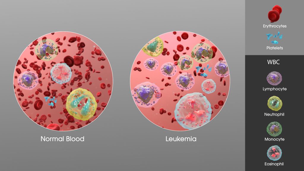
SAG Leukaemia. Credit: Scientific Animations CC0

A new scientific study identified taurine, which is made naturally in the body and consumed through some foods, as a key regulator of myeloid cancers such as leukaemia, according to a paper published in the journal Nature.

The preclinical research shows that scientists are a step closer to finding new ways to target leukaemia, which is one of the most aggressive blood cancers. The Wilmot Cancer Institute investigators at the University of Rochester were able to block the growth of leukaemia in mouse models and in human leukaemia cell samples by using genetic tools to prevent taurine from entering cancer cells.

Led by Jeevisha Bajaj, PhD, the research team discovered that taurine is produced by a subset of normal cells in the bone marrow microenvironment, the tissue inside bones where myeloid cancers begin and expand. Leukaemia cells are unable to make taurine themselves, so they rely on a taurine transporter (encoded by the SLC6A6 gene) to grab taurine from the bone marrow environment and deliver it to the cancer cells.

The discovery occurred as scientists were mapping what happens within the bone marrow and its ecosystem—a longtime focus among Wilmot researchers, who have advanced the science around the microenvironment with the goal of improving blood cancer treatments.

“We are very excited about these studies because they demonstrate that targeting uptake by myeloid leukaemia cells may be a possible new avenue for treatment of these aggressive diseases,” said Bajaj, an assistant professor in the Department of Biomedical Genetics and a member of Wilmot’s Cancer Microenvironment research program.

Researchers also discovered that as leukaemia cells drink up taurine, it promotes glycolysis (a breakdown of glucose to produce energy) to feed cancer growth. Prior to this, the authors said, it was not known that taurine might have a cancer-promoting role.

Leukaemia has several subtypes and survival rates vary. This study finds that taurine transporter expression is essential for the growth of multiple subtypes including acute myeloid leukaemia (AML), chronic myeloid leukaemia (CML), and myelodysplastic syndromes (MDS), which all originate from blood stem cells in the bone marrow. Future studies will investigate signals from the microenvironment that promote the transition of MDS, a precursor to leukaemia, to acute leukaemia.

Source: University of Rochester Medical Center

CAR-T Cell Therapy Causes ‘Brain Fog,’ Study Shows

Killer T cells about to destroy a cancer cell. Credit: NIH

After treatment with CAR-T cells, immune cells engineered to attack cancer, patients sometimes tell their doctors they feel like they have “brain fog,” or forgetfulness and difficulty concentrating.

A new Stanford Medicine-led study shows that CAR-T cell therapy causes mild cognitive impairments, independent of other cancer treatments, and that this happens via the same cellular mechanism as cognitive impairment from two other causes: chemotherapy and respiratory infections such as flu and COVID-19. The study, conducted mostly in mice, which was published in Cell, also identifies strategies for reversing the problem.

Medications that ameliorate brain fog will enable better recovery from cancer immunotherapies, the researchers said.

“CAR-T cell therapy is enormously promising,” said senior author, Michelle Monje, MD, PhD, professor in paediatric neuro-oncology. “We need to understand all its possible long-term effects, including this newly recognised syndrome of immunotherapy-related cognitive impairment, so we can develop therapeutic approaches to fix it.”

The study’s lead authors are Anna Geraghty, PhD, senior staff scientist in the Monje lab, and MD/PhD student Lehi Acosta-Alvarez.

Cognitive impairment after CAR-T cell therapy is typically mild; patients are not developing dementia, for instance. But it is frustrating and may not resolve on its own, Monje said. In mice, her team reversed the impairment using compounds similar to existing medications or medications in clinical development – meaning a treatment could be available relatively quickly, she said.

“We’re deeply interested in how cancer therapies affect cognition because it affects patients’ quality of life,” Monje said. “And this is especially important for kids because their brains are still developing.”

Investigating brain fog

CAR-T cell therapy was approved in the US for acute lymphoblastic leukaemia in 2017. The treatment involves removing some of the patient’s own immune cells, known as T cells, and engineering them to attack targets on cancer cells. The modified T cells are returned to the patient’s body, where they recognise and destroy cancer.

In addition to leukaemia, CAR-T cells are now used to treat other blood cancers, including multiple myeloma and some kinds of lymphoma, and they are being tested in clinical trials for various solid tumours. Monje and her colleagues have an ongoing trial of CAR-T cells for deadly brain stem and spinal cord tumours in children, which is beginning to show success.

Although patients report brain fog after CAR-T cell therapy, studies to measure how much cognitive impairment the therapy causes are only just emerging.

The research team wanted to get a comprehensive understanding of the situations in which CAR-T cell therapy might cause cognitive impairment. They studied mice that had tumours induced in the brain, blood, skin and bone. The researchers wanted to understand the influence on cognition of CAR-T cell treatment in combination with the tumours’ location (originating in, spreading to or staying outside the brain), as well as the degree to which the engineered cells evoked additional, accompanying immune responses. Before and after CAR-T cell treatment, the researchers used standard cognitive tests on the mice, measuring how mice responded to a novel object and navigated a simple maze.

CAR-T therapy caused mild cognitive impairment in mice with cancers originating in, metastasizing to and located completely outside the brain. The only mice tested that did not develop cognitive impairment after CAR-T treatment were those that had bone cancer that causes minimal additional inflammation beyond the cancer-fighting activity of the CAR-T cells.

“This is the first study to demonstrate that immunotherapy on its own is sufficient to cause lasting cognitive symptoms,” Monje said. “It’s also the first paper to uncover the mechanisms. We found the exact same pathophysiology we’ve seen in brain fog syndromes that occur after chemotherapy, radiation, and mild respiratory COVID-19 or influenza.”

The researchers demonstrated that the brain’s immune cells, called microglia, are key players in the problem. First, the microglia become activated by the body’s immune response. The activated, “annoyed” microglia produce inflammatory immune molecules known as cytokines and chemokines, which in turn have widespread effects throughout the brain. They are particularly harmful for oligodendrocytes, the brain cells responsible for making myelin, the fatty substance that insulates nerve fibres and helps nerves transmit signals more efficiently. Reduction in the nerves’ insulation translates into cognitive impairment.

Examining tissue samples

The scientists also analysed samples of brain tissue from human subjects who participated in the team’s ongoing clinical trial of CAR-T cells for spinal cord and brain stem tumours. Using post-mortem tissue samples, the researchers confirmed that microglia and oligodendrocytes appear dysregulated in the same way the team had observed in mice after CAR-T therapy.

In mice, the research team tested strategies to resolve the cognitive problems. They gave a compound that depleted microglia in the brains of the mice for a two-week period. After that transient depletion, the microglia  returned in the brain in a normal, non-reactive state. The mice were no longer cognitively impaired.

The researchers also gave the mice a medication that enters the brain and interferes with signals from damaging chemokines, blocking a specific receptor for these molecules.

“That alone rescued cognition,” Monje said, adding that the researchers are now exploring how to safely translate the two strategies – transiently depleting microglia or interrupting chemokine signals – in people who have had CAR-T cell therapy.

“This research further illustrates that there is a unifying principle underpinning brain fog syndromes,” said Monje, a member of the Stanford Cancer Institute. “And this particular study is so exciting because not only have we identified the cells central to this pathophysiology, we’ve found a molecular target we can investigate to treat it.”

Source: Stanford Medicine

Abdominal Ascites Fuels Immune Failure in Ovarian Cancer

Human NK cells have large nuclei stained in blue and droplets of fat stores stained in red. Image: Dr Karen Slattery, Trinity College Dublin.

New research led by Irish scientists has uncovered how lipid-rich fluid in the abdomen, known as ascites, plays a central role in weakening the body’s immune response in advanced ovarian cancer. The findings offer new insights into immune suppression in ovarian cancer and open promising avenues for future immunotherapy approaches

Over 70% of patients with ovarian cancer are diagnosed at an advanced stage, often presenting with large volumes of ascites. This ascites fluid not only supports the spread of cancer throughout the abdominal cavity but also significantly impairs the body’s immune defences.

Understanding how ascites affects the immune system is important for developing better treatments that use the immune system to fight cancer. 

In this recent study, researchers from Trinity and University College Dublin explored how ascites disrupts immune cell function, with a particular focus on natural killer (NK) cells and T cells, which are key players in the body’s ability to eliminate tumours.

By analysing the contents of ascites fluid from ovarian cancer patients, the team identified a group of fat molecules called phospholipids as key drivers of this immune dysfunction. 

Dr Karen Slattery, Research Fellow in the Trinity Translational Medicine Institute, is the first author of the research article just published in the leading international journal Science Immunology.

She said: “We found that these lipids interfere with NK cell metabolism and suppress their ability to kill cancer cells. Crucially, we also discovered that blocking the uptake of these phospholipids into NK cells using a specific receptor blocker can restore their anti-tumour activity, which offers a compelling new target for therapeutic intervention.” 

“This work adds a critical piece to the puzzle of why ovarian cancer is so aggressive and has such poor outcomes. While the immune system is naturally equipped to detect and destroy cancer cells, this function is switched off in many individuals with ovarian cancer, and we now know that this is in part due to the fat-rich environment created by ascites.” 

Prof Lydia Lynch, formerly based in Trinity and now in Princeton University, is the senior author of the research article. She said: “This study marks a significant advancement in ovarian cancer research, identifying a new mechanism underpinning immune failure and laying the foundation for new therapies that could restore immune function in these patients. By targeting the fat-induced suppression of immune cells, future treatments could empower the body’s own immune defences to fight back and in doing so, improve outcomes for ovarian cancer patients.”

Source: Trinity College Dublin

Removing Ovaries and Fallopian Tubes Linked to Lower Risk of Early Death Among Certain Breast Cancer Patients

Photo by National Cancer Institute on Unsplash

Women diagnosed with breast cancer who carry particular BRCA1 and BRCA2 genetic variants are offered surgery to remove the ovaries and fallopian tubes as this dramatically reduces their risk of ovarian cancer. Now, Cambridge researchers have shown that this procedure – known as bilateral salpingo-oophorectomy (BSO) – is associated with a substantial reduction in the risk of early death among these women, without any serious side-effects.

Women with certain variants of the genes BRCA1 and BRCA2 have a high risk of developing ovarian and breast cancer. These women are recommended to have their ovaries and fallopian tubes removed at a relatively early age – between the ages 35 and 40 years for BRCA1 carriers, and between the ages 40 and 45 for BRCA2 carriers.

Previously, BSO has been shown to lead to an 80% reduction in the risk of developing ovarian cancer among these women, but there is concern that there may be unintended consequences as a result of the body’s main source of oestrogen being removed, which brings on early menopause. This can be especially challenging for BRCA1 and BRCA2 carriers with a history of breast cancer, as they may not typically receive hormone replacement therapy to manage symptoms. The overall impact of BSO in BRCA1 and BRCA2 carriers with a prior history of breast cancer remains uncertain. 

Ordinarily, researchers would assess the benefits and risks associated with BSO through randomised controlled trials, the ‘gold standard’ for testing how well treatments work. However, to do so in women who carry the BRCA1 and BRCA2 variants would be unethical as it would put them at substantially greater risk of developing ovarian cancer.

To work around this problem, a team at the University of Cambridge, in collaboration with the National Disease Registration Service (NDRS) in NHS England, turned to electronic health records and data from NHS genetic testing laboratories collected and curated by NDRS to examine the long-term outcomes of BSO among BRCA1 and BRCA2 PV carriers diagnosed with breast cancer. The results of their study, the first large-scale study of its kind, are published today in The Lancet Oncology.

The team identified a total of 3400 women carrying one of the BRCA1 and BRCA2 cancer-causing variants (around 1700 women for each variant). Around 850 of the BRCA1 carriers and 1,000 of the BRCA2 carriers had undergone BSO surgery.

Women who underwent BSO were around half as likely to die from cancer or any other cause over the follow-up period (a median follow-up time of 5.5 years). This reduction was more pronounced in BRCA2 carriers compared to BRCA1 carriers (a 56% reduction compared to 38% respectively). These women were also at around a 40% lower risk of developing a second cancer.

Although the team say it is impossible to say with 100% certainty that BSO causes this reduction in risk, they argue that the evidence points strongly towards this conclusion.

Importantly, the researchers found no link between BSO and increased risk of other long-term outcomes such as heart disease and stroke, or with depression. This is in contrast to previous studies that found evidence in the general population of an association between BSO and increased risk of these conditions.

First author Hend Hassan, a PhD student at the Centre for Cancer Genetic Epidemiology, Department of Public Health and Primary Care, and Wolfson College, Cambridge, said: “We know that removing the ovaries and fallopian tubes dramatically reduces the risk of ovarian cancer, but there’s been a question mark over the potential unintended consequences that might arise from the sudden onset of menopause that this causes.

“Reassuringly, our research has shown that for women with a personal history of breast cancer, this procedure brings clear benefits in terms of survival and a lower risk of other cancers without the adverse side effects such as heart conditions or depression.”

Most women undergoing BSO were white. Black and Asian women were around half as likely to have BSO compared to white women. Women who lived in less deprived areas were more likely to have BSO compared to those in the most-deprived category.

Hassan added: “Given the clear benefits that this procedure provides for at-risk women, it’s concerning that some groups of women are less likely to undergo it. We need to understand why this is and encourage uptake among these women.”

Professor Antonis Antoniou, from the Department of Public Health and Primary Care, the study’s senior author, said: “Our findings will be crucial for counselling women with cancer linked to one of the BRCA1 and BRCA2 variants, allowing them to make informed decisions about whether or not to opt for this operation.”

Professor Antoniou, who is also Director of the Cancer Data-Driven Detection programme, added: “The study also highlights the power of exceptional NHS datasets in driving impactful, clinically relevant research.”

The research was funded by Cancer Research UK, with additional support from the National Institute for Health and Care Research (NIHR) Cambridge Biomedical Research Centre.

The University of Cambridge is fundraising for a new hospital that will transform how we diagnose and treat cancer. Cambridge Cancer Research Hospital, a partnership with Cambridge University Hospitals NHS Foundation Trust, will treat patients across the East of England, but the research that takes place there promises to change the lives of cancer patients across the UK and beyond. Find out more here.

Reference

Hassan, H et al. Long-term health outcomes of bilateral salpingo-oophorectomy in BRCA1 and BRCA2 pathogenic variant carriers with personal history of breast cancer: a retrospective cohort study using linked electronic health records. Lancet Oncology; 7 May 2025; DOI: 10.1016/S1470-2045(25)00156-1

The original text of this story is licensed under a Creative Commons Attribution-NonCommercial-ShareAlike 4.0 International License.

Read the original article.

Does Cancer Treatment Affect Connections in the Brain?

Photo by Fakurian Design on Unsplash

New research published in the Journal of Magnetic Resonance Imaging has uncovered changes in brain connectivity during chemotherapy in patients with breast cancer.

In the study of 55 patients with breast cancer and 38 controls without cancer, investigators conducted functional magnetic resonance imaging scans of participants’ brains over several months.

Scans from patients revealed changes in brain connectivity, particularly in the frontal-limbic system (involved in executive functions) and the cerebellar cortex (linked to memory) throughout the course of treatment. These changes got worse and spread more as chemotherapy continued.

“The findings suggest that chemotherapy can quickly disrupt brain function in breast cancer patients, potentially contributing to cognitive issues,” the authors wrote.

Source: Wiley

Hyperbaric Oxygen for Radiation-induced Injuries Provides Lasting Relief

Photo by National Cancer Institute on Unsplash

Hyperbaric oxygen treatment provides long-term relief for patients suffering from late radiation-induced injuries after treatment of cancer in the lower abdominal area. Five years after hyperbaric oxygen therapy, the positive effects remain. This has been shown in a study conducted at the University of Gothenburg, published in eClinicalMedicine.

Radiation therapy is a component of many cancer treatments in organs such as the prostate, colon, ovaries and cervix. While tumour cells are destroyed, 5-10% of patients experience severe side effects due to healthy tissue being affected by the radiation therapy.

Symptoms may include urinary incontinence, bleeding and severe pain from the lower abdomen that becomes both physically and socially disabling. These problems can occur several years after radiation therapy and cause chronic and increasing discomfort.

Researchers have previously shown that patients experience significantly less discomfort after hyperbaric oxygen treatment. The question in the current study was whether the relief would last over several years. The time aspect is important for future decisions on broader use of the method.

From severe problems to a normal life 

Initially, all participants had severe symptoms. The group that was randomly assigned to hyperbaric oxygen treatment fared significantly better than the control group in terms of incontinence, bleeding and pain. The positive effects were sustained over the five year follow-up period. 

Nicklas Oscarsson, senior consultant in anaesthesiology and intensive care, and researcher at the University of Gothenburg and Sahlgrenska University Hospital was the principle investigator of the study:

“Patients who respond to treatment go from being very distressed by their symptoms and restricted by their need to have quick access to a toilet, to being able to live a fully normal life. Now we know that this pronounced improvement last for at least five years. The treatment can therefore lead to the healing of an otherwise chronic injury,” he states.

The effects achieved are due to cells sensing and adapting to high oxygen levels. The increased levels of oxygen provided in a hyperbaric chamber increases vascular growth and stops chronic inflammation, reducing severe side effects.

For the oxygen treatments, participants spent 90 minutes a day in a hyperbaric chamber on 30-40 occasions, at a pressure of 1.4 atmospheres (equivalent to 14m underwater). The control group received the usual treatment, which normally includes medication and physiotherapy, for example.

The capacity already exists today

“We have reason to believe that there are many patients with severe symptoms who are never referred to hyperbaric oxygen therapy. Today we already have the capacity to treat more patients, but we need to be better at sharing our knowledge with our colleagues and with patient associations,” says Nicklas Oscarsson.

Severe side effects after radiation therapy are one of the main limitations on the dose of radiation that can be given in cancer treatment. The availability of a treatment that can reduce the number of people affected by these side effects opens the door to increased radiation doses and thus more curable tumours. One area for further investigation, according to the researchers, is whether early treatment with hyperbaric oxygen can prevent the occurrence of severe side effects.

The results are based on surveys and analyses of the participants who have been involved all the way, 70 adults. The treatments were conducted at five university hospitals in the Nordic countries: Rigshospitalet in Denmark, Turku in Finland, Haukeland in Norway, and Karolinska and Sahlgrenska in Sweden.

Source: University of Gothenburg

Preventing Unnecessary Pancreatic Cancer Surgery

Pancreatic cancer. Credit: Scientific Animations CC BY-SA 4.0

Pancreatic cysts are fluid-filled sacs that can form in the pancreas. Some remain benign, while others have the potential to develop into pancreatic cancer. A recent study, which followed 257 patients in Japan for an average of five years, showed that the presence or absence of invasive nodules in pancreatic cysts is key to assessing whether these cysts are benign or cancerous.

The findings, published in the journal Annals of Surgery, may help patients diagnosed with a high risk of pancreatic cancer to avoid unnecessary surgery.

Pancreatic cancer is one of the most life-threatening and rapidly growing cancers. Pancreatic cysts, known as pancreatic intraductal papillary mucinous neoplasms (IPMNs), are gaining attention as one of the precursors of the cancer that can be identified by radiological imaging. In this context, patients diagnosed with pancreatic cysts are referred for further evaluation, and if they meet the criteria for being at particularly high risk of developing cancer, called high-risk stigmata, they are often recommended for surgery.

However, it was not clear whether all patients who met the criteria would need to undergo surgery. “In fact, among patients who underwent surgery, there were a number of cases where pathological examination results showed that their IPMNs were still benign and had not progressed to cancer,” explained Ryohei Kumano from Nagoya University, the first author of the study. “Pancreatic surgery is a significant burden for patients, so we wanted to find a more accurate way to diagnose whether their IPMNs are benign or cancerous in order to avoid unnecessary surgery.”

A research group consisting of Professor Hiroki Kawashima and Dr Kumano from Nagoya University Graduate School of Medicine, Professor Eizaburo Ohno from Fujita Health University, and their colleagues focused on the presence or absence of invasive nodules in 257 IPMN patients with high-risk stigmata. The researchers evaluated the prognosis of the patients with and without these nodules.

Invasive nodules, solid growths within cysts that have begun to invade surrounding tissues, are difficult to detect with a conventional method that uses a CAT scan. Therefore, the researchers instead used contrast-enhanced endoscopic ultrasound, which is thought to detect invasive nodules more accurately.

To track the prognosis of patients with and without invasive nodules between surgical and non-surgical groups, the researchers followed them for an average of about five years (ranging from 6 months to 24 years, depending on the patient).

The results showed that the presence or absence of invasive nodules had a significant impact on their survival. For patients with invasive nodules, undergoing surgery had a positive effect on improving their survival. On the other hand, most patients without invasive nodules had a favorable outcome even without surgery. 

Endoscopic ultrasound (EUS) enables differentiation between non-invasive and invasive nodules within IPMN, providing crucial information for surgical decision making. (Credit: Ryohei Kumano) 

In this study, a total of 21 patients who did not have invasive nodules opted for clinical monitoring instead of surgery. Notably, their five-year survival rates were 84.7% for overall survival and 100% for disease-specific survival.

In addition, in patients at higher risk for surgery, such as the elderly, there was little difference in survival rates between patients who underwent surgery and those who did not, if they had no invasive nodules. “Avoiding surgery, especially in such patients, seems to be a reasonable treatment strategy, given the fact that pancreatic surgery is highly invasive, carries a high risk of complications, and requires a long recovery period,” Kumano said.  

“We expect that our findings will contribute to future clinical guidelines for IPMNs, leading to more accurate cancer diagnosis and optimised treatment selection.”

Source: Nagoya University