Category: Cancer

Statin Use May Improve Survival in Patients for Some Blood Cancers

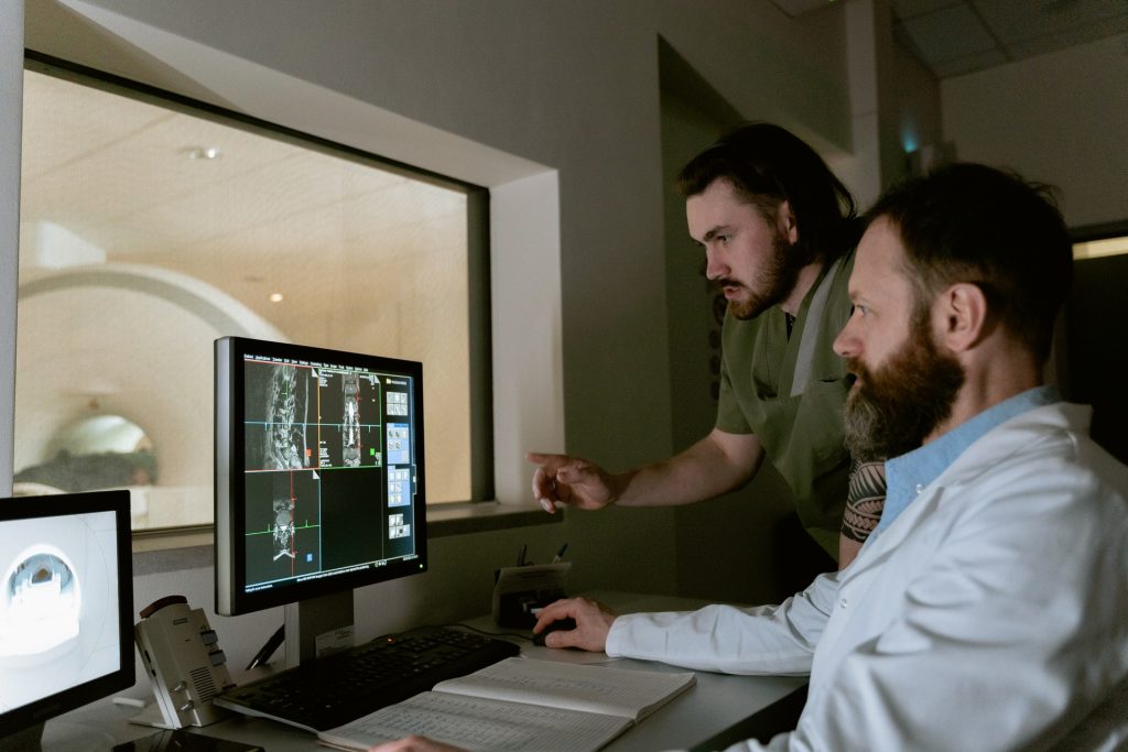
Photo by Towfiqu Barbhuiya on Unsplash

Patients with chronic lymphocytic leukaemia (CLL) or small lymphocytic lymphoma (SLL) who were taking cholesterol-lowering statin medications at the start of their cancer treatment had a 61% lower risk of dying from their cancer compared to similar patients who were not taking statins, according to a study published today in the journal Blood Advances.

“This is the first systematic evaluation of the association of statin use with survival outcomes in patients with CLL or SLL who have been treated with contemporary targeted agents such as ibrutinib,” said the study’s principal investigator, Ahmad Abuhelwa, PhD, an assistant professor of pharmacy practice and pharmacotherapeutics at the University of Sharjah in the United Arab Emirates. “Our results highlight a strong link between statin use and improved survival in this patient population.”

CLL is a slow-growing cancer that starts in the blood-forming cells of the bone marrow and is the most common form of leukaemia in adults in the United States. SLL, also a slow-growing cancer, affects the same type of cells as CLL but starts in lymphoid tissues such as the spleen instead of in the blood-forming cells.

Statins are among the most widely prescribed medications. It’s estimated that over 90 million adults in the United States take a statin drug to reduce their cholesterol levels and lower their risk for heart disease, which can lead to heart attacks or strokes. Previous studies have linked statin use to reduced death rates from several cancers, including CLL, said Dr Abuhelwa. However, those studies did not evaluate the effects of statin use in patients who were treated with newer cancer therapies such as the targeted drug ibrutinib, he said.

In the current study, Dr Abuhelwa and his colleagues analysed data from 1467 patients with CLL or SLL who participated in four international clinical trials conducted between 2012 and 2019. In these trials, patients were randomly assigned to treatment with ibrutinib either alone or in combination with other anti-cancer drugs, or to a drug regimen that did not include ibrutinib. A total of 424 patients (29%) were taking a statin at the time they started treatment across the four clinical trials. The median patient age was 65, and 66% were men; 92% had CLL, which was either newly diagnosed, had come back, or had not responded to prior treatment.

The study’s primary endpoints were cancer-specific survival (how long patients lived after starting treatment before dying specifically from their cancer), overall survival (how long patients lived after starting treatment, regardless of the cause of death), and progression-free survival (how long patients lived after starting treatment before their cancer worsened or they died from any cause). The secondary endpoint was the proportion of patients who experienced severe or life-threatening adverse events. The median follow-up time for all patients enrolled in the four trials was five years for overall survival and 22 months for progression-free survival.

To account for potential confounding factors, the investigators adjusted their analysis for variables including each patient’s diagnosis, age, sex, weight, physical functioning (as assessed by doctors), disease severity, length of time since their diagnosis, number of co-existing illnesses, use of other medications for heart conditions or high blood pressure, and the specific anti-cancer treatment regimen received.

Results showed that, regardless of any of these factors, patients who took a statin had, on average, a 61% reduced risk of dying from their cancer, a 38% reduced risk of death from any cause, and a 26% reduced risk of disease progression. Importantly, statin use did not increase the likelihood of severe or life-threatening adverse events.

“These findings don’t allow us to say for certain that statins directly improve cancer outcomes,” said Dr. Abuhelwa. “However, the fact that this association remained strong even after accounting for multiple factors makes it an important area for future research.” As next steps, he recommended conducting laboratory studies to better understand how statins may influence cancer biology, as well as prospective clinical trials in which patients with CLL or SLL are randomly assigned to take a statin or not.

The study has several limitations given its observational nature. For example, patients enrolled in clinical trials tend to be monitored more closely than those who receive treatment outside of a clinical trial, so the study findings may not be generalizable to patients treated in non-clinical trial settings. Additionally, because patients used various statins at different doses, the study could not determine the effects of specific statin types, doses, or duration of use on patients’ survival.

“While our results are very promising, we can’t recommend starting statins for CLL/SLL treatment based on this study alone,” Dr Abuhelwa said. “Future clinical trials are needed to determine definitively whether statins have a direct benefit on cancer survival.”

Source: American Society of Hematology

Research Identifies the Key to Pancreatic Cancer’s Extreme Aggressiveness

Pancreatic cancer. Credit: Scientific Animations CC BY-SA 4.0

Pancreatic cancer is one of the most aggressive cancers and has one of the lowest survival rates: only 10% after five years. One of the factors contributing to its aggressiveness is its tumour microenvironment, known as the stroma, which makes up the bulk of the tumour mass and consists of a network of proteins and different non-tumour cells. Among these, fibroblasts play a key role, helping tumour cells to grow and increasing their drug resistance.

Now, a study led by researchers from the Hospital del Mar Research Institute and other institutions has identified a new key factor contributing to this feature of pancreatic cancer: a previously unknown function of Galectin-1 protein inside the nuclei of fibroblasts. This discovery, published in the journal PNAS, offers new insights into the role of these cells in the progression of pancreatic cancer.

“The stroma is considered a key component in the aggressive nature of pancreatic cancer, as it interacts with tumour cells, protects them, and hinders the action of drugs. Moreover, stromal cells, particularly fibroblasts, produce substances that support tumour growth and dissemination,” explains Dr Pilar Navarro, coordinator of the Cancer Molecular Targets Research Group at the Hospital del Mar Research Institute and IIBB-CSIC-IDIBAPS. Until now, fibroblasts were known to secrete Galectin-1, a protein with pro-tumour properties. This study, however, shows that the molecule is also located inside fibroblasts-specifically in their nuclei-where it plays a key role in gene expression regulation.

The presence of this molecule activates fibroblasts, making them support tumour cell development. The researchers also discovered that “Galectin-1 can regulate gene expression in these cells at a highly specific level without altering the DNA sequence, through epigenetic control. One of the genes it regulates is KRAS, which plays a critical role in pancreatic tumours,” explains Dr Navarro. This gene is also present in tumour cells in 90% of patients, though in this case it is mutated. It is considered one of the main drivers of uncontrolled growth and tumour aggressiveness.

Designing new strategies

The team behind the study had previously identified the prominent role of Galectin-1 in pancreatic cancer. The newly discovered functions now pave the way for developing new strategies to tackle this type of tumour. “Until now, efforts have focused on inhibiting Galectin-1 secreted by the stroma surrounding the tumour. Now, we see that we also need to block the protein inside the fibroblast nuclei,” says Dr Neus Martínez-Bosch, researcher at the Hospital del Mar Research Institute. “We need to find new inhibitors that work inside fibroblasts, not just on the protein they secrete,” she adds.

To carry out the study, researchers worked with tissue samples from pancreatic cancer patients, allowing them to analyse the presence and function of Galectin-1 in fibroblast nuclei. They also performed in vitro experiments with human fibroblast cell lines, investigating the effects of inhibiting both the protein and the KRAS gene, and observed deactivation of these cells-effectively halting their cooperation with tumour cells.

Dr. Judith Vinaixa, also a researcher at the Hospital del Mar Research Institute and first author of the study, highlights the importance of these results: “We have confirmed the key role of Galectin-1 in the fibroblast cell nucleus, where it regulates the expression of multiple genes critical for cell behaviour.”. Dr. Gabriel Rabinovich, researcher at IBYME (CONICET) and the CaixaResearch Institute, adds: “The next steps will involve exploring therapeutic combinations that inhibit both extracellular and intracellular Galectin-1. This protein also participates in key processes such as blood vessel formation and resistance to immunotherapy. Therefore, this strategy becomes particularly relevant given the multiple antitumoral effects of Galectin-1 inhibition.”

Source: IMIM (Hospital del Mar Medical Research Institute)

Cancer Risk from CT Scans up to Four Times Higher than Previous Estimates

Photo by Mart Production on Pexels

Computed tomography (CT) scans may account for 5% of all cancers annually, according to a new study out of UC San Francisco that cautions against overusing and overdosing CTs. For children, the greatest risk comes from scans of the head.

The danger is greatest for infants, followed by children and adolescents. But adults are also at risk, since they are the most likely to get scans. In the U.S., nearly 103 000 cancers are predicted to result from the 93 million CT scans that were performed in 2023 alone. This is 3 to 4 times more than previous assessments, the authors said.

“CT can save lives, but its potential harms are often overlooked,” said first author Rebecca Smith-Bindman, MD, a UCSF radiologist and professor of epidemiology and biostatistics and obstetrics, gynaecology and reproductive sciences.

“Given the large volume of CT use in the United States, many cancers could occur in the future if current practices don’t change,” said Smith-Bindman.

“Our estimates put CT on par with other significant risk factors, such as alcohol consumption and excess body weight,” she said. “Reducing the number of scans and reducing doses per scan would save lives.”

Benefits and potential dangers

CT is both indispensable and widely used to detect tumours and diagnose many illnesses. Since 2007, the number of annual CT exams has surged by 30% in the U.S. But the ionising radiation dose from CT is a known cancer risk.

To assess the public health impact of current CT use, the study estimates the total number of lifetime cancers associated with radiation exposure in relation to the number and type of CT scans performed in 2023.

“Our approach used more accurate and individualised CT dose and utilisation data than prior studies, allowing us to produce more precise estimates of the number of radiation-induced cancers,” said co-author Diana Miglioretti, PhD, a breast cancer researcher and division chief of biostatistics at UC Davis. “These updated estimates suggest the excess risks – particularly among the youngest children – are higher than previously recognised.”

Researchers analysed 93 million exams from 61.5 million patients in the U.S. The number of scans increased with age, peaking in adults between 60 to 69 years old. Children accounted for 4.2% of the scans. The researchers excluded testing in the last year of a patient’s life because it was unlikely to lead to cancer.

Future cancers from radiation exposure

Adults 50 to 59 had the highest number of projected cancers: 10 400 cases for women, 9300 for men. The most common adult cancers were lung, colon, leukaemia, bladder and breast. The most frequently projected cancers in children were thyroid, lung and breast.

The largest number of cancers in adults would come from CTs of the abdomen and pelvis, while in children they came from CTs of the head. Projected cancer risks were highest among those who underwent CT when they were under 1 year old. They were 10 times more likely to get cancer compared to others in the study.

The researchers said some CT scans are unlikely to help patients, and are overused, such as those for upper respiratory infections or for headaches without concerning signs or symptoms. They said patients could lower their risk by getting fewer of these scans or by getting lower dose scans.

“There is currently unacceptable variation in the doses used for CT, with some patients receiving excessive doses,” Smith-Bindman said.

Co-author Malini Mahendra, MD, a UCSF assistant professor of Pediatric Critical Care, said it was important that families understand the risk of developing cancer from paediatric scans.

“Few patients and their families are counselled about the risk associated with CT examinations,” she said. “We hope our study’s findings will help clinicians better quantify and communicate these cancer risks, allowing for more informed conversations when weighing the benefits and risks of CT exams.”

Source: University of California – San Francisco

New Method Boosts Cancer Vaccine Potency

Squamous cancer cell being attacked by cytotoxic T cells. Image by National Cancer Institute on Unsplash

The concept of using vaccines to treat cancers has been around for several decades. A vaccine was first approved for prostate cancer in 2010, and another was approved in 2015 for melanoma. Since then, many therapeutic – as opposed to preventive – cancer vaccines have been in development, but none approved. One hurdle is the difficulty in finding antigens in tumours that look foreign enough to trigger an immune response.

Researchers at Tufts have now developed a cancer vaccine that makes tumour antigens more visible to the immune system, leading to a potent response and a lasting immunological memory that helps prevent tumour recurrence. Their vaccine avoids the need to hunt down a specific tumour antigen, instead relying on a digested mix of protein fragments called a lysate that can be generated from any solid tumour. 

The vaccine they produced worked against multiple solid tumours in animal models, including melanoma, triple-negative breast cancer, Lewis lung carcinoma, and clinically inoperable ovarian cancer.

Developed by a team led by postdoctoral scholar Yu Zhao and Qiaobing Xu, professor of biomedical engineering, the method builds on earlier work expressing specific antigens for an enhanced immune response by making lipid nanoparticles that carry mRNA into the lymphatic system. 

“We have significantly improved the cancer vaccine design by making it applicable to any solid tumour from which we can create a lysate, possibly even tumours of unknown origin, without having to select mRNA sequences, and then conjugating another component called AHPC that helps channel the protein fragments from the cancer cells into the immunological response pathway,” said Xu.

Unlike traditional vaccines designed to prevent infectious diseases caused by bacteria or viruses, cancer vaccines work by stimulating the body’s immune system to recognise and attack cancer cells. Unlike most vaccines against pathogens, they are designed to be therapeutic rather than preventive, acting to eliminate an existing disease. Some preventive cancer vaccines do exist, but they are generally targeted to viruses that are linked to cancers, such as HPV.

The key to the increased potency of the new cancer vaccine lies in its ability to direct tumour-derived antigens into a cellular pathway that efficiently presents the antigens to the immune system.

Rounding up the antigens and getting them into an antigen presenting cell like a macrophage or dendritic cell (the police stations, if we continue with the analogy) is generally an inefficient process for tumor antigens. This is where the Tufts research team applied a two-stage method to power up the process.

First, to make sure they round up all tumour proteins-of-interest, they modified the mix of tumour proteins with the AHPC molecule, which in turn recruits an enzyme to put a tag on the protein called a ubiquitin. It allows the cell to identify and process the protein into fragments for presentation to the immune system.

The researchers then packaged the AHPC-modified tumour proteins into tiny lipid nanoparticles, specifically designed to home in on lymph nodes, where most antigen-presenting cells can be found. 

Tested in animal models of melanoma, triple-negative breast cancer, Lewis lung carcinoma, and inoperable ovarian cancer, the vaccine elicited a strong response by cytotoxic T cells, which attack the growing tumours, suppressing further growth and metastasis.

“Fighting cancer has always been an arsenal approach,” said Xu. “Adding cancer vaccines to surgical excision, chemotherapy, and other drugs used to enhance cytotoxic T cell activity could lead to improved patient responses and longer-term prevention of cancer recurrence.”

Source: Tufts University

Court Orders Gauteng Health Department to Treat Cancer Patients

Judge rules failure to deal with backlog of patients needing radiation treatment is unconstitutional

By Liezl Human

Photo by Bill Oxford on Unsplash

The Gauteng Department of Health is appealing against a judgment by the Johannesburg High Court ordering it to provide radiation oncology treatment to a backlog of nearly 3000 cancer patients at Charlotte Maxeke Hospital and Steve Biko Hospital.

In April last year, activists from SECTION27, Cancer Alliance and the Treatment Action Campaign (TAC) joined cancer patients to march to the department’s provincial office, demanding that millions of rands set aside for radiation treatment be used.

The matter was then taken to court by the Cancer Alliance, represented by SECTION27, after years of attempts to engage with the department about radiation services. They said in a statement they wanted the court to compel the department to provide treatment to the backlog of cancer patients still waiting.

Some patients have been on the list for nearly three years, while others have died while waiting, according to the judgment by Acting Judge Stephen van Nieuwenhuizen. He noted that “irreparable harm” has occurred and continues to occur in the absence of treatment.

He said the backlog, of mostly Charlotte Maxeke patients, had grown due to a lack of radiation equipment at the hospital and a shortage of staff. This was in spite of an allocation of R784-million over three years, specifically ring-fenced for radiology oncology services. The allocation was also meant to help clear the backlog of patients.

Delays in finalising a tender for these services meant that R250-million was returned to National Treasury at the end of the fiscal year, he said.

The judge found that the provincial department had infringed on the rights of these cancer patients in that a high standard of professional ethics had not been maintained. “Efficient and effective use of resources were not promoted. Services were not provided impartially, fairly, equitably and without bias.”

Judge van Nieuwenhuizen said the provincial health department had done “nothing meaningful” since the money was allocated in March 2023 to actually provide radiation oncology treatment to the cancer patients. “On the other hand, the health and general well-being of cancer patients has significantly deteriorated. There is a clear, imminent and ongoing irreparable harm that cancer patients who are on the backlog list are suffering.”

The judge ruled that the department’s failure to provide radiation services to cancer patients on the backlog list was unconstitutional and unlawful.

He added that the provincial health officials “have conducted themselves as a law unto themselves” and ordered that measures be put in place to ensure officials are “held to account for their constitutionally imposed obligation to provide healthcare services … to cancer patients who are on the backlog list”.

He also ordered that the list of cancer patients still awaiting radiation treatment must be updated within 45 days and that a progress report and long-term plan must be submitted to the court within three months.

Salomé Meyer, director of the Cancer Alliance, told GroundUp the ruling would allow the court to get accurate information on the circumstances of each patient still waiting for treatment.

She said the judgment “confirms that civil society has a role to play to hold the government responsible for what it is supposed to do”.

In a statement on 2 April, the department confirmed that it had filed an application for leave to appeal against the ruling. The department said “there are several substantive grounds of appeal, which if left unchallenged will be greatly prejudicial to the patients undergoing radiation oncology services at the hospitals” and might set an “undesirable” precedent.

Republished from Groundup under a Creative Commons Attribution-NoDerivatives 4.0 International License.

Read the original article.

Do Lifetime Body Weight Patterns Affect the Risk of Kidney Cancer?

Study links higher body mass index at various ages across adulthood with greater risks of developing different types of kidney cancer.

Photo by I Yunmai on Unsplash

Excess weight in mid-life is a known risk factor for kidney cancer, but new research indicates that weight patterns throughout life may also affect an individual’s likelihood of developing this malignancy. The findings are published by Wiley online in CANCER, a peer-reviewed journal of the American Cancer Society.

To assess weight patterns and their associations with kidney cancer and its different subtypes, investigators analysed data from 204 364 individuals from the NIH-AARP Diet and Health Study, including body mass index (BMI) data when participants entered the study (an average age of 61.6 years), and prior BMI recordings at 18, 35, and 50 years of age. The team noted that there were 1,425 cases of kidney cancer, or renal cell carcinoma (RCC), among the study’s participants, with 583 having aggressive RCC and 339 having fatal RCC. The researchers also recorded the different subtypes of RCC, including clear cell RCC (541 patients), papillary RCC (146 patients), and chromophobe RCC (64 patients).

Higher BMI at any of the ages assessed was linked with higher risks of overall RCC and all subtypes (except chromophobe RCC), with a 10-40% higher risk for each 5-unit increase in BMI. Similar increased risks were linked to weight gain during adulthood that resulted in overweight or obesity, compared with maintaining normal BMI.

Also, long-term excess weight was associated with higher risks of overall RCC, aggressive RCC, fatal RCC, and clear cell RCC, but not papillary RCC and chromophobe RCC. Weight loss in which BMI was reduced by at least 10%, particularly later in life, was associated with a lower risk of RCC. Specifically, weight loss from age 18–35 years and after age 50 years was associated with 21% and 28% reductions in RCC incidence, respectively.

“These findings emphasise that maintaining a healthy weight across one’s lifetime is important for reducing RCC risk. More importantly, weight loss, even later in life, may offer protective benefits,” said lead author Zhengyi Deng, PhD, of Stanford University School of Medicine. “We should support initiatives that promote healthy weight maintenance and weight loss strategies. Some of these include lifestyle interventions, weight-loss programs, and emerging medical treatments for obesity; however, individuals should consult with their healthcare providers prior to initiation of any plan.” 

Source: Wiley

Could Possible Budget Cuts Cost Cancer Patients’ Lives?

Finance Minister Enoch Godongwana holding a copy of the 2025 Budget Speech. (Photo: Parliament of RSA via X)

The Department of Health’s 2025/26 Budget Vote is expected to focus on addressing the shortfall caused by the withdrawal of international funding from programmes combating communicable diseases such as HIV and TB. But as non-communicable diseases like blood cancer surpass infectious diseases, redirecting resources could further cripple screening, diagnosis, and treatment – putting more lives at risk.

Too Few Resources, Too Many Lives Lost

Palesa Mokomele, Head of Community Engagement and Communications at DKMS Africa, highlights the difficulties faced by blood cancer patients within the country’s healthcare system. “Nearly 80% of South Africans rely on a system already operating at full capacity, with many left with little more than hope due to limited access to care. Even before these new funding shifts, resources for blood cancer detection and treatment were critically scarce, contributing to the loss of more than 4000 lives each year.”

Illustrating how a lack of medical infrastructure creates additional barriers to life-saving care, she says, “The survival rate for a stem cell transplant is up to 50% with a matched unrelated donor and 61% with a matched related donor. Yet many healthcare facilities simply don’t have the resources to perform these procedures. As a result, most patients receive only medical management, which may not be enough to ensure survival. Given our population size, transplant activity remains critically low – only 139 of the required 600 transplants are performed on adults annually, and just 18 of the 250 needed for children.”

Finances Dictate Healthcare Choices

Beyond the overstretched and under-resourced public health sector, the financial burden on patients remains a major obstacle. “One in five South African households delays seeking healthcare simply because they cannot afford it,” notes Mokomele. “While the state covers the cost of a stem cell transplant from a matching donor, other essential expenses such as tissue typing, donor searches, and stem cell procurement are not covered. These out-of-pocket costs place treatment out of reach for many, leading to heartbreaking decisions and poorer outcomes.”

She adds that socio-economic challenges often make accessing care even harder for patients. “Being the sole breadwinner means some individuals struggle to take time off work for necessary treatment. In other cases, mothers face the impossible choice between continuing their own treatment or staying home to care for their children when no other support is available.”

Post-transplant Survival Challenges

Even for patients who manage to undergo a transplant, their survival remains at risk due to conditions in some public healthcare facilities. “Overcrowding and poor sanitation create dangerous environments for these highly immunocompromised patients,” warns Mokomele. “To safeguard their fragile health, they need access to clean water, proper sanitation, isolation, and balanced nutrition.”

She stresses that long recovery periods make it difficult for patients from distant areas to complete their care without proper housing at treating hospitals. “Without these accommodations, many are forced to abandon treatment, putting their survival at risk.”

“No patient should be denied life-saving treatment due to funding constraints. We urge government and the private sector to collaborate in strengthening blood cancer care, and we encourage the public to play their part by supporting fundraising initiatives that help bridge critical gaps in treatment access,” concludes Mokomele.

Go to https://www.dkms-africa.org/get-involved/donate-money to contribute to this cause.

Three out of Ten Breast Cancers are Detected Between Screenings

Photo by National Cancer Institute

In a new study, researchers from Karolinska Institutet have shown that so-called interval cancers, which are detected between two screening sessions, account for a significant proportion of breast cancer cases and that certain risk factors may increase the likelihood of developing this type of cancer. The study was published in the journal JAMA Oncology.

Mammography screening has been shown to be effective in reducing breast cancer mortality by detecting cancer at an early stage. Despite this, some cancers are not diagnosed during screening but between screening rounds, known as interval cancers. This type of cancer can be more aggressive and difficult to treat than breast cancer detected during a scheduled screening.

The study, which covers half a million women in Stockholm between 1989 and 2020, shows that interval cancer accounts for a significant proportion of all breast cancer cases.

“We found that interval cancers account for about 30 per cent of all breast cancers detected by screening and this percentage has remained constant over three decades, despite advances in screening technology,” says Yuqi Zhang, postdoctoral fellow at the Department of Medical Epidemiology and Biostatistics, Karolinska Institutet and lead author of the study.

The researchers identified several factors that increase the risk of developing interval cancer. These factors include high breast density, hormone therapy, higher education level and older age at first childbirth.

“Women with high breast density and use of hormone therapy are at increased risk of being missed at screening due to false negative results,” says Yuqi Zhang.

Rapid growth of tumour

In addition, the study showed that women with a family history of breast cancer, especially interval cancer, are at a higher risk of developing interval cancer themselves. Women with a family history of breast cancer were 1.9 times more likely to develop interval cancer, and this risk increased to 2.9 times if they had a family history of interval cancer specifically.

“This is often due to the rapid growth of tumours between screening rounds rather than missed detections. The study therefore emphasises the need for more frequent or improved screening methods specifically designed for women with these particular risk factors,” says Yuqi Zhang. 

“Adapting screening protocols to reflect individual risk profiles – whether through more frequent mammography, supplemental imaging like contrast-enhanced mammography, or incorporating genetic testing – could significantly improve early detection rates,” says last author Professor Kamila Czene at the same department. 

“By identifying cancers earlier, we can offer more effective and less invasive treatments, improve long-term survival outcomes, and reduce the emotional and physical toll on patients.”

Source: Karolinska Institutet

Radiopharmaceuticals Being Tested for Brain Tumours in Children

Credit: National Cancer Institute

Neuroblastoma is a rare disease that affects children, often before the age of two. Some are born with the disease. Paediatric surgeon Jakob Stenman is investigating whether targeted radioactive drugs can slow down the disease in those with the most severe form.

Neuroblastoma is a complicated disease, with the most aggressive variant called high-risk neuroblastoma. Children with this disease are treated very intensively. They may undergo surgery, chemotherapy, high-dose chemotherapy with stem cell transplantation, radiotherapy and antibody treatment. Treatment often lasts up to a year and a half.

Despite this, the survival rate is around 60%, according to the Swedish Childhood Cancer Foundation.

“Some relapse in their disease, and we currently lack curative treatment for them,” says Jakob Stenman, a researcher at the Department of Women’s and Children’s Health at Karolinska Institutet.

It is these children, those who have relapsed, that he is treating in a study with targeted radioactive drugs. These are molecules that attach to the surface of cancer cells. These molecules have an appendage: the radioactive substance lutetium-177. The drug first moves through the bloodstream but then attaches to the cancer cells. The emitted radiation damages the cancer cells but unfortunately also the neighbouring healthy cells.

“We have treated ten children so far. Unfortunately, the disease has not disappeared in any of these cases, but it seems to be slowing down, and some benefit more than others from the treatment. When it comes to side effects, the children have tolerated the treatment well,” says Jakob Stenman.

The hope is to be able to prevent relapse

He reports that the interest has been great from clinics in other countries where these children are treated. Hospitals from Lithuania, the Netherlands, the United Kingdom and are now involved.

In neuroblastoma, cancer cells often look very different, even in the same patient. In some metastases, there may be many cells with a surface where the drug attaches, while in other metastases there may be fewer such cells. This means that the targeted drug attaches to fewer cells in some of the metastases. As a result, the local radiation dose is too low in these metastases, which can then continue to grow and spread further.

Jakob Stenman therefore believes that the treatment could be more effective if the radioactive substance used is even more potent, which in this context means that it emits even more energy (ie, radiation). If it then attaches to fewer cells in a metastasis, it might still be able to eliminate all the cancer cells there. But it must act even more locally to protect other tissues from the higher radiation dose.

The researchers have identified several substances they believe could work in this way. These include actinium-225, astatine-211 or lead-212. The effects and side effects of actinium-225 are now being investigated in cell studies and animal experiments. The goal is to start a clinical trial with actinium in three to five years.

“If what we believe turns out to be true, we hope to be able to prevent relapse and thereby enable a cure for a larger proportion of children who have developed high-risk neuroblastoma,” says Jakob Stenman.

Text: Annika Lund for Medicinsk Vetenskap nr 4 2024 

Source: Karolinska Institutet

Shorter, Higher-dose Radiotherapy for Prostate Cancer Delivers Promising Results

Photo by Jo McNamara

A new large-scale study co-led by UCLA Health Jonsson Comprehensive Cancer Center investigators provides the strongest evidence yet that a shorter, standard-dose course radiation treatment is just as effective as conventional radiotherapy for early-stage prostate cancer, without compromising the safety of patients.

The shorter approach, known as isodose moderately hypofractionated radiotherapy (MHFRT), delivers slightly higher doses of radiation per session, allowing the total treatment duration to be over four to five weeks instead of seven to eight weeks.

According to the study, patients who received this type of MHFRT had the same cancer control rates as those who received conventional radiotherapy. Additionally, the risk of long-term side effects affecting the bladder and intestines was no higher with MHFRT, confirming its safety. 

The findings were published in the Lancet Oncology.

“We believe these data strongly support that isodose MHFRT should become the preferred standard of care MHFRT regimen for prostate cancer,” said Dr Amar Kishan, executive vice chair of radiation oncology at the David Geffen School of Medicine at UCLA and co-first author of the study. “More broadly, there appears to be little reason to consider conventional radiotherapy over MHFRT for the types of patients enrolled in these trials given these results.”

While MHFRT is now the most commonly used radiotherapy regimen for prostate cancer, concerns remain about whether delivering a higher daily dose increases the risk of urinary and bowel issues, such urinary incontinence, chronic diarrhoea and rectal bleeding.

MHFRT: isodose versus dose-escalated

To better understand whether there might be an increased risk of toxicity with the delivery of a higher dose per day of radiation, Kishan and the team of researchers examined data from more than 5800 patients across seven randomised clinical trials comparing standard therapy with two different MHFRT approaches: isodose MHFRT, which maintains the total radiation dose at a level similar to standard therapy, and dose-escalated MHFRT, which increases the total dose in hopes of enhancing tumour control.

The analysis found patients who received isodose MHFRT (60Gy in 20 fractions) had similar cancer control and side effects compared to those receiving conventional radiation therapy, with no significant difference in the five-year progression-free survival (77.0% for MHFRT vs 75.6% for conventional).

Patients who received higher dose-escalated MHFRT did not improve cancer control when compared to those receiving standard doses, with five-year progression-free survival rates being identical to conventional therapy (82.7% in both groups). Patient-reported outcomes also showed significantly higher gastrointestinal side effects (7.2% vs 4.9%), particularly bowel issues. 

While dose-escalated MHFRT was expected to improve outcomes, the data showed no additional benefit in cancer control and a higher risk of gastrointestinal side effects, noted Kishan. This underscores the advantage of isodose MHFRT, which provides the same effectiveness as conventional therapy without increasing toxicity.

“These findings reinforce isodose MHFRT as the standard of care, offering the same cancer control as conventional treatment but with fewer side effects than dose-escalated MHFRT,” said Kishan, who is also a researcher in the UCLA Health Jonsson Comprehensive Cancer Center. “Patients can safely opt for a shorter treatment schedule without compromising their outcomes, ensuring they receive effective care with fewer visits and minimal added risk. Less time in treatment can still mean the best possible results.”

Source: University of California – Los Angeles Health Sciences