Author: ModernMedia

Latest Antibiotic Discovery Offers New Way to Kill drug-Resistant Bacteria

Drug-resistant Salmonella. Credit: CDC on Unsplash

Researchers at McMaster University have discovered a new antibiotic that kills some of the world’s most dangerous and drug-resistant bacteria – and does so by targeting a previously unknown vulnerability, opening the door to an entirely new class of treatments.

The new compound, called manikomycin, was identified by a team led by McMaster Professor Gerry Wright and has shown early effectiveness against priority pathogens including Salmonella, E. coli and Klebsiella.

Unlike any antibiotic currently used in clinics, it works by blocking the exit site of the ribosome, the protein-producing machinery found inside every bacterial cell.

The discovery, published today in Nature, marks the fourth new antibiotic candidate from Wright’s lab in just over a year, underscoring a promising new approach to drug discovery at a time when antibiotic resistance is a growing global threat.

“Not a single antibiotic prescribed in clinics today does what manikomycin does,” says Wright, a member of the Michael G. DeGroote Institute of Infectious Disease Research. “Not azithromycin, not tetracycline – none of them.”

“So we’ve not only found a brand-new drug candidate, but we’ve also established a brand-new target in bacteria that could potentially be exploited with other new drugs.”

It’s the latter part of the discovery that has researchers most excited. Wright notes that because most antibiotics in use today target the same handful of vulnerabilities on the ribosome, bacteria have evolved broad defence strategies against such attacks. However, drugs that attack a different part of the ribosome – the exit site – leave them defenceless.

“Even newly discovered drugs that attack those same old targets may quickly face resistance,” says Wright, a professor in McMaster’s Department of Biochemistry and Biomedical Sciences.

“But across the history of medicine, we’ve put absolutely no selective pressure on this particular target, so bacteria have no existing resistance mechanisms for manikomycin.”

Wright likens the ribosome to a factory assembly line. Finished components, he says, must be moved off the line before the next piece can advance. Manikomycin blocks the exit lane, causing the entire assembly process to jam and eventually grind to a halt. And, without the ability to produce proteins, bacteria cannot survive.

The discovery of manikomycin builds on work that began more than 75 years ago, when scientists first discovered that the soil bacterium Streptomyces rimosus produced oxytetracycline, a powerful new drug that would help usher medicine into the antibiotic age.

While the breakthrough was one of several like discoveries made in the mid-1900s, S. rimosus and related bacteria have long since been abandoned as a potential source of new antibiotics.

“There is an overwhelming perception in science that these bacteria have been mined completely dry – that we’ve found all there is to find,” Wright says. “Our lab has found that this is not at all the case.”

Wright’s group, working with collaborators at the University of Illinois Chicago and the University of Hamburg in Germany, used an advanced laboratory technique called fractionation to uncover the new antibiotic.

By filtering out oxytetracycline and other abundant compounds from the chemical mixtures produced by S. rimosus, the researchers were able to isolate scarcer molecules that had gone unnoticed over the years.

Manpreet Kaur, a postdoctoral fellow in Wright’s lab and first author on the new study, says that finding a viable new drug candidate this way signals new opportunities for antibiotic discovery.

“There is likely so much still to be discovered through fractionation,” says Kaur. “Revisiting the extracts of even-well studied bacteria like Streptomyces may lead to similar discoveries in the future.”

Wright’s team is now advancing manikomycin toward clinical development. They have already shown that the new antibiotic is not toxic to human cells, and that it works well in a lab-controlled model of infection – both key milestones on the early development pathway.

They are now working on optimiaing the drug’s “residency time” – or how long it stays active in the body – and have produced 60 derivatives, with plans to push the best one forward.

“We’re excited about this molecule’s potential,” Wright says. “There’s a clear path forward, and we may even be able to expand its spectrum so that it eventually affects even more bacteria, too.”

Source: McMaster University

Asymptomatic Bladder Cancer Could be Unmasked by Taking Aspirin

Photo by cottonbro studio

Haematuria, the presence of blood cells in urine, is a sign of bladder cancer. Because aspirin blocks platelets from forming harmful blood clots, the medication can cause mild bleeding or worsen existing bleeding in the urinary tract. Results from a study in the Journal of Internal Medicine suggest that this may prompt a clinician to run tests that uncover an asymptomatic bladder tumour.

For the study, investigators analysed information on 50 771 Danish adults who started taking aspirin in 2005–2023, as well as 156 191 who started non-aspirin non-steroidal anti-inflammatory drugs (NSAIDs), which have less pronounced antiplatelet abilities than aspirin.

Compared with adults from the general population who have never used aspirin or NSAIDs, aspirin initiators received more cystoscopies – minimally invasive procedures that allow a doctor to view the inside of the bladder and urethra with a lighted tube equipped with a camera. Cytoscopy results showed that recent aspirin initiators had a similar bladder cancer prevalence, but a lower prevalence of invasive stage compared with never-users. This suggests that individuals initiating aspirin treatment represent a patient population with a higher incidence of bladder cancer, and that their higher cystoscopy rate reflects this and thus is clinically warranted. The combination of a larger proportion of relevant cystoscopies and a lower prevalence of invasive cancer stage at diagnosis may represent unmasking of otherwise asymptomatic bladder cancer.

NSAID initiators received more cystoscopies than never-users, but they had a lower bladder cancer prevalence after cystoscopy and a similar stage distribution as never-users. This suggests that the higher cystoscopy rate may not have been clinically warranted.

“We are very encouraged by these results. In the clinical setting, they underline the importance of acting on suspicious bladder cancer symptoms among aspirin initiators,” said lead author Malene Söth Hansen, MD, of Aarhus University. “The findings may further have implications for the question of whether aspirin can prevent bladder cancer, as detection in trials with short-term follow-up may appear as a higher incidence in the aspirin-exposed cohort.”

Source: Wiley

‘We Cannot Ban Our Way out of a Youth Mental Health Crisis’

Social media bans for teenagers lack evidence and pose risks, scientists say

Photo by Vlad Patana on Unsplash

Bans on teenagers’ social media use are gathering pace worldwide. Their proponents claim that social media bans will improve young people’s mental health, but what evidence supports these claims? In their new Frontiers in Developmental Psychology article, Dr Monika Neff Lind and her co-authors argue that there is no solid scientific evidence behind these bans, and reason to believe they could backfire. In this guest editorial, Neff Lind explains why she and her colleagues doubt that social media bans will work, and how bans should be evaluated to determine whether they have any positive effects.

By Monika Neff Lind, PhD

In December 2025, Australia banned young people under 16 from having social media accounts. France, Greece, Spain, Denmark, Malaysia, Norway, India, Egypt, Canada, Türkiye, and the United Kingdom are hot on their heels. French president Emmanuel Macron said, “Banning social media for those under 15: this is what scientists recommend.” American senator Brian Schatz, author of the Kids Off Social Media Act, said, “Studies have revealed that when children and teens reduce or eliminate exposure to social media for longer than a month, their mental health benefits.” Proponents of youth social media bans claim that we have strong scientific evidence showing that bans will improve teenagers’ wellbeing.

As a clinical psychologist and parent, I would be thrilled if this were true, but it is not. We do not know how social media bans will affect youth because we have never studied that question. Let me explain.

Searching for evidence

When we want to test claims like ‘banning social media improves youth wellbeing’, scientific experiments are one of our most powerful tools to figure out what is causing something to happen. In experiments testing the effects of social media restriction on wellbeing, we randomly assign people to at least two groups: one quits using social media for a period of time and the other is the control or comparison group, which continues to use social media as usual. Given the strength of ban proponents’ claims, my co-authors and I were curious to know how strong the experimental evidence supporting their position was. In our new study, we collected and reviewed all of the experiments that have tested whether social media restriction improves wellbeing, and we were shocked by what we found.

Not a single social media restriction experiment has included people under the age of 16. We do not know how social media bans will affect the young people being targeted by them because we have never tested this with them!

To be fair, sometimes strong evidence in adults warrants making the leap to apply the same conclusions to teenagers. But even that leap is not justified here. The experiments with adults show weak, null, and mixed effects, with 40% of experimental studies showing harmful effects (eg, decreased life satisfaction and increased loneliness) or no effects of social media restriction. So even when adults are told repeatedly that social media is bad for their mental health and that giving it up will help, we find, on average, few to no benefits.


Read and download the original article


Unintended consequences?

There is also good reason to believe that bans may backfire. First, enforcing a youth social media ban raises major ethical concerns. Enforcement efforts invade people’s privacy and are likely to hurt marginalized people more. For example, the technology that determines age based on selfie uploads makes more mistakes with young faces and people of color. Banned youth may also miss out on important resources and communications provided via social media, as schools, clubs, and most other youth-serving organizations use social media as a main form of communication.

What happens when enforcement efforts fail? Many young people will circumvent bans by creating fraudulent ‘adult’ accounts or lurking anonymously. They will retain access to social media without any of the benefits of parental controls or content filters enabled by youth accounts. The vast majority of young people oppose youth social media bans, and teens are well known for their defiance of top-down edicts that disregard their needs. Expect more conflict between teens and caregivers, not less.

To recap, we don’t know how social media bans will affect teens, and the bans may backfire. Yet the bans are still happening! Like other policies that consume resources, political capital, and time, it is imperative for governments to evaluate these actions by funding comprehensive assessments of the bans’ impacts.

What next?

The first step in measuring the impact of these bans is to determine if the bans actually change teenagers’ social media habits. Three months in, Australian authorities reported that close to 70% of social media accounts owned by people under 16 remained active.

Second, we need a careful and well-resourced plan to measure both positive well-being and mental health problems from multiple sources, including self-report, caregiver report, and objective behavioral data, to get a full picture of whether and how altered social media use affects youth.

Third, we need creative approaches to capture the real-world impacts of the bans, since true experiments are not possible and effects may be at the community as well as the individual level. For example, we could randomly assign a subset of youth (eg within a certain region) to delayed enactment of the ban. Whatever approach is taken, governments must collaborate with diverse stakeholders – including young people – to rigorously and openly evaluate potential impacts. Rushed or improvised assessment will leave room for politicization and motivated reasoning.

Big Tech has become infamous for ‘moving fast and breaking things’. Policymakers rushing to enact these bans risk repeating Big Tech’s mistakes and compounding the problems the bans are trying to solve. We cannot ban our way out of a youth mental health crisis. Rather than take things away, we should make things better.

About the author

Dr Monika Neff Lind is a clinical psychologist, science communicator, and researcher in digital mental health based at the University of California Irvine. See more of her work here.

Source: Frontiers

Proteins in Blood Could Help Predict Retinal Degeneration in Diabetics

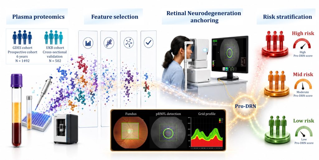
A model using 71 proteins associated with retinal degradation could predict risk in diabetics

Plasma proteomic signatures for early risk stratification of diabetic retinal neurodegeneration.
Credit: Wei Wang and Huangdong Li / Zhongshan Ophthalmic Center, Sun Yat-sen University (CC-BY 4.0, https://creativecommons.org/licenses/by/4.0/)

An AI-assisted model based on 71 different blood proteins could help doctors better predict retinal degeneration in diabetic patients before symptoms occur, according to a study published June 2nd in the open access journal PLOS Medicine by Huangdong Li from the Guangdong Provincial Clinical Research Center for Ocular Diseases in Guangzhou, China, and colleagues.

More than half a billion people around the world are now affected by diabetes. People with the disease are at risk of different neurodegenerative conditions, including the breakdown of the retina, the part of the eye that detects light, in a condition called diabetic retinal neurodegeneration (DRN). It can cause severe visual impairment and vision loss, and scientists believe that DRN is a “window” into the diabetic degeneration of other parts of the nervous system, including cognitive impairment and dementia, as well as degradation of nerves in peripheral areas like the fingers and toes.

Unfortunately, DRN is only detected after symptoms appear, when damage is already irreversible. To better predict who might suffer from DRN and when, the researchers sampled the blood plasma from 1492 patients in the Guangzhou Diabetic Eye Study with type 2 diabetes who did not yet have DRN, and examined the eyes of 1218 of them through scans over a six-year period. They compared their results with another 502 people with diabetes in the United Kingdom BioBank.

The researchers identified 71 different plasma proteins associated with DRN. The proteins were part of cell pathways for processes like inflammation and cellular maintenance. Using machine learning, the scientists used the protein levels in plasma to develop a predictive model called Pro-DRN which was able to improve on the best-performing model by 26 percent. The scientists have already put the model online to allow doctors to assess the risk. While Pro-DRN is based on plasma protein levels and relies on associations between protein levels and DRN and not direct causes, the authors hope that it could help doctors predict and potentially prevent neurodegeneration, using a simple blood test analysed by AI.

The authors add, “Our study suggests that early retinal nerve damage in diabetes leaves measurable signals in the blood. By combining plasma proteomics, longitudinal retinal imaging, and explainable AI, Pro-DRN may help move diabetic eye care from detecting established damage toward earlier, molecularly informed risk stratification, so that closer monitoring and future neuroprotective interventions can be directed to the people most likely to benefit.”

Provided by PLOS

Good Vibes. Great Music. Real Impact. Join the One Day x SANBS Community Blood Drive

The South African National Blood Service (SANBS) and J’Something Bring Blood Donation into the Culture

What if saving a life was part of your weekend plans?

On 6 June 2026, music, culture, community and purpose will come together at Artistry JHB as the South African National Blood Service (SANBS), musician and entrepreneur J’Something host One Day x SANBS Community Blood Drive a unique event designed to make giving blood more accessible, engaging and relevant to a new generation of South Africans.

Every day, blood donors help create more tomorrows for patients across the country. They make it possible for someone to celebrate another birthday, return home to their family, pursue their dreams, or simply get a second chance at life. Yet many South Africans who are eligible to donate have never taken that first step.

Through this purpose-driven partnership, SANBS and J’Something are bringing blood donation into spaces where people naturally connect, create and inspire one another. By blending culture, creativity and community impact, the One Day x SANBS Community BloodDrive aims to show that donating blood is not only life-saving it’s something everyone can be part of.

The result is more than a blood drive. It’s a day party with purpose, where every donation has the potential to save up to three lives.

Blood Drive Details:

Venue: Artistry JHB

Date: 6 June 2026

Time: 09:00 – 15:00

RSVP: tshilidzim@meropa.co.za or 076 306 1197

Please RSVP by Friday,05 June 2026

Secure your spot at the blood drive by completing the quick eligibility screening below.

Whether you’re donating blood or simply coming through to support the movement, expect good music, great energy and a community united for a meaningful cause.

Click here for the quick eligibility screening.

Be the lifeline. Give Blood. Give A Tomorrow.

Breakthrough Drug Nearly Doubles Survival with Advanced Pancreatic Cancer

– an Oncologist Explains how Daraxonrasib Overcame an ‘Undruggable’ Disease

Pancreatic cancer. Credit: Scientific Animations CC BY-SA 4.0

Christopher Lieu, University of Colorado Anschutz

For a long time, the likelihood of surviving pancreatic cancer has been extremely low. For patients who were diagnosed with metastatic pancreatic cancer between 2015 and 2021, about 97% died within five years of their diagnosis.

Pancreatic cancer is so deadly in part because there are no effective screening tests, and it rarely causes noticeable symptoms in its earliest stages. By the time a patient experiences signs, such as jaundice – a yellowing of the skin – or abdominal pain, the cancer has often already spread to other organs.

As a gastrointestinal oncologist and researcher specialising in early-phase clinical trials, I have seen the critical need for more effective therapies for patients with pancreatic cancer. For decades, successfully targeting the central mechanism that causes the vast majority of pancreatic cancers was considered impossible.

However, that narrative is rapidly changing with a new drug that can shut down the key protein that drives pancreatic cancer, nearly doubling survival rates for patients with advanced stages of the disease.

‘Undruggable’ tumours

The standard treatment for advanced pancreatic cancer has historically relied on chemotherapy, potent drugs designed to kill rapidly dividing cells. While chemotherapy can slow the progression of the disease, its effectiveness is often limited by the ability of pancreatic cancer cells to develop resistance against these drugs.

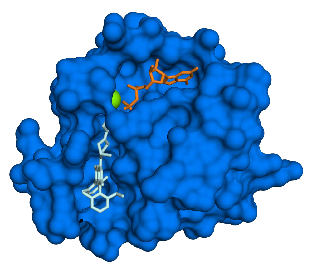
Model of the 3D structure of KRAS, resembling a rough-looking blob with a molecule tucked inside
KRAS (blue) has been difficult for drugs to target. Fvasconcellos/Wikimedia Commons

Pancreatic cancer’s success lies in its genetics. More than 90% of pancreatic tumours are driven by mutations in a gene called KRAS. This gene codes for proteins that function as switches that turn cell growth on and off. When the KRAS gene is mutated, the switch becomes permanently stuck in the “on” position, commanding cancer cells to multiply endlessly.

For decades, scientists considered KRAS to be “undruggable.” The surface of the protein is exceptionally smooth, lacking the molecular pockets that standard drugs require to bind to and turn the switch off.

Because existing drugs haven’t been able to target this protein, treatment for pancreatic cancer has primarily relied on toxic drugs that act more like blunt instruments than precise tools. Chemotherapy attempts to control the disease through widespread cell destruction, causing significant collateral damage to healthy tissues that lead to side effects.

What is daraxonrasib?

A new drug called daraxonrasib offers a critical advance in treating metastatic pancreatic cancer.

Daraxonrasib is taken daily by mouth. Instead of binding to KRAS directly, it attaches to a molecule called cyclophilin A in cells that helps fold proteins into their final 3D structures. This protein complex is then able to bind to the active KRAS protein and shut down its ability to signal cancer cells to multiply.

The company developing the drug, Revolution Medicines, presented results on May 31, 2026, from its Phase 3 clinical trial of 500 patients with metastatic pancreatic cancer who had received prior treatment. Compared to standard chemotherapy, daraxonrasib nearly doubled overall survival from 6.7 months to 13.2 months after diagnosis. Overall, daraxonrasib reduced the risk of death for metastatic pancreatic cancer patients by 60%. https://www.youtube.com/embed/sIspXSWQn1w?wmode=transparent&start=0 Daraxonrasib nearly doubled survival for patients with advanced pancreatic cancer compared to chemotherapy.

The most common side effect is a prominent skin rash, which affected more than 86% of patients in the study. Patients also frequently dealt with stomatitis – painful swelling and sores inside the mouth – as well as diarrhoea, nausea and vomiting. However, patients taking daraxonrasib were far less likely to stop treatment due to severe side effects compared to chemotherapy, and they had improved quality of life with reduced pain.

Next steps for daraxonrasib

By successfully targeting the specific genetic mutation that drives the vast majority of pancreatic cancers, researchers have demonstrated that this “undruggable” disease is treatable with targeted therapy.

The immediate next step is regulatory review of the drug’s readiness for the clinic. With data now officially published, Revolution Medicines will use these findings to seek formal approval from the Food and Drug Administration and other global regulatory bodies.

Because advanced pancreatic cancer is notoriously difficult to treat, breakthrough therapies that demonstrate this kind of significant survival benefit are often granted expedited or priority review. When daroxonrasib becomes available to patients will depend on the review timeline. Should the drug obtain approval, it could be available in clinics within months.

For the broader landscape of drug development, this milestone represents a likely shift in pancreatic cancer treatment. I expect more clinical trials exploring combination therapies pairing KRAS inhibitors with other drugs to prevent tumours from developing resistance to treatment.

Should daraxonrasib succeed, it could help set the stage for more precise, personalised and effective treatments for pancreatic cancer in the years to come.

Christopher Lieu, Professor of Medical Oncology, University of Colorado Anschutz

This article is republished from The Conversation under a Creative Commons license. Read the original article.

Brain Tumour Removal May Improve Blood Sugar Control in Diabetes Patients

Photo by Anna Shvets on Pexels

A new study published in JAMA Network Open found that removing olfactory groove meningioma, a type of brain tumour located near the base of the brain, may improve blood sugar control in patients with diabetes. 

Researchers followed patients with an olfactory groove meningioma and diabetes over five years after surgery, tracking long-term changes in haemoglobin A1c (HbA1c), which is a standard measure of blood sugar control, and body weight after tumour removal surgery. 

The research team found:

  • Blood sugar control improved after tumour removal in most patients.
  • Improvements often happened soon after surgery and lasted for years.
  • Many patients also lost weight after surgery.
  • Improvements occurred even when diabetes medications stayed the same. 

“This type of brain tumour affects both frontal lobes simultaneously and is usually thought of as causing symptoms like vision problems, personality changes or loss of smell,” said Andrew Venteicher, MD, PhD, an associate professor at the University of Minnesota Medical School and neurosurgeon with M Health Fairview. “What surprised us was how much blood sugar control improved after surgery in many of these patients. The findings may help us better counsel patients before surgery and raise new questions about how the brain influences metabolism throughout the body.”

The findings suggest that some brain tumours may affect the body’s ability to regulate metabolism and blood sugar, and that removing certain brain tumours may improve blood sugar control and weight in some patients with diabetes, in addition to improving neurological symptoms. 

Future studies will explore why these metabolic improvements occur and whether similar effects are seen in patients with other types of brain tumours. Researchers also hope to better understand how brain function, behaviour and metabolism are connected and whether these findings could help guide future treatment decisions.

Source: University of Minnesota

AI Language Models Struggle with Basic Hospital Data Tasks, Study Finds

Nine leading AI models were tested on simple administrative queries drawn from real-world emergency department records—and most failed unless paired with code-generation tools.

A new study finds that large language models (LLMs), used with straightforward prompting, perform poorly on routine number-crunching tasks that hospital administrators depend on every day to track patients and allocate resources. The findings were published this week in the open-access journal PLOS Digital Health by Eyal Klang of the Icahn School of Medicine at Mount Sinai, New York, USA, and colleagues.

Hospitals rely on structured electronic health record (EHR) data to monitor patient counts and resources and to generate administrative reports. These tasks are currently handled by data analysts using programming languages, creating delays when staff need fast answers. AI tools known as large language models, such as GPT-4o and Llama, have been proposed to simplify that process.

In the new study, researchers evaluated nine leading LLMs on two basic administrative tasks—counting patients meeting a condition and filtering records based on multiple criteria—using data drawn from 50 000 real emergency department visits at the Mount Sinai Health System.

The researchers found that straightforward prompting—asking the model a plain question like “how many patients in this table were admitted?”—produced uniformly poor results across all models. Chain-of-thought reasoning, in which the model is prompted to show step-by-step work before giving an answer, offered only modest improvements that degraded sharply as table size increased. Even GPT-4o, the top-performing model, saw accuracy drop from roughly 95% on the smallest datasets to below 60% on larger ones under chain-of-thought conditions.

A tool-based approach—where models were asked to generate code that was then executed—substantially improved accuracy for the most capable models, with GPT-4o and Qwen-2.5-72B achieving near-perfect performance. However, distilled DeepSeek models, optimised for speed and efficiency, struggled even with this approach. One model, Llama-3.1-8B, failed to produce usable output in the majority of trials and was excluded from further analysis.

“Our findings indicate that without using a tool-based strategy, current LLMs are unsuitable for standalone use even on minimally complex administrative tasks in clinical settings,” says Benjamin Glicksberg. “Structured data tasks in clinical workflows will require agentic approaches that combine LLMs with code execution to ensure accuracy and consistency.”

Provided by PLOS

Largest Study of New Parents Dismisses ‘Baby Brain’ Myth

The largest and most comprehensive study of memory and cognition in new parents has found no evidence for ‘baby brain’ in parents

Photography by Drew Hays on Unsplash

New mothers often complain about having ‘baby brain’, where memory and cognition become vague and unreliable. Now a Monash University study – the largest ever done comparing cognitive ability in new parents – has unequivocally found no evidence that ‘baby brain’ reflects underlying problems with cognition that result from becoming a parent.

In a study published in the journal Cortex, and led by PhD candidate Navyann Siddiqui and Dr Kelsey Perrykkad from the Cognitive Neuroimaging Lab at the Turner Institute for Brain and Mental Health, researchers examined cognitive differences in 150 mothers and 150 fathers up to two years after their baby’s birth.

The study found that – using a comprehensive battery of cognitive assessments – both mothers and fathers showed similar performance to non-parent (male and female) controls on all cognition measures, “suggesting the absence of so-called ‘baby brain’ effects,” Dr Perrykkad said.

As an explanation of the common stereotype of ‘baby brain’, the researchers found a gender bias, with male non-fathers self-reporting better subjective memory than all other groups. However, according to Mr Siddiqui, “this self-promotion bias appeared to be lost in fathers, driven by lack of sleep”.

Strikingly, there was no effect of time postpartum on any cognitive measure, with parents’ ability on cognitive tests remaining the same regardless of the baby’s age up to 2 years, when it is assumed that sleep deprivation would be reduced.

While there is evidence of subtle decrements in cognition during pregnancy, according to Dr Perrykkad, “the evidence is inconsistent about the presence of an objective measurable decline in cognitive function during the postpartum period with studies revealing a potential mismatch between the self-reported parental experience and objective measures of cognitive change,” she said.

Mr Siddiqui said that – before the study – they expected that mothers and fathers would show subjective and objective reduction in cognition compared to non-parents. “We expected that there would be little to no difference between the new mums and dads on account of their shared environment post birth and we also expected that cognition would improve in parents with increased time postpartum,” he said.

Dr Perrykkad continued, ‘So why do new parents, especially new mothers, commonly report experiences of baby brain? It is important not to dismiss what new parents are telling us. When we do find evidence for baby brain, it is more related to sleep and wellbeing than a true objective decline in cognition. This indicates it is just as important as ever to support new parents in these formative years. While it isn’t the end of the baby brain story, new parents can take solace in the fact that becoming a new parent doesn’t inherently impair their memory and cognition.”

Source: Monash University

Genetic Trade-off Between Youth and Longevity Uncovered by Researchers

A 3-month-old African turquoise killifish, left, and a 5-month-old killifish, right, show aging much like that in humans.

Researchers have identified a gene that directly links early-life growth and reproductive success with accelerated ageing and increased cancer risk later in life, offering new insight into a longstanding theory in evolutionary biology.

Now, an international team of researchers provides experimental evidence for the theory of antagonistic pleiotropy, the idea that certain genes can provide advantages early in life while contributing to disease and decline in old age.

What’s fascinating – and slightly terrifying – is that the cancer we see in these fish isn’t a random accident. It’s the direct shadow of their youthful vitality

Prof Itamar Harel

While widely accepted in theory, scientists have struggled to identify specific genes responsible for such trade-offs in vertebrates. Using the African turquoise killifish, a short-lived species recently pioneered by Harel and colleagues for genetic ageing research, the team focused on the gene vgll3, which has been previously linked to the timing of human puberty and maturation in other species, particularly Atlantic salmon.

By modifying this gene using CRISPR technology, the researchers observed clear effects. Fish with altered vgll3 grew faster and reached sexual maturity earlier, traits that could offer a reproductive advantage in natural environments.

However, these benefits came with significant long-term costs. The same fish showed reduced lifespans and a higher incidence of age-related tumours, including melanoma-like cancers.

“We have effectively caught evolution in the act of making a trade-off. For years, we’ve asked why our bodies can’t just maintain themselves indefinitely. This gene gives us a direct answer: nature doesn’t prioritise longevity; it prioritises continuity. We are built to sprint, not to marathon,” said Prof Itamar Harel at Hebrew University.

Further analysis showed that the gene influences key biological processes, including cell division, stem cell activity, and DNA repair. Increased cellular activity may help explain both the rapid development observed in younger fish and the accumulation of damage that leads to disease in older individuals.

The researchers also developed a new immunodeficient killifish model, enabling them to transplant and study tumour cells in ways not previously possible in this system.

“What’s fascinating – and slightly terrifying – is that the cancer we see in these fish isn’t a random accident. It’s the direct shadow of their youthful vitality. The same machinery that drives a cell to build a young body is hijacking the system to build a tumour in the old one. If we can understand this mechanism, we might finally learn how to decouple healthy growth from the disease of ageing,” Dr Harel added.

Because vgll3 is conserved in humans, the findings may have broader implications for understanding human development, ageing, and age-related diseases. While previous association studies have linked the gene to puberty timing and hormone levels, functional data were missing until now.

The discovery could contribute to future efforts in cancer prevention and research aimed at extending healthy lifespan. Researchers say the next step will be to explore whether it is possible to separate the gene’s beneficial early-life effects from its harmful consequences later in life.

Source: EurekAlert!