Data analysed by Penn researchers clarifies risks associated with general anaesthesia, giving patients more control over their delivery experience.
Photo by Anna Shvets on Pexels
Regional anaesthesia has long been favoured for caesarean births due in part to concerns about the effects that general anaesthesia may have on newborns during labour and delivery. Powerful societal pressures also push the idea that mothers need to be awake during delivery to witness the first cry and capture the ‘perfect’ birth moment. But for some women who undergo a caesarean birth, the pain can become excruciating, even after they received a spinal or epidural block.
Now, new research from a team at the Perelman School of Medicine at the University of Pennsylvania, finds that general anaesthesia may be a reasonable alternative for many patients. The findings are published today in Anesthesiology, the peer-reviewed medical journal of the American Society of Anesthesiologists.
“No patient should have to experience pain during caesarean section; as an anaesthesiologist, I never want someone to feel forced to choose between their baby’s health and not having to experience the pain of surgery,” said Mark Neuman, MD, MSc, Professor of Anesthesiology and senior author of the study. “Since regional anaesthesia is so widely used, it’s common for patients to feel that a spinal or epidural block is the only safe option for caesarean section. But as our study shows, anaesthesia type during pregnancy does not need to be one-size-fits-all.”
Reducing pain during C-section delivery
The research analysed 30 years’ worth of data from multiple clinical trials, comparing outcomes between general anaesthesia versus spinal or epidural anaesthesia for C-sections. The Penn study found that, while babies born under spinal or epidural anaesthesia had slightly higher Apgar scores than those born under general anaesthesia, the differences were small and not likely to be clinically meaningful.
While the majority of patients experience good outcomes with spinal or epidural block for caesarean delivery, recent studies show that up to one in six patients who receive an epidural or spinal may feel pain during their C-section. These experiences can be traumatic and have lasting emotional impacts.
The findings come amid growing public discourse on caesarean experiences. Recent podcasts and published news stories have featured candid patient accounts of pain under spinal or epidural anaesthesia. “This study equips women with evidence-based context about the use of general anaesthesia during c-section.” said Sarah Langer, MD, a resident in anaesthesiology at the Perelman School of Medicine and lead author the study. “Childbirth is a physically and emotionally demanding process, but we do not want patients to feel like there aren’t options when it comes to their anaesthesia for c-section,”
Broadening evidence-based choices
The study found that babies born under general anaesthesia were slightly more likely to need breathing support immediately after birth, but there was no increase in NICU admissions. The research does not suggest that general anaesthesia should replace regional techniques, but it can be a reasonable option in certain cases.
“For patients who are open to regional anaesthesia, spinal or epidural block remain great first choice options,” Neuman emphasised. “But having conversations with patients about general anaesthesia doesn’t need to be taboo. Patients deserve to know they have options, and our study helps provide the evidence to support those discussions.”
The authors note that most of the trials included in the analysis were conducted outside North America, highlighting the need for more US-based research in this area. They also point to historical barriers in studying women during pregnancy, which have limited the availability of robust data.
These two magnetic resonance imaging (MRI) scans were taken 10 months apart. On the left, the blue arrow points to the edge of a breast tumour, and the red arrow locates a biopsy clip, which appears as a black dot. The MRI on the right, which includes the biopsy clip, shows the tumour is gone after a single, targeted dose of radiation and antihormone therapy.
A single, targeted high dose of radiation delivered before other treatments could completely eradicate tumours in most women with early-stage, operable hormone-positive breast cancer, according to a study led by UT Southwestern Medical Center researchers. The findings, published in JAMA Network Open, could shift the paradigm for patients with the most common form of breast cancer, who typically undergo surgery before a regimen of radiation therapy.
“This is a major advance in the field,” said study leader Asal Rahimi, MD, Professor of Radiation Oncology. “This treatment protocol provides patients a significant time savings, spares a lot of their tissue from irradiation, and allows them to still undergo any type of oncoplastic surgery they may choose, all while very effectively treating their disease.”
Like patients with other forms of cancer, those with breast cancer are typically treated with a combination of surgery to remove tumours, medications such as hormone blockers, chemotherapy, and radiation, often in that order. In addition, many patients choose to have breast reconstructive surgeries before radiation treatment.
Having targeted radiation prior to surgery has several benefits, including a more than 100-fold smaller volume of tissue being irradiated compared with whole breast radiation; one day of radiation compared with up to 6.5 weeks of radiation, creating a huge time savings for patients; and more options for patients seeking reconstructive surgery, explained Dr Rahimi.
Early-stage, hormone-positive breast cancer accounts for 60–75% of all breast cancers. Seeking a more time-efficient way to treat these patients, Dr Rahimi and her colleagues tested a strategy in which 44 patients started treatment with a single dose of targeted radiation. While typical radiation therapy protocols call for 1.8–2.67Gy per day for 16 to 33 days, the researchers divided the study participants into three groups and gave each patient a single dose of 30, 34, or 38Gy. The volunteers then went on hormone-blocking drugs and waited a median of 9.8 months until they underwent surgery to remove any residual tumour tissue.
In 72% of study participants, the surgeons found no residual tumor left, indicating that patients had a “pathological complete response.” An additional 21% of patients had a “near complete response,” meaning that their cancer was more than 90% eliminated.
Further analysis showed that time to surgery was the best predictor of response. The longer patients waited to undergo surgery, the more likely their tumours were to disappear, regardless of the radiation dose or tumour size. These results were probably due to the time it takes cells to die or be removed by the immune system after radiation therapy, Dr Rahimi explained.
This new treatment protocol could hold significant advantages over the current gold standard, said Marilyn Leitch, MD, Professor of Surgery. For example, being able to wait to schedule surgery will allow patients to plan for the disruption it brings to their lives. The radiation course lasts a single day rather than weeks. Plus, in the future, this new approach may eliminate the need for surgery in some patients.
“Much of the current research in breast cancer is looking at ways to reduce the extent of surgery, radiation, and/or medical therapy that is required to completely treat early-stage breast cancer. It is very exciting to be part of innovative research that can improve the quality of life of our cancer patients and minimize the extent of treatment they require,” Dr Leitch said.
The research team is currently enrolling patients in a phase two clinical trial. “If the results mirror the ones from this study, an initial targeted dose of radiation could become a new treatment option for patients with small, early-stage, hormone-positive breast cancer,” Dr Leitch said.
A landmark international study finds that hospitals with better nurse staffing and work environments not only benefits nurses but is significantly associated with less physician burnout and job dissatisfaction. The research, published in JAMA Network Open, provides a clear solution to the global crisis of physician burnout.
A research team, led by Penn Nursing’s Center for Health Outcomes and Policy Research (CHOPR), surveyed more than 6400 physicians and 15 000 nurses across the United States and six European countries (Belgium, England, Germany, Ireland, Norway, and Sweden). The findings show that hospitals with better nurse staffing, supportive work environments, and effective interdisciplinary teamwork had substantially lower rates of physician burnout, job dissatisfaction, and intent to leave.
“Physician burnout is a global crisis, but few actionable solutions have been identified,” said Linda H. Aiken, PhD, RN, FAAN, FRCN, Professor of Nursing and Sociology and Founding Director, CHOPR. “Our study provides evidence that investing in nurses is a ‘two-for-one’ solution – improving both nurse and physician wellbeing while also strengthening patient care.”
Key findings include:
In US hospitals, a modest 10% improvement in the nurse work environment including staffing adequacy was associated with a 22% reduction in physician intent to leave, a 25% reduction in physicians unwilling to recommend their hospital as a place to work, a 19% reduction in physician job dissatisfaction, and a 10% reduction in physicians experiencing high burnout.
In European hospitals, a 10% increase in nurse staffing adequacy was linked to 20% lower physician intent to leave, 27% lower odds of not recommending their hospital, 15% lower physician job dissatisfaction, and 12% lower odds of high burnout.
Hospitals with stronger physician-nurse teamwork consistently reported better physician outcomes.
The results come at a critical time, as both physicians and nurses face unprecedented levels of stress, burnout, and turnover. According to the study, 20–44% of physicians surveyed reported intentions to leave their hospital positions due to dissatisfaction, and up to 45% reported high burnout.
“These findings highlight a path forward that hospital leaders can act on immediately,” said Karen B. Lasater, PhD, RN, Chair in Nursing and Health Policy, Associate Professor, and Associate Director, CHOPR. “Improving nurse staffing and creating supportive work environments are organisational reforms that are feasible, evidence-based, and capable of retaining both nurses and physicians.”
A hormone known for regulating energy balance also helps the body cope with influenza by triggering protective responses in the brain, a study led by UT Southwestern Medical Center researchers shows. The findings, published in the Proceedings of the National Academy of Sciences (PNAS), suggest that targeting this pathway could offer a new pharmacological approach for treating the flu.
“Our work demonstrates that FGF21, a stress-induced hormone that regulates whole-body metabolism, acts on the brain to protect against the hypothermia and weight loss caused by influenza infection,” said senior author Steven Kliewer, PhD, Professor of Molecular Biology and Pharmacology at UT Southwestern.
The study found that levels of fibroblast growth factor 21 (FGF21) rose in both humans and mice during flu infection. In mice, the hormone activated a brain region that regulates the noradrenergic nervous system, prompting heat production from tissues that help regulate body temperature in mice.
This thermogenic response helped stabilise body temperature and improved the response to flu infection. Mice lacking FGF21 or its receptor in these neurons recovered more slowly, while treatment with pharmacologic FGF21 improved recovery. The hormone did not change viral levels, indicating that it protects the body by mitigating the physiological stress of infection rather than directly targeting the virus. Collectively, these results suggest FGF21 could help the body respond more effectively to a range of infections, not just influenza.
“For serious cases of influenza infection, the care is mostly supportive,” Dr Kliewer said. “Our findings suggest a new pharmacological approach for treating the flu. Further studies are required to determine if these findings are applicable to other infections.”
The research builds on decades of work from the Mangelsdorf/Kliewer Lab at UTSW, which previously identified FGF21 as a hormone produced by the liver in response to metabolic stresses such as fasting and alcohol exposure. The new study extends that work to infection, showing that FGF21 uses the same liver-to-brain signalling pathway to help the body maintain metabolic balance during illness.
In a new study published in Nature Communications, researchers at the University of Chicago have discovered how prolonged exposure to ultraviolet (UV) radiation can trigger inflammation in skin cells through degradation of a key protein called YTHDF2. This protein acts as a gatekeeper in preventing normal skin cells from becoming cancerous. The finding reveals that YTHDF2 plays a crucial role in regulating RNA metabolism to keep cells in a healthy state and opens the door to developing potential new approaches to skin cancer prevention and treatment.
Uncontrolled inflammation triggers skin cancer
Each year, nearly 5.4 million people in the United States are diagnosed with skin cancer, with more than 90% of cases attributed to excessive UV exposure. UV rays can damage DNA and cause oxidative stress and inflammation in skin cells — leading to redness, pain and blistering, commonly known as sunburn.
“We’re interested in understanding how inflammation caused by UV exposure contributes to the development of skin cancer,” said Yu-Ying He, PhD, Professor of Medicine in the Section of Dermatology at the University of Chicago.
RNA or ribonucleic acid is an essential molecule that helps convert genetic information into proteins. A special class known as non-coding RNAs regulates gene expression without producing proteins. These molecules typically function in either the nucleus, where a cell’s DNA is stored or the cytoplasm, where most cellular activity occurs.
Low levels of YTHDF2 turn normal skin cells cancerous
He’s laboratory studies how environmental stressors, such as UV radiation or arsenic in drinking water, affect molecular pathways and damage cellular systems, leading to cancer. Through screening various enzymes, the researchers found that UV exposure causes a marked decrease in levels of YTHDF2, a “reader” protein that specifically binds to RNA sequences marked with a chemical tag known as N6-methyladenosine (m6A).
“When we removed YTHDF2 from skin cells, we saw that UV-triggered inflammation was much worse,” He said. “This suggests that the YTHDF2 protein plays a key role in suppressing inflammatory responses.”
Although inflammation is essential for fighting off infections, it also plays a major role in causing life-threatening diseases, including cancer. However, the molecular mechanisms that regulate this response, especially after UV damage, are not well understood.
YTHDF2 in regulation of non-coding RNA interactions
Using multi-omics analysis and additional cellular assays, the research team found that YTHDF2 binds to a specific non-coding RNA known as U6, which is modified by m6A and classified as a small nuclear RNA (snRNA). Under UV stress, cancer cells showed increased levels of U6 snRNA, and these modified RNAs were found to interact with toll-like receptor 3 (TLR3), an immune sensor known to activate inflammatory pathways linked to cancer.
Surprisingly, these interactions occurred within endosomes, where cellular compartments are typically involved in recycling materials, not where U6 snRNA is usually located.
“We spent a lot of time figuring out how these non-coding RNAs get to the endosome, since that’s not where they usually reside,” He explained. “For the first time, we showed that a protein called SDT2 transports U6 into the endosome, and YTHDF2 travels with it.”
Once both YTHDF2 and m6A-modified U6 RNA arrive at the endosome, YTHDF2 blocks the RNA from activating TLR3. However, when YTHDF2 is absent – such as after UV damage, the RNA freely binds to TLR3, triggering harmful inflammation.
“Our study uncovers a new layer of biological regulation, a surveillance system through YTHDF2 that helps protect the body from excessive inflammation and inflammatory damage,” He said.
The findings could open the door to new strategies for preventing or treating UV-induced skin cancer by targeting the RNA-protein interactions that regulate inflammation.
The study, “YTHDF2 regulates self non-coding RNA metabolism to control inflammation and tumorigenesis,” was supported by grants from the National Institutes of Health, the University of Chicago Medicine Comprehensive Cancer Center, the ChicAgo Center for Health and EnvironmenT (CACHET), and the University of Chicago Friends of Dermatology Endowment Fund.
A Penn-led team has revealed a how hydralazine, one of the world’s oldest blood pressure drugs and a mainstay treatment for preeclampsia, works at the molecular level. In doing so, they made a surprising discovery – it can also halt the growth of aggressive brain tumours.
Reseachers from the Megan Matthews lab at Penn treated human glioblastoma brain tumour cells with hydralazine, one of the oldest-known blood pressure drugs and a first-line treatment for preeclampsia, for three days. At day three (imaged), more cells become enlarged and flattened – a hallmark of senescence.
(Image: Courtesy of Kyosuke Shishikura)
Over the last 70 years, hydralazine has been an indispensable tool against life-threatening high blood pressure, especially during pregnancy. But despite its essential role, a fundamental mystery has persisted: No one knows its mechanism of action, which allows for improved efficacy, safety, and what it can treat.
“Hydralazine is one of the earliest vasodilators ever developed, and it’s still a first-line treatment for preeclampsia – a hypertensive disorder that accounts for 5-15% of maternal deaths worldwide,” says Kyosuke Shishikura, a physician-scientist at the University of Pennsylvania. “It came from a ‘pre-target’ era of drug discovery, when researchers relied on what they saw in patients first and only later tried to explain the biology behind it.”
Now Shishikura, his postdoctoral adviser at Penn Megan Matthews, and collaborators have solved this long-standing puzzle.
In a paper published in Science Advances, the team uncovered the method of action of hydralazine, and in doing so, revealed an unexpected biological link between hypertensive disorders and brain cancer. The findings highlight how long-established treatments can reveal new therapeutic potential and could help in the design of safer, more effective drugs for both maternal health and brain cancer.
“Preeclampsia has affected generations of women in my own family and continues to disproportionately impact Black mothers in the United States,” Matthews says. “Understanding how hydralazine works at the molecular level offers a path toward safer, more selective treatments for pregnancy-related hypertension—potentially improving outcomes for patients who are at greatest risk.”
Hydralazine blocks an oxygen-sensing enzyme
The team found that hydralazine blocks an oxygen-sensing enzyme called 2-aminoethanethiol dioxygenase (ADO) – a molecular switch for blood vessels contraction.
“ADO is like an alarm bell that rings the moment oxygen starts to fall,” Matthews says. “Most systems in the body take time; they have to copy DNA, make RNA, and build new proteins. ADO skips all that. It flips a biochemical switch in seconds.”
Hydralazine acts by binding to and blocking ADO – effectively “muting” that oxygen alarm. Once the enzyme was silenced, the signaling proteins it normally degrades – called regulators of G-protein signaling (RGS) – remained stable.
The buildup of RGS proteins, says Shishikura, tells the blood vessels to stop constricting, effectively overriding the “squeeze” signal. This reduces intracellular calcium levels, which he calls the “master regulator of vascular tension.” As calcium levels fall, the smooth muscles in blood vessel walls relax, causing vasodilation and a drop in blood pressure.
From preeclampsia to brain cancer: A common target
Prior to this study, cancer researchers and clinicians had begun to suspect that ADO was important in glioblastoma, where tumours often have to survive in pockets of very low oxygen, Shishikura explains. Elevated levels of ADO and its metabolic products had been linked with more aggressive disease, suggesting that shutting this enzyme down could be a powerful strategy, but no one had a good inhibitor to test that idea.
To see if hydralazine was a contender, Shishikura worked closely with structural biochemists at the University of Texas, who used X-ray crystallography to visualise hydralazine bound to ADO’s metal centre, and with neuroscientists at the University of Florida, who tested the drug’s effects in brain cancer cells.
They found that the ADO pathway that regulates vascular contraction also helps tumour cells survive in low-oxygen environments. Unlike chemotherapy, which aims to kill all cells outright, hydralazine disrupted that oxygen-sensing loop, triggering cellular senescence.
Unlocking the potential for other lifesaving treatments
Their findings highlight how long-established treatments can reveal new therapeutic potential and could help in the design of safer, more effective drugs for both maternal health and brain cancer.
They say the next step is to push the chemistry further building new ADO inhibitors that are more tissue specific and better at crossing, or exploiting weak points in, the blood-brain barrier so they hit tumour tissue hard while sparing the rest of the body.
Matthews is also working to continue engineering the next generation of medical solutions by revealing the mechanics of clinically tested, long-known treatments.
“It’s rare that an old cardiovascular drug ends up teaching us something new about the brain,” Matthews says, “but that’s exactly what we’re hoping to find more of – unusual links that could spell new solutions.”
A study conducted by University of Louvain (UCLouvain), published in Nature Communications, shows that part of the brain of babies born blind is permanently altered, while another part remains surprisingly intact. Babies’ brains are much more adaptable than previously thought: even if they cannot see at the very beginning of life, they can later learn to recognise the world around them.
Some babies are born with early blindness due to dense bilateral congenital cataracts, requiring surgery to restore their sight. This period of several months without vision can leave a lasting mark on how the brain processes visual details, but surprisingly little on the recognition of faces, objects, or words.
Using brain imaging, the researchers compared adults who had undergone surgery for congenital cataracts as babies with people born with normal vision. The results are striking: in people born with cataracts, the area of the brain that analyses small visual details (contours, contrasts, etc.) retains a lasting alteration from this early blindness. On the other hand, the more advanced regions of the visual brain, responsible for recognising faces, objects, and words, function almost normally. These “biological” results have been validated by computer models involving artificial neural networks. This distinction between altered and preserved areas of the brain paves the way for new treatments. In the future, clinicians may be able to offer visual therapies that are better tailored to each patient.
“Babies’ brains are much more adaptable than we thought,” explains Olivier Collignon, Professor at University of Louvain (UCLouvain). “Even if vision is lacking at the very beginning of life, the brain can adapt and learn to recognise the world around it even on the basis of degraded information.”
These findings also challenge the idea of a single “critical period” for visual development. Some areas of the brain are more vulnerable to early vision loss, while others retain a surprising capacity for recovery. “The brain is both fragile and resilient,” adds Olivier Collignon. “Early experiences matter, but they don’t determine everything.”
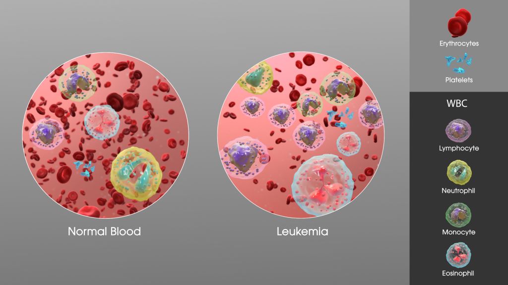
At four years old, a child’s world should be full of crayons, playgrounds, and fairy tales. For Dianty and Nqobimpi, it has become a world of chemotherapy, blood transfusions, and sterile hospital rooms. As the world observes World Children’s Day on 20 November and its theme of “My day, my rights,” their fight for the basic right to life casts a light on the most critical need of all: a second chance, which can only come from a matching stem cell donor.
A Donor for Dianty
Hailing from Randfontein, Dianty is a bright and joyful four-year-old whose smile can light up any room. Yet, behind her cheerful spirit lies a harrowing health battle. It began last year with inexplicable fevers and stomach pains, initially dismissed as a sinus issue and later as growing pains. When Dianty suddenly lost the ability to walk, her mother, Claudine, knew something was terribly wrong.
After she was admitted to the hospital, blood tests revealed the devastating truth: Dianty had leukaemia. Chemotherapy began immediately. “The ups and downs were relentless,” Claudine explains. Despite initial progress, Dianty relapsed just after her fourth birthday this year, forcing her family back to square one.
The search for a stem cell donor has been fraught with disappointment. With no suitable match found within her family, Dianty’s hope now rests on the public registry. “You never think it will happen to you until it does,” says Claudine. “I encourage every single person to go and do the cheek swab… You can save the life of a little child. You can give them a second chance.”
Nqobimpi Needs a Match
Nqobimpi from Pretoria is facing a similarly terrifying reality. Her journey began with persistent nosebleeds and terrifying episodes of vomiting blood, which left her weak and pale. Her mother rushed her to a clinic, which led to a referral and eventually a diagnosis of T-Cell Acute Lymphoblastic Lymphoma (T-Cell ALL).
Expressing the shock that rocked their family, her mother shares, “I wasn’t aware children could get cancer.” Nqobimpi was pulled out of crèche, and her life became a cycle of month-long hospital stays for treatment. The strain has rippled through their family, affecting her siblings’ ability to focus at school as they worry constantly about their baby sister. All Nqobimpi wants is to return home and play with her dolls, a simple childhood pleasure now dependent on finding a selfless stranger willing to register as a donor.
The Power to Save a Life
“For children like Dianty and Nqobimpi, a stem cell transplant from a matching donor is their only hope for a cure,” highlights Palesa Mokomele, Head of Community Engagement and Communications at DKMS Africa. Every new person who registers brings fresh hope to them and the many other patients waiting for a lifeline.”
“On this World Children’s Day, as we reflect on the rights of every child, consider giving the ultimate gift: the right to life,” she concludes.
By Thom Renwick, General Manager, Roche Pharma South Africa
Early screening and treatment. Much higher survival rates. And savings in the billions of dollars. From AI-powered medicine development to teleconsultations, technology can boost Africans’ health and livelihoods while growing economic and social impact across the continent.
Access to quality healthcare is fundamental to leading a fruitful, economically active life. Yet, breast cancer is still the number one cancer killer of women in Africa – in most cases, while they’re still in their prime. Tragically, most are diagnosed too late for curative treatment.
Across the continent, non-communicable diseases – such as treatable breast cancer – cause hundreds of thousands of preventable deaths every year[1], devastating families and hampering economic growth.
Global projections indicate a worrying 38% rise in incidence of breast cancer and a 68% increase in deaths by 2050 without urgent intervention, with the least developed countries being the most affected, according to a new white paper[2] by independent German economic think tank the WifOR Institute.
The Value of Investing in Innovative Medicines report outlines the economic burden from not treating the aggressive HER2-positive type of breast cancer over five years in seven African countries – South Africa, Kenya, Nigeria, Algeria, Tunisia, Côte d’Ivoire and Morocco. The findings are staggering, indicating a $10.3-billion loss in productivity from 2017 to 2023.[3]
The data also shows that, in Africa, 89% of the economic burden of HER2-positive breast cancer – representing 15% to 20% of all breast cancer cases around the world – falls on women of working age.[4]
It goes beyond economics. Mothers hold households together and when they die that has huge ramifications for entire families and communities.
In sub-Saharan Africa, every 100 deaths among women under 50 leave around 210 children without their mothers[5], resulting in unstable, vulnerable households and long-term developmental challenges.
These figures are a wake-up call, but in challenge lies scope for innovation. I believe we can – and must – turn the burden into opportunity.
Closing the gap through health-tech partnerships
Every woman diagnosed and treated early is not only more likely to survive but also able to remain active in her family and community[6], contributing to shared prosperity.
Through healthcare and technology partnerships, we can leapfrog traditional healthcare models and turn the tide towards survival.
Excitingly, this process has already started. Governments are increasingly setting strategies and allocating funding for digital health. Start-ups and companies are driving the uptake of digital health tools that could revolutionise care delivery.
Artificial intelligence stands out as the breakthrough technology. Pharmaceutical and biotech companies such as Roche are already using AI throughout their value chains, both in the early stage of drug development and also to correctly interpret the enormous amounts of data generated to deliver effective health solutions.
In most sub-Saharan countries, more than 20% of the population lives more than two hours from essential health services.[7] Tech’s role in Africa’s future is about much more than connectivity or commerce. It’s about lives and well-being – AI, apps, telemedicine and other digital solutions can close the gap to bring care closer to people.
But without individuals and organisations working together, innovation can’t come to life.
And since the journey for a patient experiencing a health crisis such as breast cancer involves many stakeholders, we must urgently identify opportunities for partners to come together and spur real action.
This week, Roche sponsored the 28th annual Africa Tech Festival’s first-ever health track. Policymakers, innovators, professionals and experts met to thrash out actionable solutions to the continent’s biggest health challenges.
This was part of an ambitious broader strategy to transform healthcare in Africa – investment in early intervention strategies can generate returns that far outstrip their cost.
Research by McKinsey & Company shows that the African digital health space is already seeing unprecedented growth, with $123-million in investment secured by 55 start-ups in 2021.[8]
The consultancy’s analysis showed that digital health tools – such as virtual platforms for consultations; electronic health records; mobile apps to help patients self-manage their diseases; and patient e-booking platforms – could help South Africa, Kenya and Nigeria capture efficiencies of up to 15% in total healthcare expenditure by 2030.[9]
Widespread adoption could free up an astounding $1.9-billion to $11-billion in South Africa alone.[10]
Speaking with one voice for a better future
Innovation has been the backbone of progress across any major public health disease – whether it’s HIV, cancer or ophthalmology. It takes a combination of passionate people and expert innovation to make a difference.
One existing solution and real-life example of African innovation and partnership is EMPOWER, a groundbreaking digital health platform developed to improve coordinated breast and cervical cancer care in Kenya.[11]
The initiative has grown from a single clinic in 2019 to a 76-site national platform and is integrated into Kenya’s National Cancer Registry.[12]
Empower ensures that the entire patient journey, from screening to treatment and follow-up, is digitally powered.
The current average five-year survival rate for breast cancer across Africa is roughly five out of 10 patients that are diagnosed (48%).[13] My vision is that this will increase to 80% within the next 5 years.
Realising this audacious goal will take commitment from stakeholders to drive action for the tens of thousands of African women who desperately need it.
There is no reason why someone in a Western society should have a better health outcome than here in Africa. The health of our people is the wealth of our nations. We must speak with one voice and act now.
––––––––––
Thom Renwick is general manager of Roche South Africa and the sub-region. He began his journey with the company in 2012 on its United Kingdom graduate programme, following his studies at King’s College London, Cranfield School of Management and the University of Oxford.
During his career at Roche, he has worked in global product strategy in Basel, Switzerland, as head of ophthalmology in the UK and as chief of staff for Pharma International. He won the PharmaTimes New Marketer of the Year Award in 2015 and was featured in the publication’s Smart People series in 2021.
[2] WifOR Institute, ‘The Value of Investing in Innovative Medicines: Socioeconomic Burden of HER2+ Breast Cancer and Annual Social Impact of Roche’s Treatments for the Disease in Africa’. Accessed: Nov. 7, 2025 [Online]. Available: https://africa.roche.com/stories/what-s-it-worth-the-value-of-innovation
[3] WifOR Institute, ‘The Value of Investing in Innovative Medicines: Socioeconomic Burden of HER2+ Breast Cancer and Annual Social Impact of Roche’s Treatments for the Disease in Africa’. Accessed: Nov. 7, 2025 [Online]. Available: https://africa.roche.com/stories/what-s-it-worth-the-value-of-innovation
[4] WifOR Institute, ‘The Value of Investing in Innovative Medicines: Socioeconomic Burden of HER2+ Breast Cancer and Annual Social Impact of Roche’s Treatments for the Disease in Africa’. Accessed: Nov. 7, 2025 [Online]. Available: https://africa.roche.com/stories/what-s-it-worth-the-value-of-innovation
[5] WifOR Institute, ‘The Value of Investing in Innovative Medicines: Socioeconomic Burden of HER2+ Breast Cancer and Annual Social Impact of Roche’s Treatments for the Disease in Africa’. Accessed: Nov. 7, 2025 [Online]. Available: https://africa.roche.com/stories/what-s-it-worth-the-value-of-innovation
[6] WifOR Institute, ‘The Value of Investing in Innovative Medicines: Socioeconomic Burden of HER2+ Breast Cancer and Annual Social Impact of Roche’s Treatments for the Disease in Africa’. Accessed: Nov. 7, 2025 [Online]. Available: https://africa.roche.com/stories/what-s-it-worth-the-value-of-innovation
[7] Mckinsey, ‘How digital tools could boost efficiency in African health systems.’ Accessed: Nov. 7, 2025 [Online]. Available: https://www.mckinsey.com/industries/healthcare/our-insights/how-digital-tools-could-boost-efficiency-in-african-health-systems
[8] Mckinsey, ‘How digital tools could boost efficiency in African health systems.’ Accessed: Nov. 7, 2025 [Online]. Available: https://www.mckinsey.com/industries/healthcare/our-insights/how-digital-tools-could-boost-efficiency-in-african-health-systems
[9] Mckinsey, ‘How digital tools could boost efficiency in African health systems’. Accessed: Nov. 7, 2025 [Online]. Available: https://www.mckinsey.com/industries/healthcare/our-insights/how-digital-tools-could-boost-efficiency-in-african-health-systems
[10] Mckinsey, ‘How digital tools could boost efficiency in African health systems’. Accessed: Nov. 7, 2025 [Online]. Available: https://www.mckinsey.com/industries/healthcare/our-insights/how-digital-tools-could-boost-efficiency-in-african-health-systems
[11] Roche Africa, ‘From vision to national platform: EMPOWER scales through Kenya’s National Cancer Institute’. Accessed: Nov. 7, 2025 [Online]. Available: https://africa.roche.com/stories/empower-scales-through-kenya-national-cancer-institute
[12] Roche Africa, ‘From vision to national platform: EMPOWER scales through Kenya’s National Cancer Institute’. Accessed: Nov. 7, 2025 [Online]. Available: https://africa.roche.com/stories/empower-scales-through-kenya-national-cancer-institute
[13] A. Padu-Pebrah, et al., ‘Five-Year Survival Outcomes for Breast Cancer Patients Across Continental Africa: A Contemporary Review of Literature with Meta Analysis’, eLife. Accessed: Nov. 7, 2025 [Online]. Available: https://elifesciences.org/reviewed-preprints/105488#mainMenu
Oliver Staub, 2, smiles while recovering from two complex spinal cord surgeries at UChicago Medicine Comer Children’s Hospital that reattached his head to his spinal cord. Image credit: University of Chicago Medicine
With monitors quietly beeping and multiple tubes going into his small body, Oliver Staub lay in a hospital bed as his parents tearfully started saying goodbye.
On April 17, an armoured car going 70mph (112kph) slammed into the family’s minivan during their vacation in Mexico. Everyone in the car was injured, but no one more than Oliver.
The impact disconnected the 2-year-old’s head from his spine, resulting in a transection of his spinal cord.
Doctors offered a grim prognosis. They told Oliver’s parents, Laura and Stefan, that their son’s neck was broken, he was a quadriplegic, brain dead and would die in a matter of days.
But following a surreal turn of events – which included support from German soccer star Toni Kroos, viral Instagram posts, and traveling more than 2,000 miles for two risky spinal cord surgeries at the University of Chicago Medicine Comer Children’s Hospital — Oliver is now talking, laughing, smiling, moving his fingers and toes and starting to breathe on his own.
“To see someone survive an injury like this? Nothing like this has ever been reported in neurosurgery or spinal cord injuries,” said Mohamad Bydon, MD, Chair of the Department of Neurological Surgery at UChicago Medicine and health system leader for Neurological Surgery, who performed Oliver’s surgeries in July with a multidisciplinary team of surgeons.
“We didn’t think he’d ever be able to move, and now he’s moving all four limbs,” Bydon said. “This is a unique and special case. It’s beyond our wildest expectations.”
‘We have a reason to fight’
As family members gathered at the Mexico City hospital to say goodbye, something incredible happened: Oliver began to show signs of recovery.
His eyes would follow his parents when they were in the room. Stefan and Laura raised the issue with his doctors, who ultimately determined that their son did, in fact, have brain function. They kept his life-sustaining ventilator on.
“It was at that moment that I thought, ‘We have a reason to fight,’” Laura said. “My son was there.”
When doctors could do nothing more for Oliver, they trained his parents on how to care for him and operate his ventilator. Wearing a neck collar and vest to stabilise his head – which, internally, was not connected to his body – Oliver was moved to his grandparents’ home eight hours away, near Morelia, Mexico.
With help from a daily nurse visit, Oliver survived for two months without moving and once having an incident of cardiac arrest. Bydon finds this astounding, given how unlikely it is that someone with an unstable, transected spine could survive at all, much less under the care of his parents.
“If Oliver’s parents and caretakers had made one wrong move in those two months, it could have resulted in death,” Bydon said.
A journey to Chicago
Stefan and Laura researched treatments for severe spinal cord injuries, hoping to provide a better life for their son. They contacted top spinal cord specialists around the world, including Bydon, whose groundbreaking stem cell therapy research impressed them.
They were repeatedly told that surgery, and the travel involved, would be too risky. But Bydon saw hope, in part because Oliver had survived this long.
“You should never count out a 2-year-old. They can surprise you,” Bydon said. “But it would require a complex multidisciplinary team, which is where the University of Chicago could help.”
The surgery needed to be done as soon and safely as possible, Bydon told them.
But travel to the United States for the surgery would be difficult and expensive. The Staubs received aid from family, friends and charities, but were still far short of what they needed.
Global outreach and support
A friend encouraged them to write to the Toni Kroos Foundation, the soccer player’s charity which helps seriously ill children. Stefan and Laura knew it was a long shot.
Two days later, the phone rang at midnight. It was foundation director Claudia Bartz. She’d seen Oliver’s journey on Instagram and was so moved by his story, she decided the foundation would cover the cost of Oliver’s surgery and transport to Chicago.
“We cried and cried. We couldn’t believe it,” Laura said, adding that they only posted on Instagram to keep their friends and family updated on Oliver. “None of this would have been possible without Toni Kroos.”
Oliver soon became a top-trending news story in Germany and their Instagram account blew up, going from a few hundred followers to more than 100 000. Strangers across the world continue to hold fundraisers and prayer vigils, sending the family encouraging messages and donations for his medical expenses.
“We would gladly trade all of this to go back to our normal life,” said Laura, who still has large scars on her head from the accident. “What I’m seeing here? It’s miraculous. We call it ‘The Oliver Effect.’ This is bigger than us.”
‘Harrowing’ surgery, major recovery
When Oliver arrived at Comer in July via medical jet, Bydon performed the first surgery, an occipital cervical fusion, with a team of UChicago Medicine surgeons.
This surgery for a 2-year-old is risky, not only because of how long it is, but also because a toddler cannot tolerate blood loss.
The surgery involved reconstructing Oliver’s spine, repairing his spinal cord and stabilising the back of his head to his cervical spine using titanium rods and screws.
The second surgery, two days later, stabilised the front of his spinal cord and repaired a spinal cord herniation.
“Those first few days after the surgeries were harrowing,” Bydon said. “His heart stopped at one point, and he had swelling in the brain.”
But about five days later, Oliver was making progress and smiled for the first time since the accident. One month later, he was able to grab his mom’s hand, push someone away and recognise the sensation that he needs to urinate. Most impressively, Bydon said, he can now take breaths on his own.
“We know the spine is communicating with the brain and body again,” Bydon said.
Moving forward with family
Oliver was discharged from Comer Children’s on August 15. The family will permanently move from Germany to Mexico, near Laura’s family, and now have hope for the future.
Oliver will have regular physical therapy and take medications for inflammation. In about six months, he’ll be able to remove his neck brace, Bydon said.
Laura and Stefan plan to return to Comer in spring 2026, when Bydon may be able to use novel stem cell therapy clinical trials to improve Oliver’s physical functions, pending special FDA approval.
Stefan and Laura said they’ll always be grateful to Bydon and UChicago Medicine.
“He didn’t promise us a miracle,” Laura said, “but he delivered one.”