Study finds that intestinal circadian clocks become misaligned by off-schedule eating, causing gastrointestinal issues
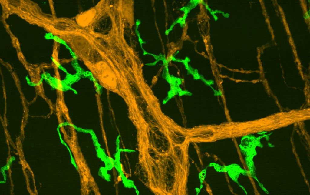
A microscopic image shows enteric neurons (orange) and macrophages (green) in the muscularis externa of a mouse small intestine. Muscularis macrophages were among the intestinal cell types that glowed green when a key circadian clock gene called Per2 was active during UT Southwestern investigators’ research.
Eating when the body is normally asleep appears to desynchronise the circadian clocks of different cell types in the intestines, a UT Southwestern Medical Center study suggests. The findings, published in PNAS, could help explain why shift work, jet lag, and other environmental stressors that affect circadian rhythms are associated with irritable bowel syndrome, inflammatory bowel disease, constipation, and other gastrointestinal disorders.
“Understanding how intestinal circadian clocks become misaligned may ultimately guide strategies involving meal timing, circadian-based therapies, or dietary interventions to improve gastrointestinal and metabolic health,” said Yuuki Obata, PhD, Assistant Professor of Immunology and Neuroscience at UT Southwestern. Dr Obata co-led the study with Shin Yamazaki, PhD, Professor of Neuroscience.
Research in the 1990s and 2000s showed that a region of the brain known as the suprachiasmatic nucleus (SCN) acts as a master timekeeper for the body, setting various cellular processes to occur rhythmically on a 24-hour period based on cycles of light and darkness. However, in 2000, Dr Yamazaki and his colleagues showed that cells throughout the body have their own autonomous circadian clocks that are influenced both by signals from the SCN and environmental cues.
In line with this idea, research has shown that the intestines have their own rhythms that can be influenced by a variety of factors, such as timing of meals. These findings were made using whole intestinal tissue, Dr Obata explained, but the intestines contain a variety of cell types – including muscle, nerve, and immune cells. It’s been unclear whether each of these populations has its own circadian clock and if they run on the same schedule.
To find out, Drs Obata and Yamazaki monitored novel mice with set 12-hour cycles of light and dark. They were engineered by Joseph Takahashi, PhD, Chair and Professor of Neuroscience at UT Southwestern, and his colleagues in the Takahashi Lab. Five intestinal cell types – enteric neurons, enteric glial cells, interstitial cells of Cajal (ICCs), smooth muscle cells, and muscularis macrophages – glowed green when a key circadian clock gene called Per2 was active. Although food was available at all times, the mice ate about 80% of their meals at night due to their nocturnal nature.
After about a week in this environment, the researchers observed intestinal cells glowing green at approximately the same times, suggesting the different cell populations had their own autonomous circadian clocks that cycled in sync. But when the researchers made food available only for four hours in the daytime – forcing the mice to eat at abnormal times – Per2 activity shifted to match this new rhythm in every cell population except for the ICCs. These cells resisted changes to their circadian clock, staying out of sync with the other cell types for weeks.
Such asynchrony may also occur in people who eat outside the body’s usual circadian rhythms, such as night shift workers or those who fly to different time zones. Because ICCs play a key role in intestinal motility, their resistance to adapt to a changed circadian clock could affect digestive and metabolic function.
Finding a way to synchronise the different intestinal cell populations through diet, probiotics, or drugs could eventually help ease the gastrointestinal problems associated with altered circadian timing, the researchers said.
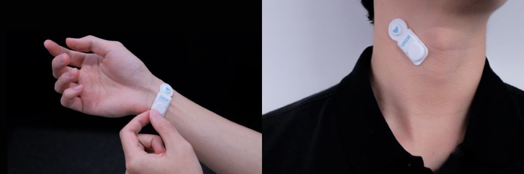
The AusculPatch is a tiny wearable sensor that weighs just 3.2 grams.
Australian researchers have developed a lightweight wearable sensor that could continuously check on people with heart and breathing problems, potentially reducing hospital visits and allowing doctors to detect problems earlier.
The flexible sensor patch, which attaches to the chest or over peripheral arteries using medical adhesive tape, is designed to continuously capture subtle vibrations produced by the heart, lungs and, blood flow, and pulse waves.
Researchers hope the technology could eventually help people with chronic heart and respiratory conditions track their health remotely and alert clinicians when something may be wrong before symptoms become severe.
The proof-of-concept work, led by researchers from UNSW in collaboration with clinicians and biomedical engineers, has been published in Nature Communications.
Lead researcher and corresponding author of the paper, Scientia Associate Professor Hoang-Phuong Phan, says the goal is to create a wearable device which patients can use themselves – as a home alternative to the traditional doctor’s stethoscope.
“What we have developed is a tiny wearable device that can attach onto the human chest and hear heart sound and respiration,” A/Prof. Phan says.
“Technically, it aims to replace the stethoscope, which is normally used in clinic centres to assess cardiovascular or respiration disease.”
Addressing a growing healthcare challenge
Heart disease and chronic respiratory illnesses remain among the leading causes of death worldwide, but many patients only receive brief assessments during occasional medical appointments. A/Prof. Phan says this can create major challenges for people living in regional and remote areas, or patients reluctant to repeatedly visit hospitals and clinics.
“Normally, when patients are assessed by a doctor, they have to go to a clinic centre, and it’s not very convenient for those who live in remote areas,” he says. “Sometimes people are hesitant to go to hospital, so they wait until symptoms are clearly developed.”
By the time symptoms become serious enough to seek medical care, diseases may already have significantly worsened.
“At that stage, the disease may already have worsened, leaving poorer outcomes for patients even when treated,” says Dr Anthony Sunjaya, a medical doctor and Program Lead for Chronic Respiratory Disease at UNSW’s School of Population Health, who co-authored in this work. “When they go to a clinic, patients often only have a 15-minute window for assessment. The danger is that the abnormalities experienced will not be fully recognised during that short period of time they are being seen.”
How the patch works
The device, known as ‘AusculPatch’, is smaller and lighter than many existing wearable monitoring systems, weighing only 3.2 grams and measuring roughly 20x47x3 millimetres. At the centre of the patch is an ultra-thin silicon sensing element that detects tiny mechanical vibrations travelling through the skin from the heart, lungs and blood vessels.
“The heart sound propagates through the body fluid and tissue generates an acoustic pressure that vibrates the sensing element,” Tran Bach Dang, the first author and a PhD candidate from the School of Mechanical and Manufacturing Engineering says. “What the patch is doing is picking up that vibration.”
The new sensor can detect extremely low-frequency vibrations that are difficult to capture with current wearable technology. The device can detect a remarkably broad range of physiological signals, including breathing patterns, pulse waves, heart sounds and blood flow vibrations.
In tests, the AusculPatch was able to continously monitor a range of physiological markers while the wearer was undertaking regular daily tasks. Importantly, researchers say the patch was designed to minimise interference from surrounding environmental noise — a major challenge for wearable acoustic sensors.
“The sensor element is designed to shield the sound coming from one direction, typically from the human body,” Dang says. “In that way, it is less susceptible to ambient sound.”
Although tested on only a small number of healthy participants, the research paper showed the device could continue capturing clear heart sounds even in noisy environments, including during conversation and under simulated background noise conditions.
Beyond smartwatches and fitness trackers
While consumer devices such as smartwatches and sleep trackers can already monitor heart rate and blood oxygen levels, the research team says AusculPatch captures more direct mechanical information about how the heart and lungs are functioning.
The researchers believe the technology could eventually have applications ranging from chronic disease management to sleep monitoring and general wellbeing.
The paper also highlights potential use in monitoring blood pressure, pulse waves and subtle heart valve abnormalities that are difficult to continuously track outside hospital settings.
In laboratory and early human testing, the device showed strong agreement with clinical tools including electrocardiograms (ECGs), ultrasound scans, blood pressure monitors and digital stethoscopes.
Researchers were also able to continuously record cardiorespiratory data over extended periods while participants walked, worked, ate meals and climbed stairs.
AI-powered monitoring
One of the most promising aspects of the technology is the possibility of combining continuous monitoring with artificial intelligence.
Because the patch collects large amounts of physiological data over time, researchers hope machine learning systems could eventually identify patterns linked to worsening disease or emerging health problems.
“We can potentially apply machine learning to identify abnormal signal and warn the patients, and also notify their doctor,” Dr. Chi Cong Nguyen, an Associate Lecturer and a corresponding author of the paper says.
“The goal is to create a system that can automatically flag concerning changes before patients experience severe symptoms.”
Potential future applications
Beyond cardiorespiratory monitoring, the researchers also demonstrated that the patch could detect vocal cord vibrations from the throat. In proof-of-concept experiments, the team used machine learning to recognise spoken words and wirelessly control a robotic arm. While those experiments are still early-stage, the researchers say the technology could eventually support people with speech disorders or physical disabilities.
Although the technology is still in the research and testing phase, larger clinical studies are already being planned.
That group is expected to include people with heart valve disease or implanted heart assist devices. Researchers then hope to scale up studies to around 1000 patients over the following years to further develop AI-assisted diagnostic tools. Regulatory approval for a medical-grade device would still take time, with A/Prof. Phan estimating a timeline of around four to five years before possible clinical deployment. However, consumer-focused wellness versions of the technology could potentially become available sooner.
People who wore a cooling cap for 30 minutes experienced multiple changes that could affect their mental health, according to a pilot study by Penn State researchers
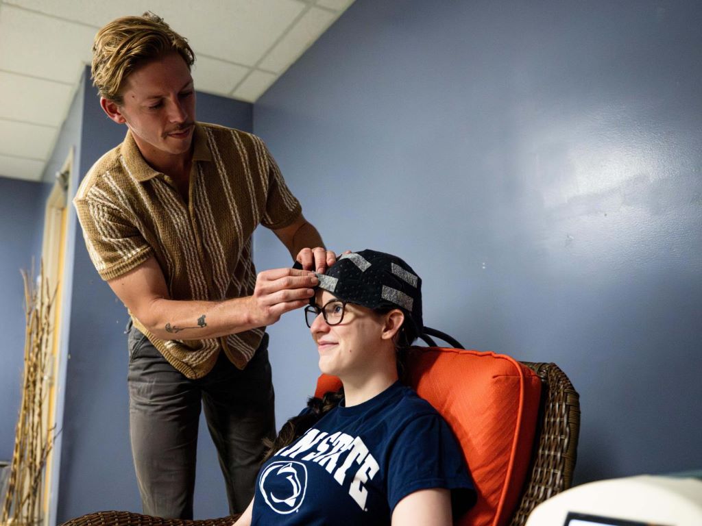
Co-authors Owen Griffith, standing, and Maddie McLaughlin demonstrate the head cooling cap used in the study. Credit: Jaydyn Isiminger / Penn State. Creative Commons
Wearing a cooling cap for 30 minutes may improve a person’s sense of well-being, according to a new study by Penn State researchers.
In a recent publication in Acta Psychologica, the researchers demonstrated that head cooling may reduce depressive symptoms and alter the types of brain waves people produce. While no medical recommendations can be derived from this small, exploratory study, the results indicate head cooling may provide mental health benefits for the general population.
The work was inspired by lead author and Penn State Professor of Kinesiology Semyon Slobounov’s prior research, which found that athletes with concussions heal faster and experience fewer symptoms when their head is regularly cooled.
“A person’s mood is tied to their cognition and general brain function,” said Owen Griffith, assistant teaching professor of kinesiology at Penn State and co-author of the study. “In this study, results suggested that people enjoy the sensation of head cooling. This, in turn, improved their mood, which altered their brain activity.”
The researchers recruited 24 college students between the ages of 18 and 26. At the beginning of the study, all participants completed questionnaires that measured their mental health and cognitive abilities and underwent an electroencephalogram (EEG) to measure brain activity.
Following the EEG, participants spent 30 minutes sitting in a dimly lit room listening to ocean sounds. Half of participants wore a fitted cooling cap, which uses liquid circulating close to the head to maintain a temperature of 33 degrees Fahrenheit. The other participants wore nothing on their heads. Immediately after the cooling or sitting session, participants repeated the questionnaires and EEG.
Participants repeated the same sitting or cooling session without testing every day for one week. The day after the last session, participants repeated the questionnaires and EEG again. This design allowed the researchers to observe both the short- and longer-term effects of head cooling.
“The brain produces different types of waves that are associated with different levels of excitement or brain activation,” said Laura Cooney, co-author of the study who graduated from Penn State’s Schreyer Honor College in 2025 and based her undergraduate thesis on the research. “Alpha waves are associated with calmness. More specifically, they are indicative of less brain activity overall, so this finding suggests that there was an immediate calming effect of head cooling.”
People in the head cooling group displayed an increase in alpha brain waves during the EEG immediately following the first cooling session. They experienced a 4% increase in alpha waves while participants whose heads were not cooled displayed a .5% decrease in alpha waves.
In contrast, there was no significant difference in the alpha wave levels of the sitting and cooling groups when measured on the day after the final cooling session, suggesting cooling does not have a longer-term impact on brain wave activity, the researchers said.
Over the course of the week, both groups of participants reported a decrease in depression symptoms, but individuals in the head cooling group reported a larger decrease than those in the sitting group.
“The reduction of depression symptoms among healthy people suggests that this might be a promising treatment,” Griffith said.
The researchers said they had hypothesised head cooling affected people through changes in neural electrical activity, but the EEGs did not show evidence of that. Now, the researchers suspect the effects are psychosomatic, meaning that mental and emotional factors, rather than physiological changes, are causing people’s reduced depression symptoms and increased alpha brain wave activity.
“Anecdotally, most people who come into the lab agree that head cooling is relaxing and enjoyable,” Griffith said. “This may not be surprising. A cold compress or a bag of ice have been home treatments for migraines for many years.”
Overall, the study suggests that widespread head cooling could be useful, the researchers said.
“Head cooling shows some potential as an acute calming therapy,” Cooney said. “Not as a replacement for any current therapy, but as another tool in the toolbox.”
Slobounov, senior author of the study, agreed.
“Our previous research demonstrated that head cooling is useful for athletes recovering from concussions,” Slobounov said. “This research suggests it may be more useful to a wide group of people. It is low risk, does not involve any drugs or chemicals, and people enjoy it.”
Other Penn State researchers who contributed to this work include Zach Napora, graduate student in kinesiology and first author of the publication, Maddie McLaughlin, graduate student in kinesiology, and Elle McNally, 2025 graduate in biobehavioral health and current physician assistant graduate student.
Children with asthma and allergies who live with one or more cats do not experience worse asthma outcomes than children without cats at home. This is according to a comprehensive Swedish registry study from Karolinska Institutet, published in Frontiers in Allergy.
Many families with asthmatic children are advised to avoid keeping furry pets in the home. At the same time, previous studies on how cats as pets affect children with asthma and allergies have often been small and produced conflicting results.
The new study covers over 30 000 children in Sweden aged 4–17 with diagnosed asthma and allergies. The researchers followed the children for two years, comparing those living in households with cats with those who did not. Data on cat ownership was obtained from the national cat register and combined with data from several Swedish health and quality registers.
“We found no evidence that living with cats worsens asthma in children who already have asthma and allergies,” says Resthie R. Putri, postdoctoral researcher at the Department of Medical Epidemiology and Biostatistics, Karolinska Institutet. “Unfortunately, however, we had no information on the specific allergies the children had, so we do not know whether they were allergic to cats or not.”
Individual advice is needed
The researchers analysed asthma exacerbations leading to emergency care, asthma severity based on medication use, asthma control, and lung function in the children. Children living with one or more cats experienced similar asthma outcomes to those without a cat. Nor could the number of cats in the home, or the age or sex of the cat, be linked to differences in asthma outcomes.
One strength of the study is the large number of participants from across Sweden. However, differences between countries may limit the applicability of the results to other contexts. As the Cat Register is relatively new, some cat exposure may not have been captured in the study. Furthermore, there was no information on how long the children had been exposed to cats for, or how much time the cats spent indoors. The researchers also cannot rule out the possibility that families with children suffering from more severe allergies may have chosen not to keep a cat.
“Clinical advice always needs to be tailored to the individual, and our study can contribute to the evidence base in discussions with families about pets and asthma,” says Catarina Almqvist Malmros, professor at the Department of Medical Epidemiology and Biostatistics, Karolinska Institutet, and paediatrician at Astrid Lindgren Children’s Hospital.
Will continue to follow the children
The researchers now plan to continue studying how different types of allergies may affect the outcome, and whether it makes a difference if the child is allergic to cats. They also plan to follow the children for a longer period to see how cat ownership may affect asthma over time.
A new study from the University of Kansas details how US tobacco corporations expanded into global food markets from the mid-1980s to the mid-2000s, using strategies honed through cigarette sales to market ultra-processed foods, which are industrially processed and contain ingredients and additives that maximise their appeal. The research appears in the American Journal of Public Health.
“We’d previously published on US tobacco company activities in relation to the US food system,” said lead author Tera Fazzino, associate professor of psychology at the University of Kansas. “In that prior study, I noted that tobacco-owned food company activities extended far beyond the United States. They had an extensive international business development strategy. Given the global conversation about ultra-processed and hyperpalatable foods saturating food systems, it felt important to investigate that international dimension. This study contributes to understanding the origin and nature of these foods and how they became widespread globally.”
For two decades, tobacco firms heavily invested in food companies, applying marketing, distribution and product-engineering strategies developed in the tobacco business.
“The scale was massive,” said Fazzino, who also serves as associate scientist at the KU Life Span Institute’s Cofrin Logan Center for Addiction Research and Treatment. “For Philip Morris, its food business, for a substantial period of time, generated sales comparable to its tobacco business – roughly a 50–50 split. A significant portion of those food revenues came from international markets.”
Meanwhile, Fazzino said, R.J. Reynolds had closer to 30% of its portfolio in food sales.
“But it also maintained a very large international food presence,” she said. “In many of the regions where these companies operated, their food subsidiaries were among the largest players in those markets at the time.”
Fazzino’s co-authors were KU graduate students Sydney Kong and Gayeon Lee, along with undergraduate research assistant Madison Stewart.
The researchers found that tobacco companies applied their long-established business strategies directly to their food operations.
“On the business development side, they aggressively expanded and acquired local companies to build infrastructure within regions,” Fazzino said. “This allowed them to distribute their primary products efficiently. They also created coordinated product distribution and sales systems that integrated both tobacco and food operations.”
On the product-development side, tobacco companies used strategies similar to those previously applied to cigarettes.
“For example, they manipulated product size,” Fazzino said. “Just as they had introduced ‘king-size’ cigarettes to increase consumption occasions, they later used similar size strategies in food products – king-size sugary drinks, cookies and other items.”
Fazzino said tobacco firms also developed products that maintained “full flavour” while being marketed as lighter or healthier.
“In tobacco, this meant ‘low-tar’ or ‘low-nicotine’ cigarettes,” she said. “In food, it meant ‘low-fat’ or ‘light’ versions of products designed to appeal to health-conscious consumers while retaining strong flavour profiles.”
The researchers found that different tobacco firms developed their food businesses in different geographic areas, even if their marketing and product-formulation strategies were similar. According to Fazzino, the data indicate tobacco companies held leading market shares in multiple regions.
“Philip Morris expanded rapidly into Canada, where it held leading market positions in many product categories as early as the late 1980s,” she said. “Its entry into the Canadian market was swift and aggressive. The company also expanded significantly into Europe and secondarily into parts of Asia and the Asia-Pacific region. R.J. Reynolds primarily focused on Central and South America, and Mexico, and also expanded into parts of Europe and Asia. Collectively, this meant that from the late 1980s through the mid-2000s, large portions of the globe were covered by one or both companies.”
The public health implications of increased ultra-processed and hyperpalatable foods in the world’s food supply are substantial, Fazzino said.
“Globally, there is ongoing discussion among scientists, policymakers and citizens about the shift toward ultra-processed and hyperpalatable foods,” she said. “There is now strong evidence that these foods carry significant health risks. Their risk profile parallels other major contributors to morbidity and mortality, including smoking. As these products were disseminated internationally using highly refined and efficient business strategies, the potential for global health harm was significant.”
Fazzino cited rising rates of obesity and related metabolic and cardiometabolic diseases in many parts of the world.
“These increases have paralleled shifts in food systems toward more ultra-processed and hyperpalatable foods,” she said.
Although tobacco companies divested from food businesses in the early to mid-2000s, Fazzino said their influence in disseminating ultra-processed foods globally leaves a lasting legacy.
“The food companies continued operating under profit-maximizing models that had already proven successful for other addictive products,” she said. “It’s likely other food companies observed these strategies and adopted similar approaches. As a result, these practices appear to have spread across the global food industry.”
According to Fazzino, today at least 70% of the US food supply is composed of hyperpalatable foods, and their presence globally is elevated. The KU researcher said policymakers could apply lessons from tobacco regulation, including labelling, marketing restrictions and litigation.
“However, food presents unique challenges because people must eat,” Fazzino said. “In food systems saturated with ultra-processed and hyperpalatable products, such as in the US, stronger regulation of the nutrient profiles and properties that drive addictive qualities may be necessary. In contrast, South American countries, which have not experienced saturation of their food supplies, have implemented warning labels and other measures with demonstrated public health benefits.”
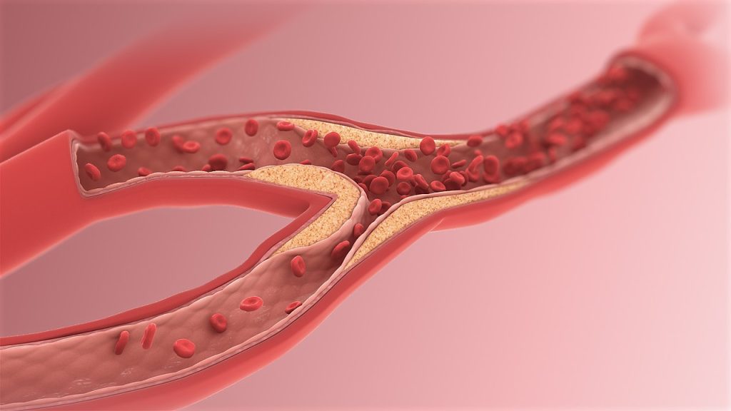
Doctors should consider treating high cholesterol much earlier and more aggressively than they have in the past, according to a new perspective by Weill Cornell Medicine, New York-Presbyterian and Yale School of Medicine investigators. The updated 2026 cholesterol management guidelines from the American College of Cardiology and the American Heart Association encourage doctors to identify risk earlier and intervene before heart attacks and strokes happen. Heart disease remains the leading cause of death in the United States.
The perspective, published May 19 in the Journal of the American College of Cardiology, highlights the role and potential for primary care clinicians to implement the guidelines, while also acknowledging the challenges they may face.
“Primary care clinicians are really the backbone of patient care, especially when it comes to cardiovascular prevention. Due to our longitudinal relationships with patients we are uniquely positioned to reinforce healthy behaviours related to diet, exercise, sleep and smoking,” said Dr Madeline R. Sterling, associate professor of medicine at Weill Cornell and an internist at NewYork-Presbyterian/Weill Cornell Medical Center, and lead author of the perspective. Dr Sterling also served as the Inaugural Chair of the Primary Care Science Committee at the American Heart Association. “Primary care clinicians can screen patients earlier, assess lifetime cardiovascular risk and consider additional risk factors to enhance their management of dyslipidaemia.”
Dr. Erica S. Spatz, associate professor of cardiology and epidemiology at Yale School of Medicine, also co-authored the perspective.
In the past, treatment decisions often focused on whether someone was likely to have a heart attack or stroke within the next 10 years. However, even younger adults with moderately elevated cholesterol may benefit from earlier intervention if they carry other hidden risks, the authors said.
To better identify those risks, the new guidelines recommend that doctors look beyond standard cholesterol tests and use newer tools including measuring lipoprotein(a), or Lp(a), a genetically inherited cholesterol-related particle linked to heart disease, as well as coronary artery calcium (CAC) scans that can detect silent plaque buildup in the heart’s arteries.
Overall, the guidelines recommend a more aggressive approach to lipid management, including a focus on the cumulative damage cholesterol can cause over a lifetime. This may include starting a statin medication earlier along with lifestyle changes.
But implementing these recommendations may be challenging in practice, Dr Sterling said
Primary care clinicians often must address competing medical concerns during office visits, which tend to be short. Explaining lifetime cardiovascular risk, advanced cholesterol testing and preventive medications can require detailed conversations that need to occur over time and in concert with cardiologists and other specialists.
Experts also worry that without careful implementation, these advances could widen existing health disparities. Some advanced tests and newer cholesterol-lowering medications may not be covered by insurance or readily available in all communities.
Drs Sterling and Spatz say successful adoption will likely require better insurance coverage, improved electronic health record tools, patient education efforts, and stronger collaboration between primary care clinicians and specialists, including research and implementation efforts.
The guidelines also promote team-based care, where nurses, pharmacists and specialists help patients manage medications and reach cholesterol targets.
Ultimately, a multi-pronged approach will be necessary for the guideline recommendations to be consistently translated into practice, Dr Sterling said.
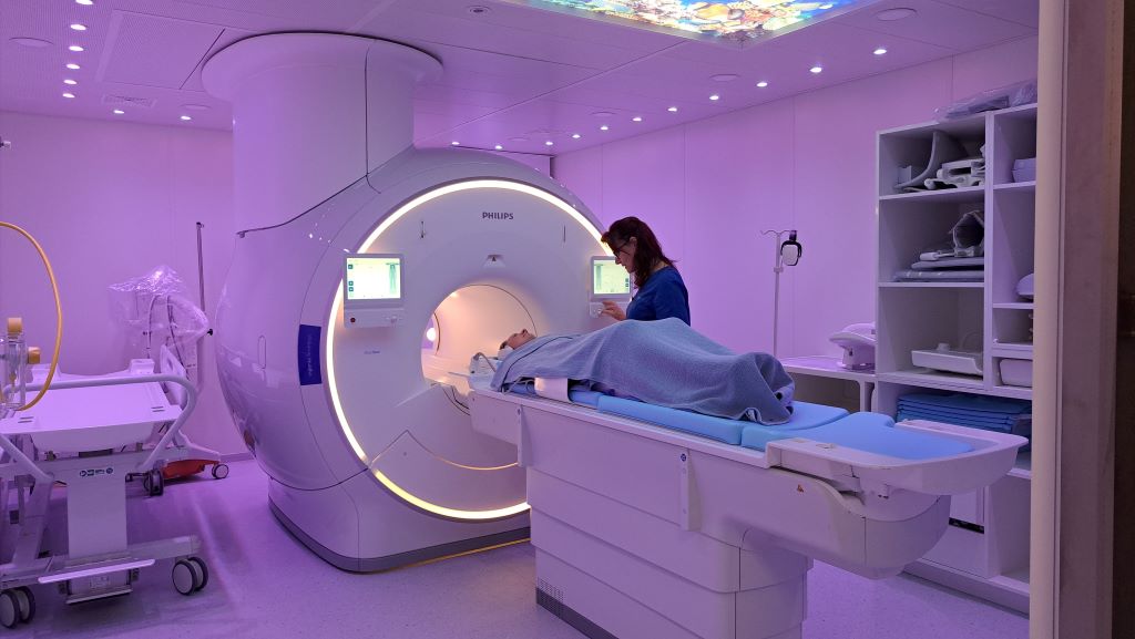
Moving a Magnetic Resonance Imaging (MRI) machine is not as simple as out with the old and in with the new. It is an engineering feat – part physics, part choreography. It is not just a machine, it is an entire system that needs structural support and specialised housing.
The MRI machine being hoisted into its new position.
Some Quite Interesting (QI) facts about moving an MRI machine
MRI machines are marvels of engineering, with their powerful magnets requiring precise handling and specialised support. The magnet, which is the heart of the MRI, can weigh several tonnes and must remain cold, often near absolute zero, maintained by liquid helium or other cooling methods.
‘When replacing an MRI machine, the process is carefully orchestrated, starting with meticulous planning and structural assessments,’ explains Tinus van Rooyen, Business Project Manager at SCP Radiology. The date of the move is carefully selected to coordinate the team of engineers, crane operators and logistics professionals.
‘It is far more complex than moving almost any other piece of hospital or healthcare equipment. Which is why,’ says van Rooyen, ‘removing our old MRI machine and replacing it is not quite ‘all in a day’s work’. And our practice is doing it across multiple sites over the coming months.’
The new MRI machines contain less than 1% of the scarce and non-renewable resource, helium, than that of a conventional MRI machine, improving operational efficiency and long-term sustainability. ‘The newer MRI systems use sealed magnets that, although having to be kept at a temperature of 4 Kelvin (-269.15° Celsius), require very little helium and no refilling over their lifetime’, explains van Rooyen. ‘The improvements in technology in the new machines also ensure improved image quality.’
What is an MRI machine?
MRI ready for its first patient.
The powerful magnetic field and radio waves create detailed images of the body, enabling radiologists to look at soft tissues like the brain, spine, joints and organs in extraordinary detail and without using any radiation. ‘Everything is controlled by advanced computer systems, which convert signals into detailed scans. If an MRI is listening for whispers from the body, a Faraday cage – using copper or aluminium – shields the room to ensure it’s completely silent, so these can be heard clearly.’
How heavy is an MRI machine?
Heavier than most people expect. A low helium MRI machine weighs in at around 3.3 tonnes (3 300kg) – about the weight of an elephant and is significantly less than the conventional machines. The magnet alone is about 60 000 times stronger than Earth’s magnetic field when it’s fully operational and can weigh several tonnes. It’s unsurprising that installing one is a major engineering exercise.
Why would an MRI machine be replaced – what is the usual lifespan of a machine?
The lifespan of an MRI is approximately 10 – 15 years. Replacing ageing equipment ensures access to the latest technology, including improved image quality and an enhanced patient experience. Machines can also reach End of Support (EOS). ‘This means manufacturers no longer support and maintain the unit and parts are unavailable,’ says van Rooyen. ‘It therefore becomes unreliable to keep it running. Patient care is paramount, as is minimising potential downtime and ensuring continuity of service.’
What happens when a machine is replaced?
The old one needs to be ramped down (gradually reducing the strength of the scanner’s main magnetic field to zero in a controlled way). It is then disconnected and removed.
Installation of new unit is basically a reverse of the ramping down process. Once the unit is in place, the magnet is cooled to a superconducting state, then ramped up by gradually increasing the electrical current in the magnet coils, causing the magnetic field to slowly rise. After the magnet has reached its specified strength, the magnetic field is then aligned to make it as uniform as possible, ahead of the unit being calibrated and tested. The entire process can take up to eight weeks. Careful planning ensures continuity of service, with alternative arrangements in place where necessary.
What are the challenges and logistics during the move?
The MRI machine at the SCP Radiology branch at Mediclinic Louis Leipoldt is housed on the first floor. The challenges included building a platform outside the building, moving and lifting the units, with the external wall being removed to create access. The equipment manufacturers oversee the installation but it required a team of riggers to assist with taking out the old unit, then lifting and installing the new unit off the delivery vehicle using a crane, onto a platform and then into the building. Additional contractors are responsible for preparing the room – this includes copper cladding, drywalling, reinforced flooring (if required), painting, joinery, etc.
‘The installation of each MRI unit is unique and depends on a number of factors. In the case of this new one at Louis Leipoldt, we had to partly close off a section of the road for the unloading and lifting. Getting the MRI into the building is a display in itself. Powerful cranes are used to lift the machine, hoisting it through a specially constructed opening. Every step demands precision to avoid damaging the magnet or the building and the installation requires coordinated planning with multiple stakeholders to ensure that the project is executed safely and efficiently,’ explains Heinie Matthysen, SCP’s Facilities Manager.
‘Everything is planned in minute detail by our facilities manager,’ says van Rooyen, ‘however, we also have to factor in the Cape Town weather that has a mind of its own’.
So, you can’t install an MRI in any room?
No, there are key requirements for the machine to work effectively and safely. These include a Faraday cage, which serves two purposes: To keep external radiofrequency signals out (so that they do not interfere with the MRI) and keep MRI radiofrequency signals in (to prevent these signals from affecting other equipment).
Copper (which this cage is made of, although aluminium can also be used) is an excellent conductor of electricity, highly effective at blocking electromagnetic waves, durable and relatively easy to install as sheets or mesh.
‘The room is engineered around the scanner. Without shielding and safety systems, the images would be unreliable and the risks much higher, ‘says van Rooyen.
The three facilities having their MRI machines replaced are:
SCP Radiology Louis Leipoldt (at Mediclinic Louis Leipoldt in Bellville): 16 April to 3 June
SCP Radiology Vredenburg (at Life West Coast Private Hospital in Vredenburg):
27 June to 3 August
SCP Radiology Worcester (at Mediclinic Worcester in Worcester): Timelines TBC
Resistance to antimicrobial agents is rising among human infections with Escherichia coli bacteria that produce the Shiga toxin, according to a study analysing data from nearly 2000 infections in the United States between 2010 and 2021.
The increase in resistance points to a need for antibiotic stewardship in the food production chain as well as in human health, says study leader Csaba Varga, a professor of pathobiology at the University of Illinois Urbana-Champaign.
“Shiga toxin–producing E. coli is a type of foodborne bacteria that can cause anything from mild diarrhoea to very serious illness. About 100 000 people in the U.S. get sick from this strain each year, and some end up in the hospital,” Varga said. “The biggest concern is for children under five years of age, who are more likely to develop serious complications, such as kidney failure.”
Varga and graduate student Tarjani Bhatt used data reported by the U.S. Centers for Disease Control and Prevention, which collects the information through a national surveillance system. They focused on the E. coli strain O157, which produces the Shiga toxin responsible for the majority of severe illnesses. They looked at 1995 samples collected between 2010 and 2021 to see whether there was any change in antimicrobial resistance rates over time, and whether there were any patterns in age or geography.
“Most of the previous studies have looked at snapshots in time, not how resistance changes year by year. We didn’t have a clear picture of long-term trends, whether resistance was increasing, decreasing or staying the same,” Varga said. “Resistance doesn’t stay in one place; it moves through people, animals and the environment. Our study helps fill those gaps by looking at when, where and in whom resistance is emerging over time.”
The group found that, while overall resistance remains low, it has steadily increased over time – especially for the common antibiotics tetracycline and sulfisoxazole. They also found that resistance varied by geographical region and by age group, with younger adults in their 20s and 30s most likely to have infections resistant to some antibiotics.
The most mystifying aspect of the findings is that antibiotics are not typically recommended for Shiga-producing E. coli infections, Varga said. Though the treatment kills the bacteria, that action triggers the release of more Shiga toxin, making the illness more dangerous and increasing the risk for serious complications. Antibiotics are avoided unless the patient has another severe infection at the same time.
“Even though we don’t usually treat this infection with antibiotics, we’re still seeing resistance emerging and spreading, which tells us these bacteria are being exposed to antibiotics somewhere along the way,” Varga said.
The researchers propose a “One Health” approach to the issue, taking into account not only human health and antibiotic use, but animals and environment as well, particularly because the illness is foodborne.
“Better antibiotic stewardship in agriculture, along with food safety and environmental controls, will be key to slowing this trend. What happens on farms, in food production and in the environment can directly impact human health. Prevention has to happen from farm to fork,” Varga said.
Pilot trial reports reduced eating disorder and depressive symptoms in difficult-to-treat psychiatric condition
Photo from Freepik.
A pilot study published today in Communications Medicine demonstrates the potential of a new approach to treating anorexia nervosa, a disorder for which effective treatments have been significantly limited. The research from UC San Diego School of Medicine reports that a ketogenic nutritional intervention – a high-fat, low-carbohydrate, moderate-protein diet – was feasible and safe for patients with weight-normalised and mildly underweight anorexia nervosa. The ketogenic intervention was well-tolerated by participants, with high adherence rates and no significant weight loss observed throughout the program.
Furthermore, significant improvements were observed in eating disorder symptoms, with nearly 3 in 4 of study completers in the recovered range at study end, no longer meeting criteria for an anorexia nervosa diagnosis, and all completers experiencing an improvement in depression scores.
Study lead Guido Frank, MD, Professor of Psychiatry at UC San Diego School of Medicine, who has been studying and treating anorexia patients for over 25 years, launched this study to broaden treatment options for this high-risk population. “We urgently need new approaches to anorexia nervosa. Our work with ketogenic therapy looks beyond standard therapies and potentially at the underlying physiology of the disorder,” states Dr. Frank. “Growing evidence links anorexia nervosa to neurometabolic dysfunction, and we are hopeful that direct metabolic intervention can regulate neural function and address the psychological symptoms patients experience.”
The outpatient, nationwide, single-arm clinical study delivered a supervised 14-week ketogenic intervention, with 18 of the 22 enrolled participants (82%) completing the study. No significant change in weight was observed throughout the program (as measured by BMI). By the end of the study, 72% of study completers reached the recovered range of eating disorder symptoms as measured by eating disorder scales (Eating Disorder Examination Questionnaire, EDE-Q, and Eating Disorder Inventory-3, EDI-3) and all showed improvements in depression scores (as measured by the Beck Depression Inventory, BDI), with 72% within normal range.
For co-author Barbara Scolnick, MD, an internal medicine physician in Waban, Massachusetts, this study is the culmination of a decade-long personal journey. “The scientific inquiry that led to this research began in search of answers for my niece, Caroline Beckwith,” Dr. Scolnick shared. “Ketogenic therapy, a standard in epilepsy care, was the major catalyst, when combined with other interventions, that allowed Caroline to achieve remission after a 15-year struggle with anorexia nervosa. I am encouraged by these preliminary findings, which indicate that this treatment may provide a path forward for others like Caroline.”
While the authors acknowledge the clinical sensitivities of dietary interventions in this patient population, this study builds on priorpreliminaryevidence to provide proof of concept. The findings indicate that, when delivered with specialized medical supervision and trained support, ketogenic therapy holds potential for those who have failed to respond to traditional treatments.
“This study highlights the promise of dietary interventions that target normalizing underlying neurometabolic function for even the most intractable psychiatric conditions like anorexia nervosa,” said Jan Ellison Baszucki, co-founder and president of Baszucki Group, who funded the study. “We hope this work drives awareness and support for researching and delivering ketogenic therapy for eating disorders, providing new hope for patients and their families.”
A current extension of this study, for patients with both anorexia nervosa and bulimia nervosa diagnoses, is underway and recruiting participants nationally. Those interested in learning more or joining the study can find more information at the study site.
Study strongly suggests that most persistent cases of viral detection, despite ideal HIV drug therapy, are not due to virus transmission or a rebound of active disease
Colourised transmission electron micrograph of an HIV-1 virus particle (yellow/gold) budding from the plasma membrane of an infected H9 T cell (purple/green).
Antiretroviral drugs for HIV infection have enabled most people living with the virus to live long and healthy lives. However, a small portion of people experience detectable – and worrisome – traces of the virus that causes AIDS despite strict adherence to long-term treatment regimens and the absence of symptoms. New findings published in Nature Communications suggest that most cases of this phenomenon, which is called non-suppressible viraemia, are explained by defective and noninfectious copies of the virus. The research was partially supported by grants from the National Institutes of Health (NIH).
The study, which involved more than 50 people, found that while traces of HIV-1 RNA can persist in blood after optimal therapy, cases of non-suppressible viraemia are driven by HIV-1 RNA with defects in a piece of the RNA known as 5’-leader.
“From a clinical perspective, this is important because people with HIV are taught that the absolute goal of their medication is to achieve undetectable viral load and they worry,” says Francesco R. Simonetti, MBChBD, PhD, the senior study author and an assistant professor of medicine in the Division of Infectious Diseases at Johns Hopkins University School of Medicine. The new findings, says Simonetti and his team, should provide relief to many people living with HIV who fear a viral rebound or who are concerned about transmitting the virus to partners despite taking effective treatment.
For the study, the investigators examined blood samples from 52 people living with HIV who had detectable loads of the virus despite taking long-term antiretroviral drug therapy.
These samples, which were assessed from 32 people and compared to an additional 20 samples, were collected between 2021 and 2025. The majority of participants were white men, between ages 58 and 68, and received care in the U.S., Canada and Denmark. The researchers found that most detectable forms of the virus, around 95%, were due to defective copies, and most defects were due to mutations or deletions in the 5’-leader region of HIV-1 RNA. This region is known to orchestrate the production of copies of the virus, but in this case the defects prevented the generation of infectious virus.
Modern antiretroviral therapies, which date back to 1996, prevent HIV from infecting new populations of immune system cells, but aren’t able to retroactively prevent previously infected cells from releasing HIV viral particles. Since those cells usually represent a small portion of infected cells after a person is on stable therapy, most people who take antiretroviral therapies are able to bring their viral loads to clinically undetectable levels in their blood.
However, in some cases, which are estimated to occur less than 1% of the time, people may experience clinically detectable levels after taking long-term antiretroviral drug therapy. This could happen years later, or, in less frequent cases, they may have never achieved undetectable levels.
This new study offers evidence that clinicians can now study the virus in blood plasma and confirm if clinically detectable levels are due to defective copies released from one or a few T-cell clones, says Simonetti. If so, he adds, this could eliminate the need for extra medications and could prevent related complications. It could also help people living with HIV have access to surgeries or other procedures, such as hip or knee replacements or organ transplants, and participate in clinical studies if they know they have HIV under control.
The assay taht the researchers created and used to confirm the defective copies of the virus for this study is cost-effective and can be broadly used in HIV clinics and research settings. Similar to using a liquid biopsy to detect cancer mutations in DNA, the assay, which is called CLAWS (Capturing 5′ Leader Anomalies Without Sequencing), uses advanced technology to identify detectable viral loads that are due to defective copies.
“We know that these defective proviruses cannot infect new cells, but they are still clinically relevant,” says Simonetti. “Think of how many extra visits, extra drugs, extra costs and tests they’ve been causing.”
“It’s also clear from the new study that, over time on treatment, intact proviruses that make virus are pruned away, while defective ones escape the immune system,” he says. “Now we want to understand these differences in immune recognition to uncover HIV’s vulnerabilities.”