Photo by Andrea Piacquadio: https://www.pexels.com/photo/young-man-in-sleepwear-suffering-from-headache-in-morning-3771115/
In a randomised clinical trial published in the Journal of Internal Medicine, ingesting capsules containing faecal microbes from healthy donors helped treat symptoms of insomnia.
One month after treatment, participants receiving faecal microbiota capsules showed significantly higher sleep efficiency and reduced wake after sleep onset, as measured by overnight polysomnography. Patient questionnaires also showed that sleep quality scores improved from 2 to 6 months in the intervention group compared with the placebo group.
“Our findings provide clinical evidence that targeting the gut microbiota may offer a promising new therapeutic strategy for chronic insomnia disorder,” said co–corresponding author Yanping Bao, PhD, of Peking University, in Beijing. “This work also strengthens our understanding of the gut–brain axis as an important regulator of human sleep.”
Cambridge researchers have shown how commonly-used sweeteners slow the growth of certain gut bacteria. Isosteviol – a compound derived from stevia (a common sweetener) – when combined with the anti-depressant duloxetine significantly impaired two important gut bacteria linked to regulating blood sugar and gut health and may affect the body’s immune responses.
Sweeteners are often marketed as metabolically neutral, but our study challenges this idea
Sonja Blasche
The scientists say more research is needed to understand the real-world health impacts of this laboratory study, one of the first to assess the direct impact of sweeteners on gut bacteria, particularly when they are combined with other substances.
Sweeteners are widely used in a range of food and drinks, including soft drinks, sweets, desserts, snacks and cereals. While marketed as healthier alternatives to sugar, there is increasing evidence of links to diseases such as type 2 diabetes, obesity and cancer.
Despite their pervasive use, there have been very few studies that look at the direct interactions between sweeteners and gut bacteria – the vast community of microorganisms that live in the digestive tract and play a crucial role in keeping our bodies healthy.
Professor Kiran Patil from the Medical Research Council (MRC) Toxicology Unit at the University of Cambridge said: “Most of what we know about the potential impact of sweeteners on our health comes from animal research or from population studies. While these studies have indicated involvement of the microbiome in mediating the effect of sweeteners, it’s difficult to know how sweeteners act in the body – is it through direct interactions with our gut bacteria?”
“Answering this is further complicated by the fact that we rarely ever take sweeteners by themselves – we take them with drinks, in snacks, or even in medication to mask bitterness,” added Dr Sonja Blasche, a lead author of the study, also the MRC Toxicology Unit.
In research published in Molecular Systems Biology, Dr Blasche and colleagues looked at how artificial and low‑calorie sweeteners affect the bacteria living in our gut, and how these effects change when sweeteners are consumed together with other common substances such as caffeine, flavourings or medicines.
The researchers grew each of 25 gut bacterial species – including beneficial, neutral, and potentially harmful bacteria – in the lab. They then exposed each culture individually to 39 common, commercially-used sweeteners, some of which are artificial, others natural, and measured how well the bacteria multiplied.
Around three‑quarters of the sweeteners changed how at least one bacterial species grew. Some sweeteners slowed down or stopped the growth of certain bacteria linked to a healthy gut.
The researchers then tested each sweetener in combination with common compounds such as caffeine, vanillin (vanilla extract), advantame (an artificial sweetener) and eight commonly-used drugs to assess whether this had any impact on the gut bacteria. They found over 100 interactions where sweeteners acted differently when combined with other substances. In 34 cases, combinations made the effects stronger, while in 68 cases the effects were weaker.
Most striking was the combination of isosteviol (a compound derived from stevia, a sweetener widely used in the food and beverage industry) and the antidepressant duloxetine. This combination strongly suppressed Roseburia intestinalis and Parabacteroides merdae, two gut bacteria that play important roles in maintaining a healthy digestive system. In the US in 2023, over 4.2 million patients were prescribed duloxetine.
As no gut bacterium exists alone, but rather as part of a ‘community’ within the gut, the researchers created a synthetic community containing all 25 bacteria. After allowing it to grow over time, they tested the community against a variety of sweetener and drug combinations, looking at which species increased or decreased and whether the overall diversity changed.
By mimicking in this simplified way what might happen in the human gut, they showed that the combination of isosteviol and duloxetine reduced microbial diversity. A diverse microbiome is considered important for good gut health. The sweetener-drug combination also altered which bacterial species thrived or declined.
Further analysis showed that the effect of the isosteviol-duloxetine combination on the community increased toxicity towards certain host cells and interfered with other cells that play a role in the body’s inflammation and immune responses.
Dr Blasche said: “Sweeteners are often marketed as metabolically neutral, but our study challenges this idea. We found that they can directly affect gut bacteria, particularly when mixed with other compounds such as medication and food additives. These common combinations could have unintended effects on our gut microbiome.”
The researchers stress that, as their experiments were carried out in the lab, not tested in humans, more research needs to be done before it is possible to conclude that there will be direct health effects in people.
Professor Patil, the study’s senior author, added: “Our study suggests that artificial sweeteners don’t just pass through the body passively — they can interact with gut microbes, and these effects can be amplified or altered by other substances like medications. These findings can help guide new studies towards understanding how sweeteners might influence health in unexpected ways.”
The research was funded by the European Union’s Horizon 2020 programme and the UK Medical Research Council.
A targeted approach using bacteriophages to disarm harmful microbes without disrupting the broader gut ecosystem
Credit: CC0
A research team at McMaster University has developed a targeted approach to treating inflammatory bowel disease (IBD) using bacteriophages, viruses that infect specific bacteria, to disarm harmful microbes without disrupting the broader gut ecosystem.
The study, published in Science Translational Medicine and featured on the cover of the journal, brings together researchers from the Faculty of Engineering and the Faculty of Health Sciences, combining expertise in microbiome science and targeted antimicrobials to tackle a complex challenge in gut health.
Although current treatments for IBD can be effective, they can fail long-term or require escalating doses, increasing the risk of serious side effects.
IBD is shaped by a combination of genetics, immune responses and the gut microbiome. The research team focused on a group of bacteria known as adherent-invasive Escherichia coli (AIEC), which have been linked to inflammation in some people with Crohn’s disease. These bacteria can be difficult to identify and selectively target, making them an important test case for more precise microbiome-based therapies.
“One challenge is that AIEC are defined by what they do, not simply by how they appear in a microbiome analysis,” says Elena Verdu, professor in the Department of Medicine, director of the Farncombe Family Digestive Health Research Institute, and an executive member of NexusHealth.
“To identify them, we need to test their behaviour, such as their ability to adhere to and invade intestinal cells and persist in immune cells.”
Working with E. coli strains isolated from patients with Crohn’s disease, the team used controlled experimental models to isolate how AIEC contribute to inflammation and explore ways to neutralise their harmful behaviour without damaging beneficial bacteria.
To target AIEC without collateral damage, the team turned to bacteriophages, or phages, which are naturally occurring viruses that infect bacteria with remarkable precision.
“Phages work like a lock-and-key system – each phage targets only certain bacteria. That precision gives us a way to intervene without wiping out the entire microbiome,” explains Zeinab Hosseinidoust, associate professor in the Department of Chemical Engineering and the School of Biomedical Engineering and a member of the Michael G. DeGroote Institute for Infectious Disease Research (IIDR).
The team identified and characterised phages that selectively target AIEC strains isolated from patients with IBD and found that this approach significantly reduced gut inflammation.
The phages did not eliminate the bacteria entirely. Instead, they altered their behaviour by supressing a molecular “grappling hook” that helps AIEC attach to the gut lining and trigger immune responses. When that virulence mechanism was turned off, inflammation subsided.
“The bacteria were still there, but they lost the traits that drive inflammation,” says Hosseinidoust.
“We like to think of it as knocking out a few teeth. The bacteria can’t do as much damage anymore.”
The researchers also found that phage therapy enhanced the effectiveness of a commonly used steroid treatment for IBD. When combined with the phage, a lower-than-standard dose produced benefits comparable to higher doses of the drug alone.
While phages have previously been shown to increase the effectiveness of antibiotics, this is the first time a positive collaboration between phage and a non-antibiotic drug has been reported.
The findings point to a precision-medicine approach for IBD. The bacterial function targeted by the phage can be measured in stool samples and was found to be higher in a subset of patients with Crohn’s disease, suggesting a potential way to identify those who could benefit most from this therapy.
“If we can identify which patients carry the harmful bacterial function, we could, in the future, intervene with a targeted therapy designed specifically to turn down that activity,” says Verdu.
“This is what personalised medicine should look like: matching the right biological tool to the right patient,” says Hosseinidoust.
Next steps for the team include evaluating broader collections of bacterial strains from IBD patients and developing combinations of phages – work that brings the approach closer to human trials.
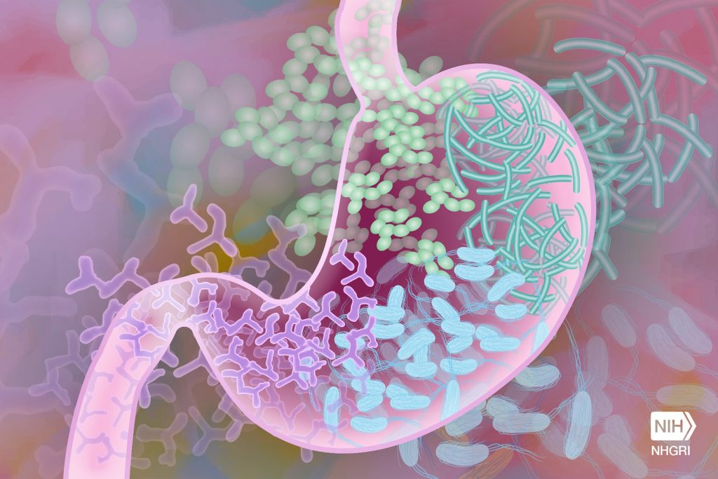
Gut Microbiome. Credit Darryl Leja National Human Genome Research Institute National Institutes Of Health
In a pilot clinical trial published in the Journal of the American Geriatrics Society that included older adults with depression receiving standard care, adding probiotic therapy produced modest but meaningful reductions in depressive and anxiety symptoms compared with adding a placebo. However, both groups demonstrated substantial overall improvements during follow-up.
For the trial, 58 participants in India aged ≥ 60 years with moderate depression were randomised 1:1 to receive daily probiotics or a placebo for 12 weeks, alongside standard antidepressant care. They were followed up for another 12 weeks.
Based on validated psychological scores, biomarker (serum brain-derived neurotropic factor level), and faecal microbiota profiling, investigators found that probiotics helped improve patients’ symptoms but did not confer clear additional gains in quality of life compared with placebo. The findings support probiotics as a safe, biologically plausible adjunct to standard care, but larger trials are needed.
“The results of our study are novel, and we are now planning a follow-up, larger-scale clinical trial due to the encouraging findings,” said co-corresponding author Dr. Saibal Das, MBBS, MD, DM, PhD, of the Indian Council of Medical Research – National Institute for Research in Bacterial Infections, Kolkata. “My vision is to develop affordable healthcare solutions and make them available to the larger population for meaningful public health impact,” added co–corresponding author Abhinaba Ghosh, MBBS, MSc, PhD, a physician-neuroscientist from Tata Medical Center, Kolkata.
Eliminating sugar from your diet may be more detrimental than previously thought, according to an animal study being presented Saturday at ENDO 2026, the Endocrine Society’s annual meeting in Chicago, Ill.
“Completely removing sucrose from a low-fat diet may unexpectedly disrupt gut health and promote inflammation and metabolic dysfunction, highlighting that balanced nutrition is more important than simply eliminating sugar,” said Rasheed Ahmad, PhD, principal scientist and head of the Immunology & Microbiology Department at the Dasman Diabetes Institute, in Kuwait City, Kuwait. The institute was founded by Kuwait Foundation for the Advancement of Sciences. Researchers investigated the effects of a sucrose-free low-fat diet compared to a sucrose-containing low-fat control diet in two groups of mice for 16 weeks.
They evaluated glucose tolerance, insulin sensitivity, circulating metabolic hormones, the gut microbiome and inflammation in the colon and liver.
Mice fed the sucrose-free diet developed impaired glucose control, insulin resistance, gut microbial imbalance, intestinal inflammation and fatty liver changes, despite having no significant differences in body weight compared with control mice.
“The findings suggest that complete removal of sucrose from a low-fat diet may negatively affect gut microbiota and metabolic health,” Ahmad said. “The study highlights the importance of maintaining balanced dietary carbohydrates to support gut and immune homeostasis.”
Until now, the consequences of restrictive diets that eliminate sugar from a low-fat diet were unknown.
“This research may influence future dietary recommendations by emphasizing the importance of maintaining a healthy gut microbiome rather than focusing only on sugar restriction,” Ahmad said. “In the long term, these findings could help improve strategies for preventing and managing metabolic disorders, fatty liver disease and chronic inflammatory conditions.”
“Studies such as this reflect our institute’s commitment to advancing evidence-based scientific discoveries that improve public health outcomes and deepen our understanding of metabolic disease,” said Faisal Hamed Al-Refaei, MD, Acting Director General of Dasman Diabetes Institute.
Houston Methodist researchers find antibiotics aid recovery from traumatic brain injury
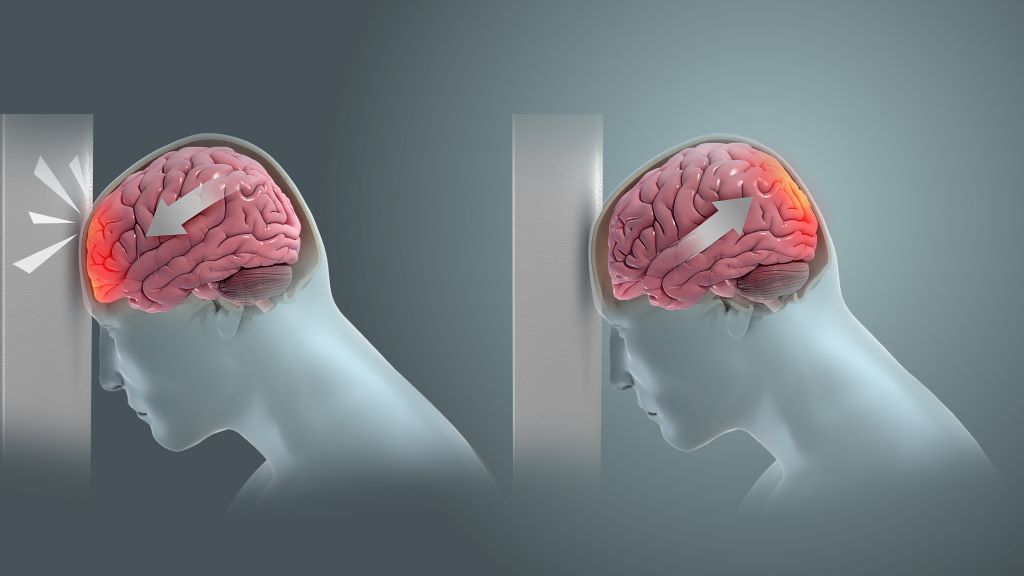
Coup and contrecoup brain injury. Credit: Scientific Animations CC4.0
In a new study published in Nature Communications Biology, Houston Methodist researchers led by Sonia Villapol, PhD, found that short-term antibiotic treatment significantly reduced neuroinflammation and neurodegeneration following traumatic brain injury (TBI) by altering the gut microbiome in animal models.
“We found that antibiotic treatment following TBI can reduce harmful gut bacteria, decrease lesion size and limit cell death,” said Villapol, an associate professor in the Department of Neurosurgery at Houston Methodist. “Our results support a gut–brain mechanism in which microbiome changes influence peripheral immunity and, in turn, neuroinflammation after TBI.”
Administering antibiotics cleans the gut of harmful bacteria, allowing beneficial bacteria to flourish. The study found that two helpful bacteria, Parasutterella excrementihominis and Lactobacillus johnsonii, are key to driving cell repair. According to Villapol, they could also be major regulators for peripheral inflammation in the body.
Notably, 70% of immune system regulation is generated by the gut microbiome. During gut imbalance, the bidirectional nature of the brain-gut axis can wreak havoc throughout the entire body.
“Our brains are constantly sending signals to the rest of our bodies. Following a traumatic brain event, those signals can get scrambled and disrupt other organs, including our digestive system,” Villapol said. “If the gut stays out of balance, the brain may have a harder time healing.”
There are an estimated 4 million traumatic brain injuries a year in the U.S. Recent studies indicate that TBI-induced gut microbiome imbalance may even contribute to the development of neurodegenerative diseases like Parkinson’s, Alzheimer’s and dementia.
Villapol’s lab is focused on investigating and developing new neuroprotective treatments to fight inflammation linked with neurodegenerative disease. “If we can break neuroinflammation in the acute or chronic stage, we can reduce the risk of developing Alzheimer’s or dementia,” said Villapol.
The next phase of the research will focus on bioengineering Parasutterella excrementihominis and Lactobacillus johnsonii to further develop precision therapies to reduce neuroinflammation.
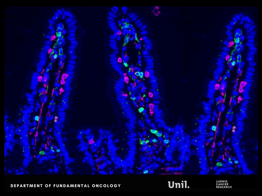
Microscopy image of intestinal lining in mice, shows CD4 (green), CD8 (magenta) and DAPI (blue). Ludivine Bersier 2025
In Nature Communications, researchers from Lusanne University reveal that chemotherapy alters gut microbes and bone marrow immune cell development, unexpectedly reprogramming systemic immunity in ways that help restrict metastatic progression.
Chemotherapy commonly damages the intestinal lining, a well-known side effect. But this injury does not remain confined to the gut. It reshapes nutrient availability for intestinal bacteria, forcing the microbiota to adapt.
The researchers report that chemotherapy-induced damage to the intestinal lining alters nutrient availability for gut bacteria, reshaping the microbiota and increasing the production of indole-3-propionic acid (IPA), a tryptophan-derived microbial metabolite.
Rather than acting locally, IPA functions as a systemic messenger. It travels from the gut to the bone marrow, where it rewires immune cell production. Elevated IPA levels reprogram myelopoiesis, reducing the generation of immunosuppressive monocytes that facilitate immune evasion and metastatic growth.
“We were surprised by how a side effect often seen as collateral damage of chemotherapy can trigger such a structured systemic response. By reshaping the gut microbiota, chemotherapy sets off a cascade of events that rewires immunity and makes the body less permissive to metastasis.” says Ludivine Bersier, first author of the study.
This immune reconfiguration enhances T-cell activity and remodels immune interactions within metastatic niches, particularly in the liver, resulting in a metastasis-refractory state in preclinical models.
Experimental findings are mirrored in patients. Clinical relevance is supported by patient data obtained in collaboration with Dr Thibaud Koessler (Geneva University Hospitals, HUG). In patients with colorectal cancer, higher circulating IPA levels following chemotherapy are associated with reduced monocyte levels, a feature of improved survival outcomes.
“This work shows that the effects of chemotherapy extend far beyond the tumor itself. By uncovering a functional axis linking the gut, the bone marrow and metastatic sites, we highlight systemic mechanisms that could be harnessed to durably limit metastatic progression.” says Tatiana Petrova, corresponding author of the study.
This research was supported by multiple funders, including the Swiss National Science Foundation and Swiss Cancer League. An ISREC Foundation Tandem Grant supported close collaboration between clinical and fundamental research, led at Unil by Professor Tatiana Petrova and Dr Thibaud Koessler at HUG. The project posits that chemotherapy can induce a form of biological “memory”, mediated by gut microbiome–derived metabolites that durably inhibit metastatic growth.
Together, these findings reveal a previously underappreciated gut–bone marrow–liver metastasis axis through which chemotherapy can exert durable systemic effects, opening new avenues to harness microbiota-derived metabolites as adjuvant strategies to limit metastasis.
First-of-its-kind study offers evidence that microbes from different primate species influence physiology in ways linked to brain size and function
Source: Pixabay
Humans have the largest relative brain size of any primate, but little is known about how mammals with larger brains evolved to meet the intense energy demands required to support brain growth and maintenance.
A new study from Northwestern University provides the first empirical data showing the direct role the gut microbiome plays in shaping differences in the way the brain functions across different primate species.
“Our study shows that microbes are acting on traits that are relevant to our understanding of evolution, and particularly the evolution of human brains,” said Katie Amato, associate professor of biological anthropology and principal investigator of the study, which was published in PNAS.
The study builds upon previous findings from Amato’s lab that showed the microbes of larger-brained primates, when introduced in host mice, produced more metabolic energy in the microbiome of the host – a prerequisite for larger brains, which are energetically costly to develop and function. This time, the researchers wanted to look at the brain itself to see if the microbes from different primates with different relative brain sizes would change how the brains of host mice functioned.
What they found
In a controlled lab experiment, the researchers implanted gut microbes from two large-brain primate species (human and squirrel monkey) and one small-brain primate species (macaque) into microbe-free mice.
Within eight weeks of making changes to the hosts’ microbiomes, they observed that the brains of mice with microbes from small-brain primates were indeed working differently than the brains of mice with microbes from large-brain primates.
In the mice with large-brain primate microbes, the researchers found increased expression of genes associated with energy production and synaptic plasticity, the physical process of learning in the brain. In the mice with smaller-brain primate microbes, there was less expression of these processes.
“What was super interesting is we were able to compare data we had from the brains of the host mice with data from actual macaque and human brains, and to our surprise, many of the patterns we saw in brain gene expression of the mice were the same patterns seen in the actual primates themselves,” Amato said. “In other words, we were able to make the brains of mice look like the brains of the actual primates the microbes came from.”
Another surprising discovery the researchers made was a pattern of gene expression associated with ADHD, schizophrenia, bipolar and autism in the genes of the mice with the microbes from smaller-brained primates.
While there is existing evidence showing correlations between conditions like autism and the composition of the gut microbiome, there is a lack of data showing the gut microbes contribute to these conditions.
“This study provides more evidence that microbes may causally contribute to these disorders —specifically, the gut microbiome is shaping brain function during development,” Amato said. “Based on our findings, we can speculate that if the human brain is exposed to the actions of the ‘wrong’ microbes, its development will change, and we will see symptoms of these disorders, i.e., if you don’t get exposed to the ‘right’ human microbes in early life, your brain will work differently, and this may lead to symptoms of these conditions.”
Implications and next steps
Amato sees clinical implications for further exploration of the origins of some psychological disorders and for taking an evolutionary perspective on the way microbes affect brain physiology.
“It’s interesting to think about brain development in species and individuals and investigating whether we can look at cross-sectional, cross-species differences in patterns and discover rules for the way microbes are interacting with the brain, and whether the rules can be translated into development as well.
Gut Microbiome. Credit Darryl Leja National Human Genome Research Institute National Institutes Of Health
Nitrogen metabolism of gut bacteria can provide health benefits. Specifically, gut microbes metabolise dietary nitrates and nitrites and prevent the formation of cancer-causing compounds called nitrosamines. New research published in The FEBS Journal sheds light on these processes and pinpoints which types of bacteria are most important.
Investigators found that Escherichia coli – and to a lesser extent, species of the genera Lactobacillus, Bacteroides and Phocaeicola – can efficiently metabolise different forms of nitrogen, thus preventing carcinogenic nitrosamine formation. They also demonstrated that this bacterial processing is critical to enable microorganisms to survive and colonise the intestinal tract, likely preventing harmful changes in the composition of the gut microbiota.
The findings highlight the importance of the gut microbiota in preventing the formation of harmful nitrogen metabolites, potentially decreasing the risk of certain cancers. The study also illustrates how the microbiota facilitates crosstalk between our diet and the gut, thus having important implications for both health and disease.
“The discovery that specific gut bacteria rapidly metabolise nitrite suggests a protective mechanism through which the microbiota contributes to the maintenance of intestinal and systemic health,” said corresponding author Prof Uwe Deppenmeier, of the University of Bonn, in Germany.
There’s no scientific evidence that the gut microbiome causes autism, a group of scientists argue in an opinion paper published in the international Cell Press journal Neuron.
They say conclusions from past research that supported this hypothesis – including observational studies, mouse models of autism, and human clinical trials – are undermined by flawed assumptions, small sample sizes, and inappropriate statistical methods.
“Despite what you’ve heard, read, or watched on Netflix, there is no evidence that the microbiome causally contributes to autism,” says first author and developmental neurobiologist Prof Kevin Mitchell from Trinity.
The hypothesis that autism is caused, at least partially, by the gut microbiome stems from the fact that many people with autism suffer from gastrointestinal symptoms.
In addition, the recent rise in autism diagnoses has led some to believe that environmental or behavioural changes are driving an increase in autism, though the authors note there is strong evidence that the rise in diagnoses reflects increased awareness and broadened diagnostic criteria rather than a biological mechanism.
Nevertheless, researchers have pursued the microbiome-autism hypothesis by comparing the gut microbiomes of people with and without autism, by studying mouse models of autism, and by conducting clinical trials involving people with autism. The authors argue that in all of these studies, the results are flawed and unconvincing.
“There’s variability in all three of those areas, and the studies just don’t form a coherent story at all,” says senior author and developmental neuropsychologist Dorothy Bishop of the University of Oxford.
In the most highly cited studies comparing the gut microbiomes of people with and without autism, researchers used sample sizes ranging from 7 to 43 individuals per group, whereas statistical recommendations call for sample sizes in the thousands.
“Autism is not rare, so there’s no reason to be having studies with only 20, 30, or 40 participants,” says co-author and biostatistician Darren Dahly of the University College Cork.
These studies also used varying methods to characterise microbiome composition, which makes their results difficult to compare. And although some studies found differences between the microbiomes of people with autism and controls, these differences were often contradictory—for example, some studies found lower microbial diversity in the guts of people with autism, while others found the opposite.
These differences also disappeared when the studies accounted for other variables, such as diet, or when they compared the microbiomes of children with autism with their neurotypical siblings.
“If anything, there is stronger evidence for a reverse causal effect, in that having autism can affect someone’s diet, which can affect their microbiome,” says Prof Mitchell.
Mouse models of autism that have claimed to show a link between the gut microbiome and autism are also unconvincing, the researchers say, because of behavioural, cognitive, and physiological differences between humans and mice.
“There’s no evidence that ‘autistic-like’ behaviours in mice models have any relevance to autism, and the experiments themselves had methodological and statistical flaws that undermine their claims,” says Prof Mitchell.
Several human clinical trials have tested the microbiome-autism hypothesis by performing faecal transplants or by administering probiotic therapies to people with autism and then monitoring changes in their characteristics. Again, the researchers say that most of these studies used inadequate sample sizes and inappropriate statistical methods that undermine their findings, and many didn’t use a control group or randomisation.
“The consensus across the studies that we surveyed is that when you do the trials properly, you don’t see anything,” says Dahly.
Based on the lack of convincing evidence, and the lack of progress in the field, the researchers argue the hypothesis that the microbiome causes autism has reached a dead end.
“If you accept our message, there’s two ways you can go. One is to just stop working on this area, which is something that we would be quite happy to see,” says Bishop. “But given that realistically, people are not going to stop, they need to at least start doing these studies in a much more rigorous way.”