Day: July 27, 2026

Researchers Find Dormant Melanocytes in Vitiligo, Suggesting a New Treatment Strategy
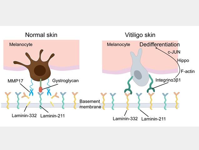
How changes in melanocyte attachment to the extracellular layer drive vitiligo
Under normal conditions (left), melanocytes adhere to laminin-211 in the basement membrane through dystroglycan, maintaining their mature pigment-producing state. In vitiligo (right), remodelling of the basement membrane increases laminin-332 and promotes integrin α3β1-mediated adhesion, activating signalling pathways, including c-Jun, that drive melanocyte dedifferentiation and loss of pigment production. Credit: Osaka Metropolitan University


Vitiligo is an autoimmune disease in which the body’s immune system attacks and destroys melanocytes, the cells that produce the skin pigment melanin. Once the melanocytes are gone, the skin turns white, leading to the characteristic skin patches of the disease.

However, the standard model of vitiligo doesn’t explain some of its unusual features. Some vitiligo lesions re-pigment and treatments sometimes restore pigmentation even in areas that lack melanocytes, suggesting that the melanocytes may not be fully destroyed.

A team from Osaka Metropolitan University team led by Specially Appointed Professor Ichiro Katayama and Specially Appointed Associate Professor Lingli Yang has found evidence that melanocytes do not completely disappear from vitiligo skin and instead enter a “dedifferentiated-like state” in which the mature cells revert to a more primitive form, losing many of their specialised functions including the production of pigment.

“This study uncovers a new mechanism underlying the development of vitiligo, which could change how we treat the disease,” Dr Yang explained.

Their research adds to a body of research that shows that cells do not just respond to chemical signals, but also to what they are physically attached to. As melanocytes are located on the basement membrane, the thin layer that separates the epidermis from the dermis, the membrane provides instructions that help them remain functional pigment-producing cells. 

However, the extracellular matrix surrounding melanocytes is remodelled in vitiligo patients. Usually, melanocytes bond with laminin-211; however, in vitiligo patients, the basement membrane becomes enriched in laminin-332. Because their preferred binding partner is no longer available, melanocytes change how they attach. Instead of using their preferred attachment agent, dystroglycan, they attach through integrin α3β1 instead.

These changes activate pathways that are commonly activated when cells are remodelling. In this process, the actin cytoskeleton of the cell is reorganised, gene expression is altered, including that of genes associated with an immature melanocyte state. The findings suggest a self-reinforcing cycle in which changes to the basement membrane drive melanocyte dedifferentiation, while the dedifferentiated melanocytes become less able to maintain a healthy basement membrane, further promoting the disease process.

“This was an exciting discovery for us, as most current treatments largely focus on suppressing autoimmune attacks and reducing inflammation, but if dormant melanocytes are still present in lesions, that might change the way we treat the disease,” Professor Katayama said. “New treatment avenues such as reactivating existing cells or restoring their normal attachment to the basement membrane may be possible.” 

When the researchers used pharmacological inhibitors targeting the signalling pathways activated by this adhesion switch, they were able to restore expression of mature melanocyte markers, recover pigmentation-related gene expression, and reverse many features of the dedifferentiated-like phenotype.

“This was an exciting aspect of our research, as it suggests that changing gene expression isn’t permanent and this process may be reversible,” Dr Yang explained. “We found that drugs were able to restore melanocyte function and pigmentation-related characteristics. The next step will be to perform clinical studies to see if this approach is a viable way to manage the disease.”

The findings were published in Nature Communications.

Source: Osaka Metropolitan University

New WHO Guidelines: Up to 45% of Dementia Risk Could Be Prevented or Delayed

Photo by Jusfilm on Unsplash

The World Health Organization (WHO) has released updated guidelines on reducing the risk of cognitive decline and dementia, providing countries with evidence-based recommendations to help prevent or delay the onset of dementia across the life course.

Dementia is a condition caused by brain diseases and affects memory, thinking and the ability to function. More than 57 million people live with dementia worldwide and nearly 10 million people get newly diagnosed every year. Alzheimer disease is the most common form of dementia and is estimated to account for 60–70% of cases.

While there is no cure for dementia, up to 45% of the risks can be attributed to modifiable risk factors such as tobacco, alcohol use, social isolation, physical inactivity, air pollution and noncommunicable diseases (NCDs), including high blood pressure and diabetes. Beyond health, dementia affects a person’s independence, dignity and safety.

“We know more today than ever before about what drives dementia risk, and these guidelines translate that knowledge into action,” said Dr Tedros Adhanom Ghebreyesus, WHO Director-General. “Countries now have clear, evidence-based recommendations they can put into practice immediately to protect people’s cognitive health.”

WHO’s new guidelines reflect the latest evidence and innovations in dementia risk reduction providing proven interventions that can effectively lower dementia risk through early awareness and timely action. They represent an important opportunity to reduce the burden of dementia in the coming decades through stronger integration of services for noncommunicable diseases, mental health and brain health.

Reducing risk, preventing illness

The updated guidelines reflect significant growth in the evidence base since WHO first issued recommendations on dementia risk reduction in 2019. They provide consolidated recommendations on addressing unhealthy behaviours, managing medical conditions, and reducing exposure to environmental factors that may contribute to cognitive decline and dementia.

The guidelines recommend several healthy behaviours and lifestyle interventions to reduce dementia risk, including cognitive training and cognitive stimulation and engagement in social activities for adults who have normal cognition or are experiencing mild cognitive impairment.

The updated advice also includes interventions that reduce risk of NCDs, including increasing physical activity, stopping tobacco use, reducing alcohol consumption, adopting a healthy diet, and a new recommendation to reduce exposure to air pollution.

Management of cardiometabolic conditions such as hypertension, diabetes, and high cholesterol can also help reduce dementia risk. Further, hearing aids may be offered as part of risk-reduction strategies.

As an intervention to reduce the risk of cognitive decline and/or dementia, the guidelines do not recommend supplementation with vitamins B and E, omega-3 polyunsaturated fatty acids (PUFA) and multivitamins/minerals in the absence of a diagnosed deficiency, due to the lack of evidence of any potential benefits to outweigh unexpected harmful effects. 

Human and economic cost

Dementia affects an individual’s ability to live independently, work and function, while placing substantial burdens on families and carers. It carries a major economic loss, costing the global economy an estimated US$ 1.3 trillion annually. About half of this cost is driven by unpaid care provided by families and friends. Understanding risk factors and taking action to prevent dementia can improve health and quality of life, helping people live longer, healthier and more independent lives.

Source: World Health Organization

Kids Do Feel the Cold. So Why Won’t They Wear a Jumper?

Zachary Kadolf/Unsplash

Joshua Pate, University of Technology Sydney

It happens just when you need to leave. Bags are packed. Shoes are on. Then your child decides a jumper is impossible.

You say, “put your jumper on”.

They say, “I’m not cold”.

Do kids really not feel the cold like adults do? Or are they just expressing their independence? And when should you insist?

A clue from pain research

I study how children experience pain, and pain research offers one clue about this jumper battle.

A child may scrape their knee during a game and barely notice until the game stops. The scrape was there throughout, but chasing a friend or reaching the next base kept winning their attention.

Cold can slip into the background in a similar way. A child’s fingers may be cooling while the playground remains far more compelling. Then the game ends.

Their attention returns to their hands at around the same time their moving muscles stop producing so much heat. Suddenly, the jumper may seem like a better idea.

So “I’m not cold” can mean, “I can feel it and I’m comfortable”.

It can also mean, “the jumper feels worse” or “I want to keep playing”. Sometimes it means, “I am four years old and this has become a matter of principle”.

Similarly, when parents ask, “but aren’t you cold?” we’re often asking several questions at once.

Will you still be warm when you stop running? What if the wind picks up? How about later, when we’re standing still on the sideline at the soccer field?

Children report the present moment, and parents factor in the forecast.

What cold actually feels like

Your skin contains sensory nerve endings that respond as its temperature changes.

One cool-sensitive channel, called TRPM8, helps convert cooling at the skin into electrical activity in sensory nerves.

This is the same channel that menthol activates, which is why mint can make your mouth feel cool even when there’s no real temperature change.

And as you know with mint, a strong cooling sensation can sometimes become uncomfortable or even painful. Other factors such as wind, wetness, contact with cold surfaces, movement, and how much time we’re outside can all influence how we experience temperature.

For example, a parent who is standing still in a playground, clutching a coffee, may be acutely aware of the gap in their coat where the icy wind is sneaking in.

But children tend to run, climb and jump in bursts – and moving muscles produce heat.

Children also differ from adults in body size, body composition, metabolism and how their circulation responds to cold.

One 2024 laboratory study, done indoors, looked at children aged six to nine. It found their sedentary metabolic rates (how much energy you’re burning when you’re resting) were around 39% higher compared to adults in the study.

Their skin was also warmer over parts of their torso, and the skin on their hands recovered temperature and bloodflow faster than adults after being exposed to the cold.

So it’s not that kids don’t feel cold at all, but they may have a quite different experience from an adult standing in the same air.

Bodies prepare for what comes next

We often learn about thermoregulation – how the body maintains its core temperature – as though the body were simply a thermostat. The body detects a temperature change, then bloodflow changes, and sweating or shivering bring it back towards the middle.

But our movement and behaviour also play an important role in maintaining this balance. When we’re cold we may walk into sunshine, curl up, or add a layer; when we’re hot we take one off. A child who keeps running may already be generating the warmth they need.

Bodies also prepare for expected demands. Researchers use the term allostasis to describe this wider process of how the body maintains stability through change.

Some adjustments happen automatically. For example, before we exercise, our heart rate and breathing begin adjusting for the work ahead. Others involve choices, such as moving into the sun, seeking shelter or reaching for warmer clothing.

But young children outsource some of this forecasting to adults.

Kids supply the live report from inside their body. We add the weather forecast and the schedule. We pack snacks for hunger that has yet to arrive, spare clothes for puddles yet to be found, and jumpers too.

What the jumper itself feels like

A jumper creates its own sensations. It may feel scratchy, bulky or restrictive. It can make climbing harder, then become hot and sweaty as soon as the child starts running.

Tags, seams and some fabrics can feel intensely unpleasant, especially for children with tactile sensitivities. Clothing tags and light touch, for example, can cause marked discomfort for some autistic children.

A child may genuinely prefer mildly cold skin over an irritating texture.

So it’s worth asking whether “aren’t you cold?” is the right question. Others may work better:

are you comfortable?

will you be running or sitting still?

would you rather wear the jumper or carry it?

These questions help children connect what they feel now with what they may need later.

When should parents insist?

Parents should be firmer when a child is very young, wet, unwell, exposed to strong wind or likely to remain outside for a long time.

Persistent shivering or numbness means it is time to get warm. Increasing clumsiness, unusual drowsiness, confusion or reduced responsiveness can indicate hypothermia, where the body’s core temperature has fallen dangerously low. Hypothermia is a medical emergency.

But for ordinary winter outings, flexible layers allow the plan to change. A jumper can be carried, added when activity slows, and removed when the child warms up again.

Your child reports the weather inside their body. You keep an eye on the weather outside it. A jumper in the bag lets your child feel heard, lets you keep the forecast in view, and lets everyone finally get out the door.

Joshua Pate, Associate Professor of Physiotherapy, University of Technology Sydney

This article is republished from The Conversation under a Creative Commons license. Read the original article.

Newer Obesity Drugs Linked to Fewer Alcohol-related Hospitalisations

Use of newer GLP-1 receptor agonists for obesity or diabetes was associated with a reduction in hospital admissions suggesting a potential role for the treatment of alcohol-use disorder

Photo from Pixabay CC0

Use of newer GLP-1 receptor agonists (semaglutide or tirzepatide) for obesity or diabetes by people with alcohol-use disorder was associated with a reduction in alcohol-related admissions to hospital, finds a study published online in the open access journal BMJ Open.

The findings suggest a potential role for the drugs semaglutide and tirzepatide in the treatment of alcohol-use disorder.

While GLP-1 receptor agonists are used primarily for the treatment of type 2 diabetes and obesity, there have been reports of reduced alcohol consumption among patients taking the drugs, prompting the authors to investigate the potential impact on alcohol-related hospitalisations among adults with alcohol-use disorder.

The study compared alcohol-related hospitalisations in 40 703 adults with alcohol-use disorder and type 2 diabetes or obesity who started a newer GLP-1 receptor agonist (semaglutide or tirzepatide) or a comparator drug between 1 January 2018 and 31 December 2024. Participants were split across four trials involving clinically distinct populations – the anti-diabetic medication (ADM) trial, anti-obesity medication (AOM) trial, medications for alcohol use disorder with type 2 diabetes (MAUD- T2D) trial, and medications for alcohol-use disorder with obesity (MAUD-obesity) trial.

Compared to participants taking an active comparator drug, those taking GLP-1 receptor agonists had a lower risk of alcohol-related admission to hospital during all four trials.

Use of GLP-1 receptor agonists was associated with a 26% lower risk of alcohol-related hospital admission than other diabetes medicines during the diabetic medication (ADM) trial, and a 32% lower risk of alcohol-related hospital admission than other obesity medicines during the anti-obesity medication (AOM) trial.

In the MAUD trials, the active comparators were drugs for alcohol-use disorder including acamprosate, disulfiram and naltrexone. Compared with taking drugs for alcohol-use disorder, use of GLP-1 receptor agonists by adults with type 2 diabetes was associated with a 63% lower risk of alcohol-related admission to hospital during the trial, and for adults with obesity use of GLP-1 receptor agonists was associated a 65% lower risk of alcohol-related hospitalisations.

The authors acknowledge several limitations to their study. Most importantly, alcohol-use disorder is under-captured due in part to stigmatisation, and when documented, it may also be recorded variably with lower reporting in areas of high social deprivation. Alcohol-related outcomes may have been under captured as they were defined using diagnosis codes and laboratory testing for alcohol exposure, and the study captured hospitalisations from treatment initiation to discontinuation in a trial environment, so treatment effects in an average clinical setting may differ.

Finally, there may have been some confounding in relation to socioeconomic status, underlying clinical stability or alcohol-use disorder severity, and healthcare engagement, as newer GLP-1 receptor agonists are higher-cost therapies and patients with access to these medications may differ from comparator groups.

The risk of residual confounding was greatest in the MAUD trials, as reflected by the reduced risk of non-alcohol-related hospitalisations with use of GLP-1 receptor agonists. The authors say the results of the MAUD trials should be interpreted with greater caution as there were also high rates of treatment discontinuation increasing the potential for bias.

Nevertheless, the authors conclude, “Initiation of newer GLP-1 receptor agonists among patients with alcohol-use disorder was associated with a lower observed risk of alcohol-related hospitalisation, with similar associations across populations with type 2 diabetes and obesity.

“These findings may suggest a potential role for GLP-1 receptor agonists in the context of alcohol-use disorder.”

Source: The BMJ Group

Can a Faecal Microbe Transplant Improve Chronic Insomnia?

Photo by Andrea Piacquadio: https://www.pexels.com/photo/young-man-in-sleepwear-suffering-from-headache-in-morning-3771115/

In a randomised clinical trial published in the Journal of Internal Medicine, ingesting capsules containing faecal microbes from healthy donors helped treat symptoms of insomnia.

One month after treatment, participants receiving faecal microbiota capsules showed significantly higher sleep efficiency and reduced wake after sleep onset, as measured by overnight polysomnography. Patient questionnaires also showed that sleep quality scores improved from 2 to 6 months in the intervention group compared with the placebo group.

“Our findings provide clinical evidence that targeting the gut microbiota may offer a promising new therapeutic strategy for chronic insomnia disorder,” said co–corresponding author Yanping Bao, PhD, of Peking University, in Beijing. “This work also strengthens our understanding of the gut–brain axis as an important regulator of human sleep.”

Source: Wiley