

Day: July 23, 2026

Sweeteners Shown to Slow Growth of Important Gut Bacteria in Lab Tests

Photo by Towfiqu barbhuiya

Cambridge researchers have shown how commonly-used sweeteners slow the growth of certain gut bacteria. Isosteviol – a compound derived from stevia (a common sweetener) – when combined with the anti-depressant duloxetine significantly impaired two important gut bacteria linked to regulating blood sugar and gut health and may affect the body’s immune responses.

Sweeteners are often marketed as metabolically neutral, but our study challenges this idea

Sonja Blasche

The scientists say more research is needed to understand the real-world health impacts of this laboratory study, one of the first to assess the direct impact of sweeteners on gut bacteria, particularly when they are combined with other substances.

Sweeteners are widely used in a range of food and drinks, including soft drinks, sweets, desserts, snacks and cereals. While marketed as healthier alternatives to sugar, there is increasing evidence of links to diseases such as type 2 diabetes, obesity and cancer.

Despite their pervasive use, there have been very few studies that look at the direct interactions between sweeteners and gut bacteria – the vast community of microorganisms that live in the digestive tract and play a crucial role in keeping our bodies healthy. 

Professor Kiran Patil from the Medical Research Council (MRC) Toxicology Unit at the University of Cambridge said: “Most of what we know about the potential impact of sweeteners on our health comes from animal research or from population studies. While these studies have indicated involvement of the microbiome in mediating the effect of sweeteners, it’s difficult to know how sweeteners act in the body – is it through direct interactions with our gut bacteria?”

“Answering this is further complicated by the fact that we rarely ever take sweeteners by themselves – we take them with drinks, in snacks, or even in medication to mask bitterness,” added Dr Sonja Blasche, a lead author of the study, also the MRC Toxicology Unit.

In research published in Molecular Systems Biology, Dr Blasche and colleagues looked at how artificial and low‑calorie sweeteners affect the bacteria living in our gut, and how these effects change when sweeteners are consumed together with other common substances such as caffeine, flavourings or medicines.

The researchers grew each of 25 gut bacterial species – including beneficial, neutral, and potentially harmful bacteria – in the lab. They then exposed each culture individually to 39 common, commercially-used sweeteners, some of which are artificial, others natural, and measured how well the bacteria multiplied.

Around three‑quarters of the sweeteners changed how at least one bacterial species grew. Some sweeteners slowed down or stopped the growth of certain bacteria linked to a healthy gut.

The researchers then tested each sweetener in combination with common compounds such as caffeine, vanillin (vanilla extract), advantame (an artificial sweetener) and eight commonly-used drugs to assess whether this had any impact on the gut bacteria. They found over 100 interactions where sweeteners acted differently when combined with other substances. In 34 cases, combinations made the effects stronger, while in 68 cases the effects were weaker.

Most striking was the combination of isosteviol (a compound derived from stevia, a sweetener widely used in the food and beverage industry) and the antidepressant duloxetine. This combination strongly suppressed Roseburia intestinalis and Parabacteroides merdae, two gut bacteria that play important roles in maintaining a healthy digestive system. In the US in 2023, over 4.2 million patients were prescribed duloxetine.

As no gut bacterium exists alone, but rather as part of a ‘community’ within the gut, the researchers created a synthetic community containing all 25 bacteria. After allowing it to grow over time, they tested the community against a variety of sweetener and drug combinations, looking at which species increased or decreased and whether the overall diversity changed.

By mimicking in this simplified way what might happen in the human gut, they showed that the combination of isosteviol and duloxetine reduced microbial diversity. A diverse microbiome is considered important for good gut health. The sweetener-drug combination also altered which bacterial species thrived or declined.

Further analysis showed that the effect of the isosteviol-duloxetine combination on the community increased toxicity towards certain host cells and interfered with other cells that play a role in the body’s inflammation and immune responses. 

Dr Blasche said: “Sweeteners are often marketed as metabolically neutral, but our study challenges this idea. We found that they can directly affect gut bacteria, particularly when mixed with other compounds such as medication and food additives. These common combinations could have unintended effects on our gut microbiome.”

The researchers stress that, as their experiments were carried out in the lab, not tested in humans, more research needs to be done before it is possible to conclude that there will be direct health effects in people. 

Professor Patil, the study’s senior author, added: “Our study suggests that artificial sweeteners don’t just pass through the body passively — they can interact with gut microbes, and these effects can be amplified or altered by other substances like medications. These findings can help guide new studies towards understanding how sweeteners might influence health in unexpected ways.”

The research was funded by the European Union’s Horizon 2020 programme and the UK Medical Research Council.

Reference

Blasche, S. et al. Common xenobiotics modulate gut microbial responses to low‑calorie sweeteners in vitro. MSB; 25 Jun 2026; DOI: 10.1038/s44320-026-00225-6

Republished from Cambridge University under a Creative Commons licence.

Read the original article.

Researchers Uncover Possible Cause of Muscle Pain from Statins

The discovery may explain side effects from taking statins, opening the door to future therapies that could make statins easier to tolerate.

Photo by Towfiqu Barbhuiya on Unsplash

Millions of people rely on statins to lower cholesterol and reduce the risk of heart attack and stroke. But for some, the drugs come with an unwelcome trade-off: muscle pain, weakness and exercise intolerance that can make it difficult to continue treatment.

Now, researchers at McMaster University have uncovered a biological pathway that may explain why those side-effects occur, opening the door to future therapies that could make statins easier to tolerate while maintaining their life-saving cardiovascular benefits.

Published in Science Advances, the study identifies an immune and metabolic mechanism that drives statin-induced muscle damage, challenging longstanding assumptions about how these side-effects develop.

“Statins are among the most effective medications we have for reducing cardiovascular disease risk and preventing early death,” said senior author Jonathan Schertzer, a professor in McMaster’s Department of Biochemistry and Biomedical Sciences.

“Unfortunately, muscle side-effects lead some people to reduce their dose or stop taking the medication altogether. We wanted to understand why this happens and whether it might be possible to separate the side effects from the benefits.”

Statin-associated muscle symptoms affect an estimated seven to 29 per cent of people who take the drugs. While researchers have long known that statins can sometimes cause muscle problems, the biological mechanisms behind those effects have remained unclear.

Led by first authors Nazli Robin and Nicole Barra of the Schertzer Lab at McMaster, the team found statins can disrupt how muscle cells produce energy, triggering an immune response that damages muscle tissue. In experiments using muscle cells and mouse models, researchers were able to prevent much of that damage by blocking the immune response.

“One of the most exciting findings of the research is that the mechanism causing muscle side-effects appears to be separate from the mechanism that lowers cholesterol,” said Schertzer. “That suggests it may one day be possible to target the side-effects without interfering with the cardiovascular benefits that make statins so valuable.”

The study also revealed an unexpected link between metabolism and immunity. Researchers found that changes in muscle-cell metabolism triggered an immune response within the cells themselves, providing new insight into how inflammation can contribute to drug side-effects.

Although additional research will be needed before the findings can be translated into therapies for patients, the discovery identifies several potential targets for future drug development aimed at preventing statin intolerance.

“These findings give us a clearer understanding of why some patients experience muscle symptoms and provide promising directions for making these important medications safer and more effective in the future,” added Schertzer.

Source: McMaster University

Hitting a Nerve – in a Good Way: Vagus Nerve Calms Lung Inflammation

Stimulating a nerve in the ear may alleviate lung inflammation in mice

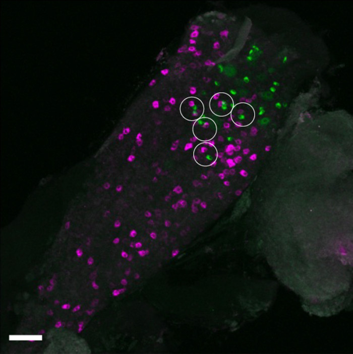
In the mouse vagal ganglion, sensory neurons labeled from the auricular skin (green) and the lung (magenta) are located in close proximity (white circles). These findings suggest a possible anatomical basis for how sensation from the auricular skin may influence airway immune responses via nerves that directly supply the lung. (Rintaro Shibuya, Kim Lab, Icahn School of Medicine at Mount Sinai.)

Neuroimmunology, the study of interactions between the nervous and immune systems, is a rapidly growing field enabling new approaches for monitoring and treating inflammatory diseases. In a recent study, scientists showed that in mice, stimulating a nerve in the external ear may help to ease inflammation in the lungs. Based on these findings, which are published in Immunity, the researchers are designing a clinical trial to test a novel device for treating asthma.

“We are always looking for new therapeutics and devices that can kickstart the body and get it back to doing what it needs to do,” says senior and corresponding author Brian S. Kim of the Icahn School of Medicine at Mount Sinai in New York. “This research suggests a new way to target the body’s inflammatory pathways.”

Neuroimmunology took off after research revealed how the vagus nerve – the ‘information superhighway’ that connects the brain to major organs and controls their functions – helps to regulate the immune system.

“The vagus nerve is hardwired to be a homeostatic organ,” says Kim. “You can think of it like a rheostat that integrates everything and keeps it in check.”

For this study, the authors leveraged the unique characteristics of the auricular vagus nerve, the only branch of the vagus nerve that reaches the surface of the skin. This nerve is found in the cymba conchae, the small, bowl-shaped depression in the upper part of the outer ear.

“Given the strong connection between the lungs and the vagus nerve, we sought to use the lungs as a test case to study these mechanisms and see whether manipulating the auricular branch could modulate inflammation,” says Kim.

The team studied these connections in mouse models using several approaches, including chemogenetics and optogenetics, to see what happened when the auricular vagus nerve was stimulated in the presence of an allergen. Their work showed that stimulation of the nerve increased levels of a neurotransmitter protein called CGRPβ in the airway. This, in turn, reduced inflammation in the lung. When the nerve fibres were instead inhibited, airway disease was exacerbated.

“Our findings reveal a previously unrecognised neuroimmune reflex linking the skin and the lung,” says first author Rintaro Shibuya of Kyoto University. “I hope this work inspires new ways of thinking about vagus nerve biology and future bioelectronic and neuroimmune therapies for inflammatory diseases.”

Although the research is in early stages, the team says this approach has many potential applications for treating diseases characterized by inflammation, including pulmonary fibrosis, inflammatory bowel disease, and rheumatoid arthritis.

“We still don’t know the extent to which the effects of stimulating the auricular vagus nerve go beyond the airway, but it’s something we will continue to study in the lab,” says Kim.

Source: Kyoto University

Certain Mental Disorders Are Associated with Faster Brain Ageing

Increased brain ageing was associated with dementia, addiction and psychiatric disorders like schizophrenia

Generated with Gencraft AI

People with dementia, mild cognitive impairment, alcohol addiction, or psychiatric disorders such as schizophrenia show increased brain ageing, each in specific patterns within the brain, according to a study published July 21st in the open access journal PLOS Medicine by Shile Qi from the Nanjing University of Aeronautics and Astronautics, China, and colleagues.

Some conditions can make the brain age faster. Scientists calculate how old the brain is relative to the body using the predictive age difference (PAD), the difference between chronological age and the age predicted by brain imaging, where a positive PAD indicates that ageing is accentuated or increased. To better understand how brain disorders and divergences might affect brain ageing, the authors of this study collected structure magnetic resonance imaging (MRI) data from 45 900 controls across several brain imaging banks, and compared them with those of 2698 patients with different brain conditions and differences, including attention-deficit/hyperactivity disorder (ADHD), autism spectrum disorder (ASD), alcohol or tobacco addiction, Alzheimer’s disease (AD), mild cognitive impairment (MCI), schizophrenia, bipolar disorder or major depressive disorder.

The authors found that neurodegenerative disorders of AD and MCI had the largest association with a high PAD. Addiction and psychiatric disorders were also associated with increased PAD. In contrast, there were no differences in PAD between people with ADHD or ASD and controls.

The researchers also looked at PAD values in specific areas of the brain, and examined which genes showed increased expression in people with different brain conditions. The prefrontal cortex showed higher PAD across brain disorders. Higher PAD in the frontal and temporal lobes was associated with psychiatric disorders, while high PAD in the frontal and occipital cortex was associated with dementia. Addiction was connected with high PAD in the default mode network, and in the salience network and the putamen and thalamus. There were also differences in gene transcription that associated with specific conditions and divergences. While the results are correlational, and not causal, and while some conditions such as psychiatric disorders and addiction have high co-occurrence, the author suggest that understanding more about PAD could help provide biomarkers for commonly occurring brain disorders.

The authors add, “Different neurological disorders appear to leave different signatures on the brain ageing clock, which may help researchers better understand the neural and biological pathways involved in these conditions.”

Provided by PLOS