Category: Cancer

Faster Detection of Pancreatic Cancer

Glycopeptide probes detect tumour-associated antibodies in blood samples

Pancreatic cancer. Credit: Scientific Animations CC BY-SA 4.0

Pancreatic cancer is one of the most lethal forms of cancer, primarily because it is usually diagnosed very late. Current markers are too insensitive and unspecific for early detection screenings. In the journal Angewandte Chemie, a research team has now introduced a new method that could lead to a significantly more precise and reliable diagnosis. It is based on the selective detection of specific antibodies in blood samples.

Tumours produce certain proteins (tumour-associated antigens) that draw the attention of our constantly “patrolling” immune system and trigger an immune response. As a consequence, antibodies directed against the tumours (tumour-associated autoantibodies) are formed, circulating in the blood at very early stages of the disease – which makes them useful for early detection. An international team led by Roberto Fiammengo and Giovanni Malerba at the University of Verona (Italy) as well as Alfredo Martínez at the Center for Biomedical Research of La Rioja (Logroño, Spain) and Francisco Corzana at the Universidad de La Rioja, has now developed an approach to diagnostic testing for pancreatic cancer that is based on the detection of such special tumour-associated autoantibodies.

They chose to use autoantibodies directed against the tumour-associated form of mucin-1 (TA-MUC1). Mucin-1 is a heavily glycosylated protein (a protein with sugar components) that occurs, for example, in glandular tissue. In many types of tumours, including pancreatic cancer, it is found in significantly elevated concentrations. In addition, the pattern of glycosylation is different from the normal form. The team’s goal was to detect autoantibodies that are directed specifically against TA-MUC1 and are a clear indicator of pancreatic cancer.

Based on structural analyses and computer simulations of known antibodies against TA-MUC1 (SM3 and 5E5), the team designed a collection of synthetic glycopeptides that mimic different segments (epitopes) of TA-MUC1. They also made unnatural modifications to increase the chances of identifying autoantibody subgroups indicative of the disease. The team immobilised these model antigens on gold nanoparticles achieving probes suitable for a serological assay (dot-blot assay). The diagnostic assay was validated with real samples from patients with pancreatic cancer and a healthy control group. Some of the nanoparticle probes could differentiate very well between samples from diseased and healthy individuals demonstrating they detected tumour associated autoantibodies. Notably, these specific autoantibodies displayed significantly better correct positive/false positive ratios than current clinical biomarkers for pancreatic cancer.

Probes with smaller glycopeptide antigens that correspond to only a single epitope, gave better results than larger probes that mimic multiple epitopes – an advantage for easier synthetic production. A short glycopeptide with an unnatural modification to its sugar component was found to be particularly effective for the detection of discriminating autoantibodies. This new structure-based approach could help in the selection of autoantibody subgroups with higher tumour specificity.

Source: Wiley
Unique Genetic Pattern can Predict Severe Side Effects of Melanoma Immunotherapy

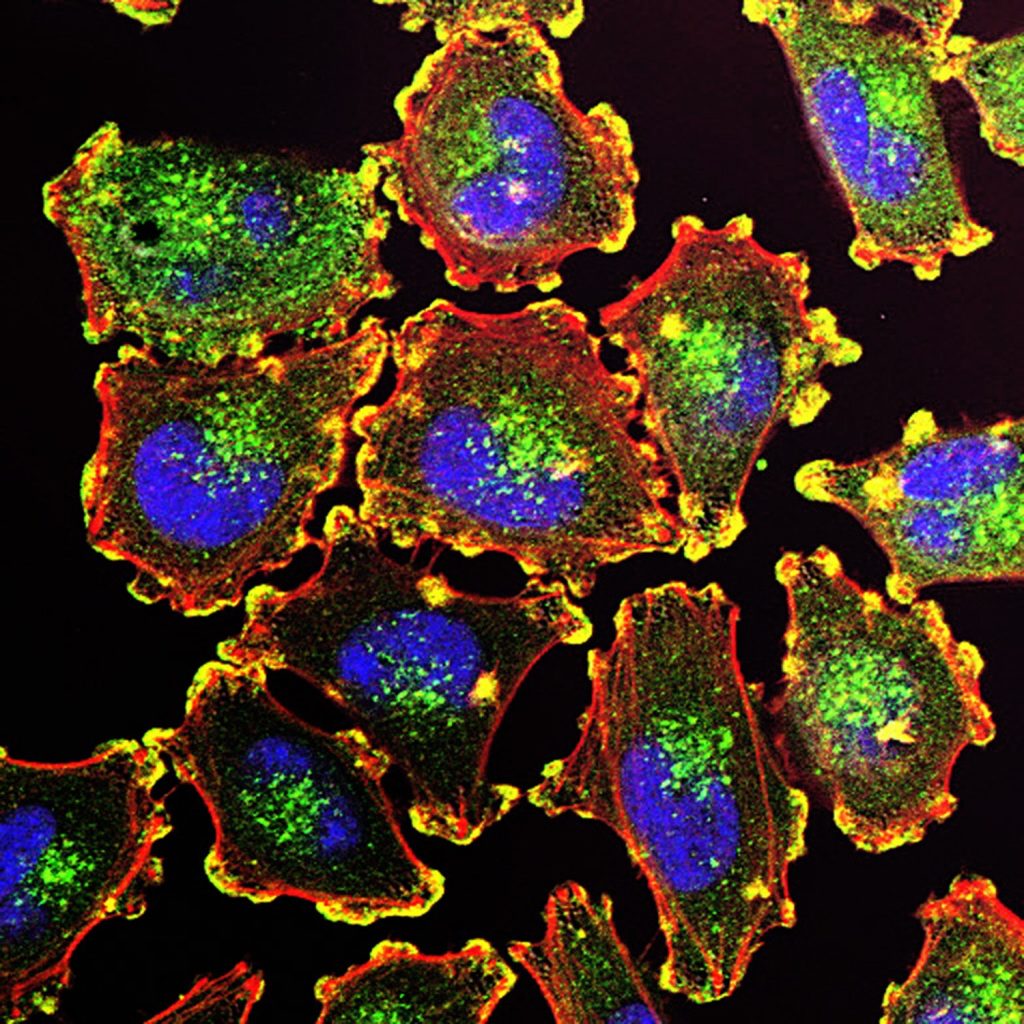
Melanoma Cells. Credit: National Cancer Institute

An activity pattern in certain genes responsible for building proteins known as spleen tyrosine kinases can predict which melanoma patients are likely to have severe side effects from immunotherapy designed to treat the most deadly skin cancer, as shown by a new study published in the journal Clinical Cancer Research.

Led by researchers at NYU Langone Health and its Perlmutter Cancer Center, the latest experiments focused on checkpoint inhibitors, drugs that have in the last decade become a mainstay of treating melanoma. This form of skin cancer kills nearly 10 000 Americans annually.

The drugs work by blocking molecules (checkpoints) that sit on the surface of immune T which the immune system uses to recognise and protect healthy cells. Cancer cells are able to hijack and turn off immune cell surveillance, evading detection. Immunotherapy drugs like nivolumab and ipilimumab are designed to block checkpoints, making cancer cells more “visible” again to T cells.

More than a third of melanoma patients given checkpoint inhibitors develop side effects so severe that they compromise their quality of life and ability to continue therapy. Side effects most often involve some form of inflammation, a sign of an overactive immune response. Patients may experience severe skin rashes, diarrhoea, or hyperthyroidism. More-severe side effects can include liver toxicity, colitis, and rheumatoid arthritis.

In the new study researchers found that even before treatment began in their test subjects, the activity of genes controlling the production of spleen tyrosine kinases predicted 83% of melanoma patients who eventually developed severe side effects from combined immunotherapy with nivolumab and ipilimumab.

Moreover, the researchers found that this heightened gene signature, as evidenced by the production of spleen tyrosine kinases, or the SYK pathway, did not interfere with the effectiveness of therapies in preventing recurrence of melanoma. The impact was connected only to side effects.

“Predictive information of this kind is critically important to oncologists and patients to help guide their immunotherapy decisions, to either minimize these side effects by taking additional precautions or to choose alternative immunotherapies,” said study co-senior investigator Tomas Kirchhoff, PhD.

“Our study results show that increased gene activity in the spleen tyrosine kinase pathway could be the basis of a possible blood test that identifies those melanoma patients most susceptible to having severe side effects from immunotherapy, and well before they start treatment,” said study co-lead investigator Kelsey Monson, PhD. 

For the study, researchers analysed immune system cell samples from 212 men and women with melanoma participating in the CheckMate-915 trial. The trial was designed to test whether combined therapy with nivolumab and ipilimumab worked better than single therapy with nivolumab in preventing postsurgical recurrence of melanoma. All immune cell samples were taken prior to the start of immunotherapy. Both drugs are manufactured by the pharmaceutical company Bristol Myers Squibb, which sponsored the CheckMate-915 trial, and provided the patient specimens and data used in the analysis.

When researchers looked at what genes were more active than others in patients who experienced side effects from their immunotherapy, they found a specific pattern among 24 genes tied to the production of spleen tyrosine kinases. Further statistical analyses showed that increased or decreased activity (transcription) of only five of these genes – CD22, PAG1, CD33, HNRNPU, and FCGR2C – along with patients’ age and the stage severity of their melanoma served as the best predictors of who would experience side effects from immunotherapy.

Study co-senior investigator Jeffrey S. Weber, MD, PhD, says that the SYK pathway has previously been linked to other autoimmune diseases, including lupus, rheumatoid arthritis, and colitis. He also points out that immunotherapy side effects were also most common in areas affected by these autoimmune diseases, including the skin, colon, and liver.

Dr Weber says the team next plans to investigate if an activated SYK pathway is predictive of side effects in patients treated with ipilimumab alone or with other combination immunotherapies.

“If our future research can explain how an activated spleen tyrosine kinase pathway leads to increased risk of side effects from immunotherapy, then it could also potentially help us to design better cancer immunotherapies and potentially other treatments for autoimmune diseases,” said Dr Kirchhoff.

Source: NYU Langone Health / NYU Grossman School of Medicine

Treating Radiation-induced Skin Injuries with Aspirin Hydrogels

Photo by National Cancer Institute on Unsplash

Radiation is a powerful tool for treating cancer, but prolonged exposure can damage the skin. Radiation-induced skin injuries are painful and increase a person’s chances of infection and long-term inflammation. Now, researchers in ACS Biomaterials Science & Engineering report an aspirin-containing hydrogel that mimics the nutrient-rich fluid between cells and accelerates healing of skin damaged by radiation in animals. With further development, the new salve could provide effective and rapid wound healing for humans. 

Most people undergoing radiotherapy for cancer will experience radiation-induced skin injury that can include redness, pain, ulcers, necrosis and infection. There are few treatments for these wounds, with the most common methods being debridement and hyperbaric oxygenation. Wound dressings made from hydrogels are gaining popularity because they are easy to apply and provide a wet environment for healing that is similar to the inside of the body. Glycopeptide-based hydrogels are especially promising: In laboratory and animal studies, the nanofibre structures have promoted cellular growth and regulated cell adhesion and migration. A research team led by Jiamin Zhang, Wei Wang, Yumin Zhang and Jianfeng Liu proposed loading aspirin, a common anti-inflammatory drug, into a glycopeptide-based hydrogel to create a multifunctional wound dressing for radiation-induced skin injuries.       

In lab tests with cultured cells, the researchers found that the aspirin-contained hydrogel scavenged reactive oxygen species, repaired DNA double-strand breaks and inhibited inflammation caused by radiation exposure without affecting cellular growth. In mouse models of radiation-induced skin injury, the researchers found that dressing wounds for three weeks with the salve reduced acute injuries and accelerated healing – results that the team says point to its potential as an easy-to-administer, on-demand treatment option for reducing radiation damage and promoting healing in humans.

Source: American Chemical Society

Gen X and Millennials at Higher Risk for 17 Cancers Compared with Previous Generations

Photo by Vitaly Gariev on Unsplash

A new large study led by researchers at the American Cancer Society (ACS) suggests incidence rates continued to rise in successively younger generations in 17 of the 34 cancer types, including breast, pancreatic, and gastric cancers. Mortality trends also increased in conjunction with the incidence of liver (in females only), uterine corpus, gallbladder, testicular, and colorectal cancers. The report appears in the journal The Lancet Public Health.

“These findings add to growing evidence of increased cancer risk in post-Baby Boomer generations, expanding on previous findings of early-onset colorectal cancer and a few obesity-associated cancers to encompass a broader range of cancer types,” said Dr Hyuna Sung, lead author of the study and a senior principal scientist of surveillance and health equity science at the American Cancer Society. “Birth cohorts, groups of people classified by their birth year, share unique social, economic, political, and climate environments, which affect their exposure to cancer risk factors during their crucial developmental years. Although we have identified cancer trends associated with birth years, we don’t yet have a clear explanation for why these rates are rising.”

In this analysis, researchers obtained incidence data from 23 654 000 patients diagnosed with 34 types of cancer and mortality data from 7 348 137 deaths for 25 types of cancer for individuals aged 25–84 years for the period Jan 1, 2000, to Dec 31, 2019, from the North American Association of Central Cancer Registries and the U.S. National Center for Health Statistics, respectively. To compare cancer rates across generations, they calculated birth cohort-specific incidence rate ratios and mortality rate ratios, adjusted for age effect and period effect, by birth years, separated by five-year intervals, from 1920 to 1990.

Researchers found that incidence rates increased with each successive birth cohort born since approximately 1920 for eight of 34 cancers. In particular, the incidence rate was approximately two-to-three times higher in the 1990 birth cohort than in the 1955 birth cohort for pancreatic, Kidney, and small intestinal cancers in both male and female individuals; and for liver cancer in female individuals. Additionally, incidence rates increased in younger cohorts, after a decline in older birth cohorts, for nine of the remaining cancers including breast cancer (oestrogen-receptor positive only), uterine corpus cancer, colorectal cancer, non-cardia gastric cancer, gallbladder cancer, ovarian cancer, testicular cancer, anal cancer in male individuals, and Kaposi sarcoma in male individuals. Across cancer types, the incidence rate in the 1990 birth cohort ranged from 12% for ovarian cancer to 169% for uterine corpus cancer higher than the rate in the birth cohort with the lowest incidence rate. Notably, mortality rates increased in successively younger birth cohorts alongside incidence rates for liver cancer (female only), uterine corpus, gallbladder, testicular, and colorectal cancers.

“The increase in cancer rates among this younger group of people indicate generational shifts in cancer risk and often serve as an early indicator of future cancer burden in the country. Without effective population-level interventions, and as the elevated risk in younger generations is carried over as individuals age, an overall increase in cancer burden could occur in the future, halting or reversing decades of progress against the disease,” added Dr Ahmedin Jemal, senior vice president, surveillance and health equity science at the American Cancer Society and senior author of the study. “The data highlights the critical need to identify and address underlying risk factors in Gen X and Millennial populations to inform prevention strategies.”

“The increasing cancer burden among younger generations underscores the importance of ensuring people of all ages have access to affordable, comprehensive health insurance, a key factor in cancer outcomes,” said Lisa Lacasse, president of the American Cancer Society Cancer Action Network (ACS CAN). “To that end, ACS CAN will continue our longstanding work to urge lawmakers to expand Medicaid in states that have yet to do so as well as continue to advocate for making permanent the enhanced Affordable Care Act tax subsidies that have opened the door to access to care for millions.”

Source: American Cancer Society

Advanced Lung Cancer Mortality Plunged Since Standardisation of Immunotherapy

The largest population-based study to date supports the survival benefits of immunotherapy for people with metastatic non–small cell lung cancer.

Squamous cancer cell being attacked by cytotoxic T cells. Image by National Cancer Institute on Unsplash

Since the first immunotherapy drug to boost the body’s immune response against advanced lung cancer was introduced in the United States in 2015, survival rates of patients with the disease have improved significantly. That’s the conclusion of a recent real-world study published by Wiley online in CANCER, a peer-reviewed journal of the American Cancer Society.

For the research, a team led by Dipesh Uprety, MD, FACP, of the Barbara Ann Karmanos Cancer Institute and the Wayne State University School of Medicine, analysed data from the National Cancer Institute Surveillance, Epidemiology, and End Results database, which compiles cancer-related data covering approximately 48% of the US population. The investigators’ analysis focused on non–small cell lung cancer (NSCLC), which accounts for up to 90% of all cases of lung cancer and is the leading cause of cancer-related death among both men and women in the United States.

In a comparison of 100 995 patients with metastatic NSCLC treated in 2015–2020 (after immunotherapy was deemed the standard of care) and 90 807 patients with metastatic NSCLC in the pre-immunotherapy era of 2010–2014, patients in the immunotherapy era were less likely to die from any cause. The overall survival rates at one, three, and five years were 40.1% versus 33.5%, 17.8% versus 11.7%, and 10.7% versus 6.8%. The median overall survival was eight months in patients in the immunotherapy era and seven months in those in the pre-immunotherapy era.

Similarly, patients treated after immunotherapy was available were less likely to die specifically from cancer than those treated before immunotherapy. The one-, three-, and five-year cancer-specific survival rates were 44.0% versus 36.8%, 21.7% versus 14.4%, and 14.3% versus 9.0%, with a median survival of 10 months versus eight months.

Survival rates remained significantly better in the immunotherapy era even after accounting for factors including age, sex, race, income, and geographical area.

“By utilizing a large national database, our study provided real-world evidence of the positive impact of immunotherapy in patients with lung cancer,” said Dr Uprety. The investigators stressed that additional studies are needed, however. “Immunotherapy provides long-term benefits. Since the durable benefits of immunotherapy are limited to a small subset of patients, future research should aim to optimize immunotherapy with new agents that can benefit a broader population,” said lead author Yating Wang, MD, of Ascension Providence Hospital.

Source: Wiley

Pancreatic Cancer Drug is Promising Against Most Aggressive Medulloblastoma Subtype

Pancreatic cancer. Credit: Scientific Animations CC BY-SA 4.0

A drug that was developed to treat pancreatic cancer has now been shown to increase symptom-free survival in preclinical medulloblastoma models – all without showing signs of toxicity. Survival rates for medulloblastoma vary according to which one of the four subtypes a patient has, but the worst survival rates of about 40%, are for Group 3. The research, published in the Journal of Clinical Investigation, focused on this most aggressive subtype.

Jezabel Rodriguez Blanco, PhD, an assistant professor at Medical University of South Carolina, led the research. Her work focused on the drug triptolide, which is extracted from a vine used in traditional Chinese medicine, and its water-soluble prodrug version, Minnelide. A prodrug is an inactive medication that the body converts into an active drug through enzymatic or chemical reactions.

MYC is an oncogene, or gene that has the potential to cause cancer. MYC is dysregulated, or out of control, in about 70% of human cancers, and it shows up in much higher levels in Group 3 medulloblastoma than in the other medulloblastoma subgroups. Despite its well-known role in cancer, this oncogene historically has been considered impossible to target with drugs.

Despite its poor druggability, previous research in other cancers had shown that triptolide and its derivatives had the ability to target MYC. When Blanco was still a postdoctoral fellow at the University of Miami, her mentor, David Robbins, PhD, attended a presentation by the research team that showed that the more copies of MYC that a tumour has, the better that triptolide works.

“He came to me, and he told me, ‘You know, as Group 3 medulloblastoma has many MYC copies, you should get some research models and try the drug,” Blanco recalled. She started the project from scratch. “I started talking to people, getting cell lines and animal models, learning how to propagate them, getting the drug, using it.” 

Blanco received initially received grants to on the Group 3 research, and continued it as a side project. She knew how well triptolide was working in these hard-to-treat tumours, and she did not want her initial results to fall through the cracks.

Determining the mechanism of action has been the most challenging part of the project, she noted, due to the drug’s multiple effects, and there could still be additional mechanisms beyond those that Blanco identified.

“It was affecting MYC gene expression by affecting the RNA pol II activity, and then it was affecting how long the protein lasts. So, the fact that it’s working through two different mechanisms on this oncogene may explain why it’s so effective in tumours that have extra copies of MYC,” she said, explaining that RNA polymerase II is a protein that helps to make copies of DNA instructions, which are used to produce proteins in the cell.

Despite the challenges of narrowing down the mechanism of action specific to the cancer, it was quite clear that however it worked, it did work, she said.

The efficacy was 100 times higher in the Group 3 tumours with extra MYC copies than in the Sonic Hedgehog tumours with normal levels of MYC, she said. She found that Minnelide reduced tumour growth and the spread of cancer cells to the thin tissues that cover the brain and spinal cord, called leptomeninges. It also increased the efficacy of the chemotherapy drug cyclophosphamide, which is currently used in treatment.

Blanco decided to move forward with publication rather than waiting to write a manuscript that answered all possible questions. Knowing that most parents whose children receive a Group 3 medulloblastoma diagnosis will lose their child in less than two years was the incentive she needed to push this work out.

“There was a point at which I could not hold these data anymore because it was working so well that it needed to go out,” she said. “The preclinical models were showing such a nice efficacy that it was like, ‘OK, I cannot keep on holding this work, digging deeper into the mechanism of action because the kids that have Group 3 medulloblastoma are dying while we are doing those experiments.”

Minnelide has been tested or is currently in testing in phase I and phase II clinical trials of adults with different types of cancer, including pancreatic cancer, where it showed some efficacy.

Blanco is hopeful that, with this new research on Group 3 medulloblastoma, a clinical trial for children with this disease can be launched.

Her paper is dedicated to the memory of Insley Horn, a 9-year-old Charleston girl who succumbed to one of these aggressive brain tumours. Research, Blanco said, is the only tool we have to prevent the loss of lives like Insley’s.

Source: Medical University of South Carolina

Transplant Beats Other Therapies for Colorectal Cancer Metastasised to the Liver

Human colon cancer cells. Credit: National Cancer Institute

Colorectal cancer often metastasises to the liver, and for some patients, surgical removal of their liver tumours is not an option. A new study led by researchers at the Wilmot Cancer Institute and University of Rochester Medical Center (URMC) shows that a select group of patients with colorectal cancer that has spread to the liver tend to fare better if they receive a liver transplant as opposed to other common therapies.

In the study, published in JAMA Surgery, patients who had liver transplants tended to live longer without cancer progression than patients who opted for other treatments. While previous studies have shown the benefits of liver transplants for these patients, this is the first study to compare liver transplants to other treatment options.

“In any cancer treatment, it’s very easy to describe the outcomes of the patients who received the intervention, but similar patients that did not undergo the intervention can serve as a good comparison,” said Matthew Byrne, MD, a surgery resident at URMC and author of the study. “Without randomised, controlled trial data, this study offers the best evidence that is available to understand whether liver transplant provides better outcomes over other treatments.”

The study followed 33 patients whose colorectal cancer was under control, but who had liver tumours that could not be surgically removed. All 33 patients were eligible for liver transplantation, but only 20 chose to have a transplant, while 13 opted for other classical therapies, like removal of part of the liver, chemotherapy, or liver-directed therapies.

Compared to the classical therapy group, the liver transplant group had significantly higher progression-free survival rates across three years of follow-up. One year after liver transplant, 90% of patients showed no signs of cancer progression. That number dropped to 73% after two years and to 36% at three years. On the other hand, only 42% of patients who opted for other therapies were cancer-progression-free after one year, which dropped to roughly 10% after two and three years.

The transplant group also had higher overall survival rates than the standard therapy group, though the difference wasn’t statistically significant. At the three-year follow-up, 90% of transplant patients had survived, compared to 73% of patients who received other therapies.

Though this study provides solid evidence, larger clinical trials will be needed to fully understand the added benefit of liver transplant compared to other treatments for these patients, and to better refine which patients benefit most.

Source: University of Rochester Medical Center

The Truth About Bone Marrow Stem Cell Donation

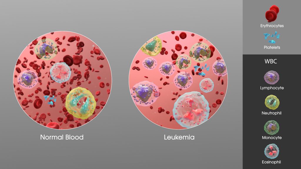
SAG Leukaemia. Credit: Scientific Animations CC0

While thousands of South Africans have registered as potential bone marrow stem cell donors, a critical challenge looms: donor attrition. These dropout rates, ranging from 23% to 56%, can significantly delay finding a suitable match for blood cancer patients in desperate need of a potentially life-saving transplant. This can unfortunately impact their chances of survival.

The good news is that donating stem cells is a safe and relatively simple process. With Bone Marrow Stem Cell Donation and Leukaemia Awareness Month taking place between 15 August and 15 October 2024, DKMS Africa aims to address some misconceptions that might deter registered donors from following through with donations.

Palesa Mokomele, Head of Community Engagement and Communications, unpacks these below:

Myth 1: Donating stem cells is a painful surgical procedure.

Fact: For over 90% of donors the process entails Peripheral Blood Stem Cell (PBSC) collection, a non-surgical procedure similar to donating blood. During PBSC, donors will rest comfortably while a needle is placed in each arm. Blood is drawn from one arm, passed through a machine that separates the stem cells, and the remaining blood is returned to the body through the other arm. While not painful, some donors may experience mild side effects like headaches, fatigue, or muscle aches, which typically resolve quickly.

For a small percentage of donors (around 2%), stem cells might be collected directly from the bone marrow in the pelvic bone. This minimally invasive procedure is performed under general anaesthesia. Although some donors experience temporary discomfort or soreness at the extraction site, the feeling is usually comparable to a bruise.

Myth 2: Donating takes too long and disrupts my life too much.

Fact: While the donation process involves some steps, it’s designed to be manageable. You’ll likely have a briefing call to explain the process, a health check to confirm your suitability, and an informative session about donation itself. The actual donation typically takes less than a day (4-6 hours) for the PBSC method.

For the bone marrow donation method, a hospital stay is involved, but it’s usually just three days. This includes check-in on day one, the procedure on day two, and discharge on day three.

Myth 3: Donating stem cells means missing a lot of work. 

Fact: The good news is that most donors can get back to work quickly. For PBSC donation, donors will likely be able to return within two days. If they donate bone marrow, a bit more recovery time is needed, so they should plan for about one week of leave.

Myth 4: My boss won’t be okay with me taking time off to donate.

Many employers are incredibly supportive of staff who donate stem cells. In our experience, most react positively to this selfless act. If your company doesn’t offer paid leave for donation, DKMS has a financial assistance programme that deals with lost wage compensation.

Myth 5: Donating stem cells will cost me money.

Fact: Donation is completely free of charge for the donor. DKMS covers all donation-related expenses, including travel, meals, and accommodation if needed. Financial support is also provided for a companion to join them at the hospital. The donor’s health insurance will never be involved, and DKMS handles the costs of any follow-up care that might be necessary.

“Seventeen-year-old Anele who was diagnosed with Acute Lymphoblastic Leukaemia (ALL), a type of cancer that affects the production of healthy blood cells, is just one of many patients in need of a stem cell transplant from a matching donor,” says Mokomele. “His father, Lawrence, is devastated, with his son now hanging on for dear life, waiting for that one person to be a match.”

“Every registered donor brings hope to a patient battling blood cancer. By staying committed to the cause, you help to ensure a readily available pool of potential matches, increasing a patient’s chance of receiving a transplant. Let’s give them a second chance at life!” she concludes.

Register today at https://www.dkms-africa.org/register-now

For more information, contact DKMS Africa on 0800 12 10 82.

US Fast-Tracks a Promising New Therapy for Metastatic Prostate Cancer

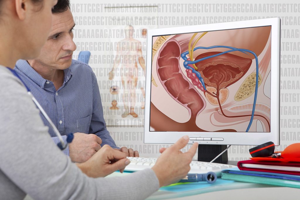
Credit: Darryl Leja National Human Genome Research Institute National Institutes Of Health

The US Food and Drug Administration (FDA) has granted Fast Track designation for SYNC-T SV-102 therapy for the treatment of patients with metastatic castrate-resistant prostate cancer (mCRPC). SV-102 is part of Syncromune Inc.’s SYNC-T platform, an in situ personalised therapy that uses a combination multi-target approach to cancer treatment, aiming to improve outcomes and quality of life for patients.

The Fast Track designation was granted based on the potential of SYNC-T SV-102 therapy to address the significant unmet need in treating patients with mCRPC. This advanced form of prostate cancer affects over 40 000 men in the US alone and is associated with a very poor prognosis. The Fast Track process is designed to facilitate the development and expedite the review of therapies that treat serious conditions and fulfil an unmet medical need, with the goal of getting important new treatments to patients sooner. Fast Track designation provides Syncromune with several key benefits, including more frequent FDA interactions, eligibility for accelerated approval, and priority review.

“The Fast-Track designation for SYNC-T SV-102 therapy signifies another step forward in bringing our potentially groundbreaking therapy to patients who need it most,” said Eamonn Hobbs, Chief Executive Officer and co-founder of Syncromune. “This accomplishment builds upon the foundation of positive Phase 1 clinical data and recent IND clearance.”

Syncromune’s lead candidate, SYNC-T SV-102, is a platform therapy that combines an in situ vaccine via partial oncolysis of a tumour followed by intratumoural infusion of the SV-102 fixed-dose multi-target biologic drug into the lysed tumour. This combination is designed to provide both immune stimulation and block immune suppression to activate and proliferate T cells to elicit a systemic anti-tumour response. Interim data from a Phase 1 study of SV-102 in males with mCRPC demonstrated an overall response rate of 85% with a favourable safety profile and tolerability. The Fast-Track designation comes on the heels of clearance of the company’s investigational new drug (IND) application, with studies expected to begin in the US this year.

Charles Link, M.D., Executive Chairman of Syncromune added, “We believe that Fast-Track designation for SYNC-T SV-102 will significantly aid our development goals for this therapy for men with difficult to treat prostate cancer. We look forward to initiating trials at multiple US sites later this year to expand our efforts to develop the SYNC-T SV-102 Therapy.”

Source: Syncromune

Surprising Finding in Glioblastomas: Islands of Potent Immune Cells in Bone Marrow

Photo by Anna Shvets

Researchers from Germany have made a surprising discovery about the aggressive, lethal glioblastomas: in the vicinity of glioblastomas, they found islands of highly potent immune cells in the neighbouring bone marrow of the skull, which play a central role in defending against cancer. The new data may open up prospects for innovative therapies. On the other hand, they cast a shadow over conventional strategies.

“What we have found is surprising and fundamentally new,” says Björn Scheffler, German Cancer Consortium (DKTK) researcher at the Essen site. Until now, the body’s own defences have always been thought of as a holistic system that sends its troops to different parts of the body as required. “However,” says Scheffler, “our data show that highly potent immune cells gather in regional bone marrow niches close to the tumour and organise the defence from there. At least this is the case with glioblastomas.”

Immune system on site

Based on new findings from animal experiments, the Essen team took tissue samples from the bone marrow near the tumor in the skull from untreated patients with glioblastoma. “However, the methods for this first had to be established,” reports first author Celia Dobersalske, emphasizing the fact that the new research results were obtained from human tissue samples.

The researchers hit the bull’s eye in their search: Bone marrow niches in close proximity to the glioblastoma appear to be the reservoir from which the anti-tumour defence is recruited. Apart from active lymphoid stem cells that develop into immune cells, the researchers also found mature cytotoxic T lymphocytes (CD8 cells) in the bone marrow close to the tumour. “These are highly effective immune cells that play a central role in the defence against cancer,” adds Celia Dobersalske. They can recognize and destroy malignant cells.

The CD8 cells in the bone marrow near the tumour had an increased number of receptors on their surface, which control the proliferation of mature T lymphocytes. In line with this, descendants of the same cell clones – one clone originates from one and the same cell – were detected both in the bone marrow and in the tumour tissue. This is clear evidence that the immune cells gathered on site are fighting the glioblastoma. “And they are successful – at least for a while,” says Björn Scheffler. “We were able to show that the course of the disease correlates with the activity of the local CD8 cells.”

Valuable immune cells destroyed?

This finding not only turns conventional ideas about how the immune system works on their head. The treatment concepts for glioblastoma must also be reconsidered in light of the new data. “Until now, we hadn’t even considered the skullcap in our considerations. How could we, since there was no evidence that highly potent immune cells could be hiding there,” says senior author Scheffler.

“When we opened the skull, we may have destroyed important immune cells in the process,” confirms Ulrich Sure, Director of the Department of Neurosurgery and member of the Essen research team. “In view of the new findings, we find ourselves in a dilemma: we have to gain access to the tumour in order to remove it and also to be able to confirm the diagnosis. There is currently no other way than through the skull. But we are thinking about how we can minimise damage to the local bone marrow in the future.”

On the other hand, the discovery of the local immune system opens up opportunities for innovative therapies. In particular, so-called checkpoint inhibitors are coming back into play. These are immunotherapeutic agents that aim to boost the body’s own cancer defenses. However, checkpoint inhibitors tested to date have shown little effect on glioblastomas.

“Various explanations have been suggested as an explanation, but perhaps we also need to rethink things in this respect,” says Björn Scheffler. “We now know that highly potent immune cells are indeed present on site. We were able to prove that they are fit to fight tumours, but they are not capable of destroying the tumour on their own. This is where we can start. One challenge will be to deliver drugs in sufficient concentration to the regional bone marrow niches at the right time. If we succeed, we may have a chance of controlling the growth of glioblastomas and improving our patients’ chances of survival.”

Source: German Cancer Research Center (Deutsches Krebsforschungszentrum, DKFZ)