A recent analysis of all relevant published studies reveals clear benefits of Cognitive Behavioural Therapy (CBT) for improving mental health and quality of life in cancer survivors. The findings, which are published in Cancer Medicine, extend CBT’s effects beyond what has long been known in the general population.
For the analysis, investigators uncovered 132 clinical trials comparing CBT with controls, including standard therapy, waitlist control, or active/alternative therapy.
Across the trials, CBT moderately improved mental health and quality of life in people with past or current cancer, regardless of cancer type. It seemed to have stronger effects in younger individuals. In-person CBT also appeared more effective than delivering CBT through technology such as web-based videoconference platforms.
“In addition to confirming the general benefit of CBT for individuals with cancer, this study unveiled important nuances of how CBT can be most effective and for which populations. This has major clinical implications for supportive oncology providers,” said corresponding author Anao Zhang, PhD, of the University of Michigan.
Researchers at Baylor College of Medicine and the Jan and Dan Duncan Neurological Research Institute at Texas Children’s Hospital have uncovered a new cell type in human brain cancers. Their study, published in Cancer Cell, reveals that a third of the cells in glioma, fire electrical impulses. Interestingly, the impulses, also called action potentials, originate from tumour cells that are part neuron and part glia, supporting the groundbreaking idea that neurons are not the only cells that can generate electric signals in the brain.
The scientists also discovered that cells with hybrid neuron-glia characteristics are present in the non-tumour human brain. The findings highlight the importance of further studying the role of these newly identified cells in both glioma and normal brain function.
“Previous studies have shown that patient survival outcomes are associated with tumour proliferation and invasiveness, which are influenced by tumour intrinsic and extrinsic factors, including communication between tumour cells and neurons that reside in the brain,” said Dr Benjamin Deneen, professor in the Department of Neurosurgery at Baylor.
Researchers have previously described that glioma and surrounding healthy neurons connect with each other and that neurons communicate with tumours in ways that drive tumour growth and invasiveness.
“We have known for some time now that tumour cells and neurons interact directly,” said first author Dr Rachel N. Curry, postdoctoral fellow in paediatrics – neuro oncology at Baylor, who was responsible for conceptualising the project. “But one question that always lingered in my mind was, ‘Are cancer cells electrically active?’ To answer this question correctly, we required human samples directly from the operating room. This ensured the biology of the cells as they would exist in the brain was preserved as much as possible.”
To study the ability of glioma cells to spike electrical signals and identify the cells that produce the signals, the team used Patch-sequencing, a combination of techniques that integrates whole-cell electrophysiological recordings to measure spiking signals with single-cell RNA-sequencing and analysis of the cellular structure to identify the type of cells.
The electrophysiology experiments were conducted by research associate and co-first author Dr Qianqian Ma in the lab of co-corresponding author associate professor of neuroscience Dr Xiaolong Jiang. This innovative approach has not been used before to study human brain tumour cells. “We were truly surprised to find these tumour cells had a unique combination of morphological and electrophysiological properties,” Ma said. “We had never seen anything like this in the mammalian brain before.”
“We conducted all these analyses on single cells. We analysed their individual electrophysiological activity. We extracted each cell’s content and sequenced the RNA to identify the genes that were active in the cell, which tells us what type of cell it is,” Deneen said. “We also stained each cell with dyes that would visualise its structural features.”
Integrating this vast amount of individual data required the researchers to develop a novel way to analyse it.
“To define the spiking cells and determine their identity, we developed a computational tool – Single Cell Rule Association Mining (SCRAM) – to annotate each cell individually,” said co-corresponding author, Dr Akdes Serin Harmanci, assistant professor of neurosurgery at Baylor.
“Finding that so many glioma cells are electrically active was a surprise because it goes against a strongly held concept in neuroscience that states that, of all the different types of cells in the brain, neurons are the only ones that fire electric impulses,” Curry said. “Others have proposed that some glia cells known as oligodendrocyte precursor cells (OPCs) may fire electrical impulses in the rodent brain, but confirming this in humans had proven a difficult task. Our findings show that human cells other than neurons can fire electrical impulses. Since there is an estimated 100 million of these OPCs in the adult brain, the electrical contributions of these cells should be further studied.”
“Moreover, the comprehensive data analyses revealed that the spiking hybrid cells in glioma tumours had properties of both neurons and OPC cells,” Harmanci said. “Interestingly, we found non-tumour cells that are neuron-glia hybrids, suggesting that this hybrid population not only plays a role in glioma growth but also contributes to healthy brain function.”
“The findings also suggest that the proportion of spiking hybrid cells in glioma may have a prognostic value,” said co-corresponding author Dr Ganesh Rao, professor and chair of neurosurgery at Baylor. “The data shows that the more of these spiking hybrid glioma cells a patient has, the better the survival outcome. This information is of great value to patients and their doctors.”
“This work is the result of extensive equal collaboration across multiple disciplines – neurosurgery, bioinformatics, neuroscience and cancer modelling – disciplines strongly supported by state-of-the-art groups at Baylor,” Deneen said. “The results offer an enhanced understanding of glioma tumours and normal brain function, a sophisticated bioinformatics pipeline to analyse complex cellular populations and potential prognostic implications for patients with this devastating disease.”
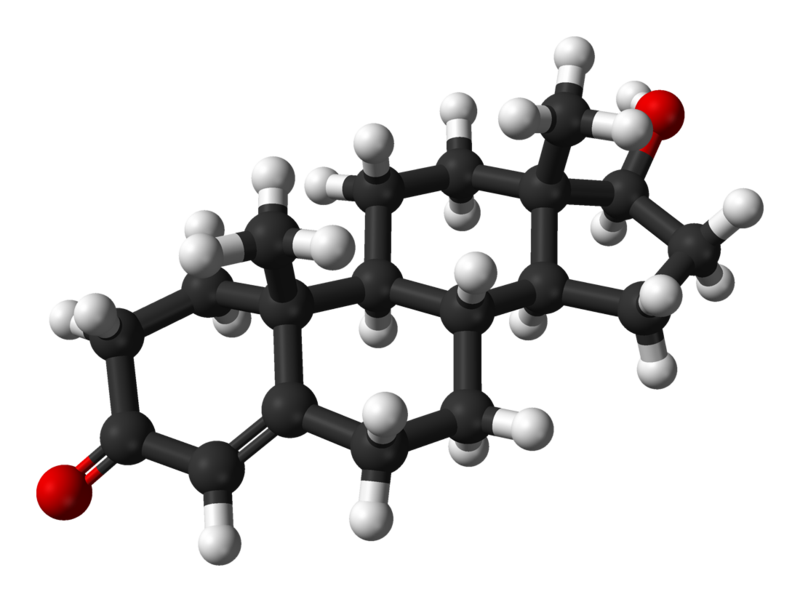
Ball and stick 3D model of testosterone. Source: Wikimedia CC0
A treatment paradox has recently come to light in prostate cancer: Blocking testosterone production halts tumour growth in early disease, while elevating the hormone can delay disease progression in patients whose disease has advanced.
The inability to understand how different levels of the same hormone can drive different effects in prostate tumours has been an impediment to the development of new therapeutics that exploit this biology.
Now, a Duke Cancer Institute-led study appearing in Nature Communications, provides the needed answers to this puzzle.
The researchers found that prostate cancer cells are hardwired with a system that allows them to proliferate when the levels of testosterone are very low. But when hormone levels are elevated to resemble those present in the normal prostate, the cancer cells differentiate.
“For decades, the goal of endocrine therapy in prostate cancer has been to achieve absolute inhibition of androgen receptor function, the protein that senses testosterone levels,” said lead investigator Rachid Safi, PhD, research assistant professor in the Department of Pharmacology and Cancer Biology, at Duke University School of Medicine.
“It’s been a highly effective strategy, leading to substantial improvements in overall survival,” he said. “Unfortunately, most patients with advanced, metastatic disease who are treated with drugs to inhibit androgen signaling will progress to an aggressive form of the disease for which there are limited therapeutic options.”
Using a combination of genetic, biochemical, and chemical approaches, the research team defined the mechanisms that enable prostate cancer cells to recognise and respond differently to varying levels of testosterone, the most common androgenic hormone.
It turned out to be rather simple. When androgen levels are low, the androgen receptor is encouraged to “go solo” in the cell. In doing so, it activates the pathways that cause cancer cells to grow and spread. However, as androgens rise, the androgen receptors are forced to “hang out as a couple,” creating a form of the receptor that halts tumour growth.
“Nature has designed a system where low doses of hormones stimulate cancer cell proliferation and high doses cause differentiation and suppress growth, enabling the same hormone to perform diverse functions,” McDonnell said.
In recent years, clinicians have begun treating patients with late-stage, therapy resistant prostate cancers using a monthly, high-dose injection of testosterone in a technique called bi-polar androgen therapy, or BAT. The inability to understand how this intervention works has hindered its widespread adoption as a mainstream therapeutic approach for prostate cancer patients.
“Our study describes how BAT and like approaches work and could help physicians select patients who are most likely to respond to this intervention,” McDonnell said. “We have already developed new drugs that exploit this new mechanism and are bringing these to the clinic for evaluation as prostate cancer therapeutics.”
Researchers from Charité are developing new methods to use the internal clock inside tumour cells to optimise cancer therapies
One of the factors determining the effectiveness of certain medications depends on various factors, including the time of day when they are administered. This is due to circadian rhythms, which vary across individuals and makes it difficult to tailor medication schedules. Researchers at Charité – Universitätsmedizin Berlin have now developed a method for determining the optimum time of cancer treatment based on certain breast cancer cell lines. They describe their approach in the journal Nature Communications.
As well as bodily functions and metabolic processes, such as sleep and digestion, individual cells also follow a circadian rhythm. This is hugely important to chemotherapy. Previous studies have shown that chemotherapy is most effective when the tumour cells are dividing. But this finding has been hardly used at all in clinical treatment to date.
An interdisciplinary team at Charité headed by Dr. Adrián Enrique Granada from the Charité Comprehensive Cancer Center (CCCC) set out to close this gap. The team began looking for the optimum time to administer medication, based on the individual circadian rhythms of the tumors.
Triple-negative breast cancer as an example
“We cultured cells from patients with triple-negative breast cancer to observe how they respond at different times of day to the medications administered,” explains Carolin Ector, a research associate in Granada’s working group. Triple-negative breast cancer is a highly aggressive form of breast cancer, with few effective treatments available. “We used live imaging, a method of continuously monitoring living cells, and complex data analysis techniques to monitor and evaluate the circadian rhythms, growth cycles, and medication responses of these cancer cells in detail.”
In this way, the researchers identified certain times of the day at which cancer cells are most responsive to medication-based treatments. For example, the chemotherapeutic drug 5-fluorouracil (5-FU) turned out to have peak efficacy against a certain cancer cell line between eight and ten a.m. As the study also shows, the crucial aspects here are certain cellular and genetic factors. The scientists were even able to identify which genes are key to the circadian effects of certain medications. “We call them ‘core clock genes’. They have a significant impact on how responsive cancer cells are to treatments administered at different times of day,” Granada explains.
Profiles show how cancer cell types respond to medications
This approach can be used to create detailed profiles showing how different types of cancer cells respond to different medications at various times. “This can help to identify the most effective combinations of drugs,” Granada says. “Overall, our findings indicate that personalized treatment plans based on individual circadian rhythms could substantially improve the efficacy of cancer treatment”, he concludes. Moreover, undesirable side effects could also be reduced.
For these findings to contribute to clinical practice soon, the results should be validated in studies involving larger groups of patients. “We’re also planning to study the molecular mechanisms behind the circadian influences on medication sensitivity to further optimize treatment times and identify new therapeutic targets,” Granada says.
A study published in Nature reveals the functional relevance of tumour-neuron interactions that regulate the growth of ependymoma brain tumours, one of the most common types in children. The study, conducted by researchers at Baylor College of Medicine and St. Jude Children’s Research Hospital, highlights how neuronal signalling, modifications in DNA-associated proteins and developmental programs are intertwined to drive malignancy in brain cancer.
“Ependymomas are the third most common type of paediatric brain tumours,” said co-corresponding author, Dr Benjamin Deneen professor in the Department of Neurosurgery. “These tumours are aggressive, resistant to chemotherapy and lack tumour-specific therapies, leading to poor survival.”
“We have not made an impact on patient survival in the last three decades. A major factor has been a poor understanding of the disease. The motivation of our collaborative work with the Deneen lab is to dissect the biology of these tumours as a basis for developing new therapies,” said co-corresponding author Dr Stephen Mack, associate member at St. Jude Children’s Research Hospital and member of the Department of Neurobiology, Neurobiology and Brain Tumor Program and Center of Excellence in Neuro-Oncology Sciences.
Previous studies have shown in other types of brain tumours that brain activity surrounding the tumour can influence its growth. “In the current study, we investigated whether brain activity played a role in ependymoma growth, specifically in a very aggressive type driven by a protein called ZFTA-RELA,” said first author Hsiao-Chi Chen, a graduate student in the Deneen lab. “In collaboration with the Mack lab, we developed an animal model to study this rare paediatric brain tumour and validated these findings in human tumour samples.”
The researchers discovered evidence of abnormal neuronal activity in ependymoma’s environment and investigated whether it affected ependymoma growth. They found that while hyperactivity of some neural circuits promoted tumour growth, hyperactivity of other neural circuits surprisingly reduced tumour growth, which had not been described before. Their study revealed a novel chain of events at play that regulates tumour growth, which may hold therapeutic applications.
“First, we found that normal neurons located in the brain region called dorsa raphe nucleus (dRN) project towards the cortex, where ependymoma grows. These neurons secrete serotonin, a brain chemical that carries messages between nerve cells, which surprisingly slows tumour growth,” Chen said.
Interestingly, ependymoma cells carry a serotonin transporter, a molecule that imports serotonin within the cell. “We were surprised to discover that serotonin enters ependymoma cells and binds to histone H3, a protein that is tightly associated with DNA,” Chen said. “Histone serotonylation, the addition of serotonin to histone, regulated tumour growth. Promoting it enhanced tumour growth while preventing it slowed down ependymoma growth in animal models.”
“Discovering histone serotonylation in ependymoma piqued our interest because a previous study from our lab had revealed that adding serotonin to histones affects which genes the cell turns on,” Deneen said.
The team discovered that histone serotonylation in ependymoma increases the expression of transcription factors, genes that regulate the expression of other genes,” Chen said. “We focused on transcription factor ETV5 whose overexpression accelerated tumour growth. But how does it do it?”
The next experiments showed that ETV5 expression triggers changes in the 3D structure of chromatin, the combination of DNA and proteins that forms chromosomes. The 3D changes prevent the activation of genes encoding neurotransmitters, molecules that mediate neural activity. The team focused on a neurotransmitter called neuropeptide Y (NPY) and found that growing tumours have little NPY. Restoring the levels of NPY in tumours slowed down tumour progression and tumour-associated neural hyperactivity through the remodeling of surrounding synapses or neuron-to-neuron communication.
“We knew that brain tumours release factors that remodel synapses towards hyperactivity. Here we found the opposite also can happen, that ependymoma tumours can release factors that suppress excitatory synaptic remodeling and that repressing this mechanism is essential for tumour progression,” Deneen said.
“I am excited that this work has redefined our understanding of how brain tumour cells grow, and how they take advantage of factors in their surrounding environment to initiate tumours,” Mack said. “I am equally excited that this work has revealed many new avenues for research that may in the future lead to new therapies, which is desperately needed for this devastating disease.”
As debate rages around the feasible application of NHI on a national scale, seemingly ad infinitum, the escalating cancer crisis in South Africa underscores the need for immediate action on the ground. Recent reports shed light on the distressing reality that individuals diagnosed with cancer, dependent on an overburdened public health system, often face extended waiting periods or impossible distances that prevent them from accessing life-saving treatment.
Yet in the Northern Cape, a remarkable success story has quietly unfolded over the last five years, impacting the lives of hundreds of cancer patients and demonstrating that the way to bring better health to the public on a macro scale may be to focus on practical micro solutions that, once proven, can be replicated around the country.
It arises out of a collaboration between the Northern Cape Department of Health, Kimberley’s Robert Mangaliso Sobukwe Hospital and private sector oncology service provider, Icon Oncology, with the shared goal of delivering the best possible care for patients needing radiotherapy services – which were previously far from home.
Jennifer Fuller, Regional Manager for Icon Oncology explains: “The average radiotherapy treatment journey spans between two to six weeks. Previously, the profound socio-economic and psycho-social toll of Northern Cape patients traveling far from their homes and families was immeasurable. During this period there was no radiation facility in the province, so patients had to travel to Bloemfontein for treatment. We collaborated with the Department of Health to treat radiation patients here in the Northern Cape. The result is a true example of how government and the private sector can work together when there is a shared focus on patient outcomes.”
Today, the province provides transport from far-off areas for treatment at Icon’s radiotherapy facility in Kimberley. If needed, accommodation is provided by the RMS Hospital for the duration of the radiotherapy treatment, which can sometimes last for six weeks.
Since the implementation of the project in October 2019, 511 cancer patients have completed radiation treatment. Previously all these patients would have had to travel to Bloemfontein to receive treatment.
“It’s a major success”, says Dr Alastair Kantani, Clinical Manager for hospital services in the Northern Cape. “It’s actually more than a success; it’s a lifesaving partnership. Personally, as a clinician, I’m proud of the fact that we, as a tertiary hospital, can give access to therapy services. Since this partnership, we’ve saved a lot of lives.”
Dr Esme Olivier, acting CEO of Robert Mangaliso Sobukwe Hospital says, “For me this collaboration between Icon and the Department of Health, specifically this hospital, is one of the best things that could happen for the sake of our oncology patients.”
“This is truly a complete collaboration” adds Fuller. “It shows that it can work – and I do believe that this can be replicated further into other provinces.”
Dr Olivier agrees: “I would really encourage every province to get involved with this. Even if they have their own radiation therapy units, just the collaboration and the expertise that Icon brings on board – they can even assist with nursing, whatever need there is that is not immediately available in public hospitals.”
Governance is key and monthly meetings are held between the two management teams to discuss patient experience, statistics, billing and other operational matters.
“This has resulted in continuous improvement of the project delivery over the past five years. This sharing of responsibility has led to the development of a very strong relationship and the interdependence has meant both parties have worked hard to make sure it works,” says Dr Olivier.
How it works – the patient journey:
The patient journey starts at the tertiary state facility, Robert Mangaliso Sobukwe Hospital, where the resident oncologist will consult with the patient once diagnosed by a surgeon or radiologist. If radiotherapy is indicated, the patient is referred to the Icon Radiotherapy unit in Kimberley.
A planning CT-scan is done by an Icon radiotherapist at Robert Magaliso Sobukwe Hospital
The treating oncologist and the Icon planning department, draws a plan on the CT-scan to determine the dose and area of radiotherapy that the patient will receive, using a sophisticated, state of the art planning system. The oncologist can access Icon’s planning system remotely and can do this work from anywhere.
This constitutes a 15 min radiotherapy session every day on Icon’s linear accelerator, until the required dose is delivered, and treatment is completed.
“The Northern Cape initiative exemplifies the potential inherent in bridging the gap between public and private healthcare sectors. It showcases how collaboration can transcend obstacles and provide specialised healthcare services and treatment to all citizens. As the country grapples with the challenges and concerns raised by the National Health Insurance (NHI) Bill, this collaborative achievement stands as a testament that the seemingly daunting task of implementing universal healthcare coverage can indeed be navigated. We must commend the vision of the Northern Cape DoH, management from the Robert Mangaliso Sobukwe Hospital and all other stakeholders who have made this project such a success,” explains Dr Ernst Marais, COO of Icon Oncology.
“It is through collaborations like this one, that we excel in providing the best possible treatment to our patients. Like Hellen Keller said: ‘Alone we can do so little, but together we can do so much’,” concludes Dr Olivier.
Credit: Darryl Leja National Human Genome Research Institute National Institutes Of Health
Men with metastatic castration-resistant prostate cancer should be treated primarily with second-generation hormone drugs, which offer better treatment response and longer life expectancy than chemotherapy. But the effect depends on which mutations the patient’s tumour carries. This is shown by results from the ProBio study, led by researchers at Karolinska Institutet in Sweden. The findings are published in Nature Medicine.
Every year, around 2500 men in Sweden are diagnosed with metastatic prostate cancer. Initially, all are treated with testosterone blockade to prevent testosterone from activating the androgen receptor, the gene that mainly fuels the growth of cancer cells. Over time, the cancer cells develop resistance and become so-called castration-resistant. This requires the use of new drugs – usually chemotherapy or second-generation hormone drugs (abiraterone/enzalutamide) that inhibit the androgen receptor. These are called Androgen Receptor Pathway inhibitors, or ARPi. Although these drugs have been available for over a decade, there is no direct comparison from a randomised trial until now.
Personalised treatment
“For the first time, we have compared these treatments with each other and also analysed the DNA of the cancer cells to find out which drug that works best for different individuals,” says Johan Lindberg, senior researcher at the Department of Medical Epidemiology and Biostatistics (MEB), Karolinska Institutet.
The bloodstream contains so-called cell-free DNA from cells that have died, something that happens all the time in healthy individuals and is perfectly normal. In patients with cancer, a fraction of the cell-free DNA originates from the cancer cells and is called circulating tumour DNA (ctDNA). By analysing ctDNA, it is possible to see what changes, or mutations, are present in a person’s tumour. The ProBio study aims to use knowledge of the tumour’s genetic signature to provide the best treatment. The idea is to be able to identify patients whose tumours are particularly sensitive or resistant to certain treatments through ongoing analyses.
“It creates a self-learning system to continuously improve treatment for men with metastatic prostate cancer,” says Martin Eklund, Professor of Epidemiology at the same department. “We are also gathering knowledge about which regions of the genome are important in prostate cancer.”
Longer life expectancy
The current sub-study included 193 patients with metastatic castration-resistant prostate cancer. They were randomly chosen to receive either chemotherapy or ARPi, which was compared to a control group where the doctor decided on the best treatment. The ARPi group responded the longest to treatment (a median of 11.1 months compared with 6.9 for chemotherapy and 7.4 for the control group). Survival for the ARPi group was also significantly longer – a median of 38.7 months compared with 21.7 months and 21.8 months respectively.
The effectiveness of ARPi varied depending on the patient’s genetic profile. For example, there was no significant difference between the treatments in the short term in patients whose tumours had mutations in the p53 gene, which occurs in around 45% of men with metastatic prostate cancer. However, data from the study suggest that also this group may have better survival if they receive ARPi rather than chemotherapy.
Results indicate the need for efforts to improve cancer outcomes equitably.
Credit: Darryl Leja National Human Genome Research Institute National Institutes Of Health
In an analysis of 30 cancer types among men, investigators uncovered substantial disparities in cancer cases and deaths by age and countries’ economic status – disparities that are projected to widen by 2050. The study is published by Wiley online in CANCER, a peer-reviewed journal of the American Cancer Society.
Men face higher rates of cancer and cancer-related deaths than women, likely due to various factors including lower participation in cancer prevention activities; underuse of screening and treatment options; increased exposure to cancer risk factors such as smoking, alcohol consumption, and occupational exposure to carcinogens; and biological differences.
To assess the burden of cancer in men of different ages and living in different regions of the world, investigators analyzed 2022 information from the Global Cancer Observatory, which encompasses national-level estimates for cancer cases and deaths for 185 countries/territories worldwide. The projected cancer cases and deaths in 2050 were derived through demographic projections: the researchers multiplied the 2022 age-specific rates with their corresponding population projections for 2050.
In 2022, poorer survival was observed among older men; for rare cancer types such as pancreatic cancer; and in countries with low human development index, which measures health, education, and standard of living.
Between 2022 and 2050, cancer cases are projected to increase from 10.3 million to 19 million, an 84% increase. Deaths are projected to increase from 5.4 million to 10.5 million, a 93% increase, with a greater than two-fold increase among men aged 65+ years and for countries/territories with low and medium human development index.
The research reveals an urgent need to address these trends and ensure equity in cancer prevention and care among men globally.
“A national and international collaboration, as well as a coordinated multisectoral approach, are essential to improve current cancer outcomes and to reverse the anticipated rise in cancer burden by 2050. Implementing and expanding universal health coverage and expanding health infrastructure and establishing publicly funded medical schools and scholarships for training medical and public health staff can improve cancer care and equity,” said lead author Habtamu Mellie Bizuayehu, PhD, of the University of Queensland, in Australia. “Emphasis should be placed on low and medium human development index countries with high unmet cancer service needs despite a significant cancer burden.
Dr Bizuayehu added that improving access to and use of cancer prevention, screening, diagnosis, and treatment options, especially for older men, could also improve cancer outcomes and equity.
A new Cleveland Clinic study has identified diet-derived molecules called metabolites as main drivers of young-onset colorectal cancer risk, especially those associated with red and processed meat. The NPJ Precision Oncologyreport, which analysed metabolite and microbiome datasets, highlighted that one of the best ways a younger ( < 60 years) adult can prevent colorectal cancer is to discuss their diet with their doctor.
Increased monitoring and screening for colorectal cancer is an extremely helpful tool. Despite the success of these methods, these data indicate physicians can take a different approach with their younger patients, says senior author and gastrointestinal oncologist Suneel Kamath,MD.
“At the end of the day, it’s impractical to apply our care models for those over 60 to younger adults simply because we cannot give everyone in the system yearly colonoscopies,” he explains. “What is much more feasible is to give everyone in the system a simple test to measure a biomarker that determines their colorectal cancer risk. Then we can give the most at-risk individuals appropriate screening.”
Former clinical fellow Thejus Jayakrishnan, MD, and Naseer Sangwan, PhD, director of the Microbial Sequencing & Analytics Resource Core co-led the work. Researchers in Cleveland Clinic’s Center for Young-Onset Colorectal Cancer provided large-scale analyses of patient data from individuals who received care for either young- or average-onset colorectal cancer at Cleveland Clinic.
One previous study from this team identified differences in the metabolites (diet-derived molecules) of young – versus average-onset colorectal cancer, while another identified differences in gut microbiome between younger and older adults with colorectal cancer. These studies provided many potential directions for studying young-onset CRC. However, when more factors are involved in cancer risk, it becomes more complicated to understand what’s going on and plan future research, Dr Sangwan says. Interactions between these factors, like when our gut bacteria consume our metabolites and produce their own, make it even more complex.
Dr Sangwan and his team then developed an AI algorithm to combine and analyse the existing studies’ datasets and clarify what factors are most relevant for future study. Surprisingly, Dr Sangwan’s analysis revealed that differences in diet (identified through analysing metabolites) accounted for a significant proportion of the differences observed between the young-onset and older-onset patients.
“Researchers – ourselves included – have begun to focus on the gut microbiome as a primary contributor to colon cancer risk. But our data clearly shows that the main driver is diet,” Dr Sangwan says. “We already know the main metabolites associated with young-onset risk, so we can now move our research forward in the correct direction.”
The team was excited to see diet play such a large role in cancer risk, because it is much easier to identify at-risk patients by counting the metabolites in their blood than it is to sequence the bacterial DNA in their stool for different microbes.
“It can actually be very complicated and difficult to change your microbiome,” explains Dr Kamath. “While it’s not always easy, it is much simpler to change your diet to prevent colon cancer.”
Addressing factors in our diet to prevent colon cancer
Younger colon cancer patients had higher levels of metabolites associated with the production and metabolism of an amino acid called arginine, and with the urea cycle compared to their older peers. These differences may be tied to long-term consumption of red meat and processed meat. The team is now analyzing national datasets to validate their Cleveland Clinic-specific findings in patients across the country.
After they show that arginine and urea cycle metabolites (and, by proxy, red and processed meat overconsumption) are elevated across younger adults with colon cancer nationwide, they plan to test whether certain diets or commercially available drugs that regulate arginine production and the urea cycle can help prevent or even treat young-onset colorectal cancer.
Dr Kamath says that even though more research is needed to understand exactly how dietary factors cause colon cancer, his current findings have already changed the way he delivers patient care.
“Even though I knew before this study that diet is an important factor in colon cancer risk, I didn’t always discuss it with my patients during their first visit. There is so much going on, it can already be so overwhelming,” says Dr Kamath. “Now, I always make sure to bring it up to my patients, and to any healthy friends or family members they may come in with, to try and equip them with the tools they need to make informed choices about their lifestyle.”
Human colon cancer cells. Credit: National Cancer Institute
In a first, a team in Germany has produced a substance capable of sending cancer cells into ferroptosis, a form of cell death discovered only in recent years. This could pave the way for the development of new drugs.
Conventional cancer drugs work by triggering apoptosis, that is programmed cell death, in tumour cells. However, tumour cells have the ability to develop strategies to escape apoptosis, rendering the drugs ineffective. In the journal Angewandte Chemie, a research team from Ruhr University Bochum, Germany, describes a new mechanism of action that kills cancer cells through ferroptosis. Ferroptosis is another form of programmed cell death that wasn’t discovered until the 2010s. The Bochum group synthesised a metal complex, demonstrated its effectiveness in cell cultures and on microtumours and identified the chemical processes underlying the mechanism of action.
Two types of programmed cell death
In programmed cell death, certain signaling molecules initiate a kind of suicide program to cause cells to die in a controlled manner. This is an essential step to eliminate damaged cells or to control the number of cells in certain tissues, for example. Apoptosis has long been known as a mechanism for programmed cell death. Ferroptosis is another mechanism that has recently been discovered which, in contrast to other cell death mechanisms, is characterised by the accumulation of lipid peroxides. This process is typically catalysed by iron which is where the name ferroptosis derives from.
“Searching for an alternative to the mechanism of action of conventional chemotherapeutic agents, we specifically looked for a substance capable of triggering ferroptosis,” explains Johannes Karges. His group synthesized a cobalt-containing metal complex that accumulates in the mitochondria of cells and generates reactive oxygen species, more precisely hydroxide radicals. These radicals attack polyunsaturated fatty acids, resulting in the formation of large quantities of lipid peroxides, which in turn trigger ferroptosis. The team was thus the first to produce a cobalt complex designed to specifically trigger ferroptosis.
Effectiveness demonstrated on artificial microtumours
The researchers from Bochum used a variety of cancer cell lines to show that the cobalt complex induces ferroptosis in tumour cells. On top of that, the substance slowed down the growth of artificially produced microtumours .
“We are confident that the development of metal complexes that trigger ferroptosis is a promising new approach for cancer treatment,” as Johannes Karges sums up the research, adding: “However, there’s still a long way to go before our studies result in a drug.” The metal complex must first prove effective in animal studies and clinical trials. What’s more, the substance doesn’t currently selectively target tumour cells, but would also attack healthy cells. This means that researchers must first find a way to package the cobalt complex in such a way that it damages nothing but tumour cells.