Researchers at Karolinska Institutet and Lund University have identified a new treatment strategy for neuroblastoma, an aggressive form of childhood cancer. By combining two antioxidant enzyme inhibitors, they have converted cancer cells in mice into healthy nerve cells. The study is published in the journal Proceedings of the National Academy of Sciences (PNAS).
Neuroblastoma is a type of childhood cancer that affects the nervous system and is the leading cause of cancer-related death in young children. Some patients have a good prognosis, but those with metastatic tumours often cannot be cured despite modern combinations of surgery, radiation, chemotherapy and immunotherapy.
“The children who survive often have lifelong cognitive difficulties due to the harsh treatment, so there is a great need for new forms of therapies for children with neuroblastoma,” says Marie Arsenian Henriksson, professor at the Department of Microbiology, Tumour and Cell Biology at Karolinska Institutet.
Transform cancer cells
Differentiation therapy is a treatment method used in neuroblastoma that aims to transform cancer cells into more mature and healthy cells. The problem with the current retinoic acid differentiation therapy is that many patients do not respond to treatment, and about half develop resistance.
In collaboration with researchers at Lund University, Marie Arsenian Henriksson’s research team has shown that inhibition of two specific enzymes, PRDX6 and GSTP1, could be an alternative to retinoic acid treatment.
Mature into healthy neurons
Neuroblastoma is characterised by high oxidative stress due to the active metabolism in the cancer cells. Tumours are therefore dependent on antioxidant enzymes such as PRDX6 and GSTP1 to manage the stress and avoid cell death. High levels of these enzymes are associated with a poorer prognosis.
“When we inhibit these enzymes in cell cultures as well as in mouse models, some of the tumour cells die while others mature into active, healthy neurons, impairing tumour growth,” says Judit Liaño-Pons, researcher at the Department of Microbiology, Tumour and Cell Biology.
Needs to be tested in children
In the next step, the treatment will need to be tested in a clinical trial to investigate its safety and efficacy in children. One of the inhibitors has received orphan drug designation from the US Food and Drug Administration for the treatment of a different diagnosis in adults, making it a particularly promising drug candidate, according to the scientists.
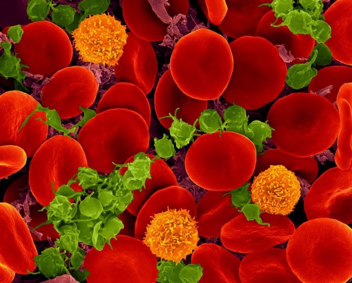
Scanning electron micrograph of red blood cells, T cells (orange) and platelets (green). Source: Wikimedia CC0
Northwestern Medicine scientists have uncovered key details about a group of rare but serious blood disorders, which may help inform potential treatments, according to a study published in the Journal of Clinical Investigation.
Myeloproliferative neoplasms (MPNs) are rare blood cancers characterised by the abnormal growth of blood cells. They have long been linked to a key signalling pathway called JAK2/STAT, but the specific details of how they develop have remained unclear.
“These diseases are often driven by abnormal activation of a protein called JAK2,” said Peng Ji, MD, PhD, ‘15 GME, Professor of Pathology and senior author of the study. “In earlier research, we discovered that another protein, PLEK2, acts downstream of JAK2 and plays a critical role in mediating JAK2’s effects, helping to drive the progression of MPNs.”
In the current study, Ji and his collaborators aimed to better understand the proteins that work alongside PLEK2, also known as the PLEK2 “signalosome.”
By analysing protein expression in cultured human blood stem cells, the investigators identified a new contributor, PPIL2, that appears to help cancer cells grow by disabling the tumour suppressor protein p53.
Under normal conditions, p53 works as a tumour suppressor protein that prevents excessive cell growth. PPIL2 effectively marks p53 for degradation, weakening its ability to control cell growth and allowing the disease to advance, according to the findings.
Investigators found that blocking PPIL2 using cyclosporin A, an immunosuppressant drug commonly used for organ transplant patients, led to an increase in p53 levels, restoring its ability to regulate cell growth. In experiments using MPN models — including mice with a mutated JAK2 gene and lab-grown human bone marrow — cyclosporin A significantly reduced the abnormal proliferation of blood cells, according to the findings.
“Even better results were seen when cyclosporin A was combined with another type of drug that also boosts p53,” said Ji. “This shows that targeting PPIL2 might be a powerful new way to treat MPNs using drugs that are already available.”
While more research is needed to fully understand how cyclosporin A works in MPN patients, this study highlights a promising new target for treatment, Ji said.
Now, Ji and his laboratory are planning to work on developing drugs that more specifically block PPIL2, since cyclosporin A affects many proteins and can have unwanted effects.
“Clinical studies will be needed to test whether this approach works in people, possibly starting by looking at how MPN patients respond to cyclosporin A if they’ve already been treated with it,” Ji said.
A Korean population-based cohort study investigated the risk of Alzheimer’s disease (AD) among breast cancer survivors compared to age-matched controls without cancer. The study, published in JAMA Network Open, found that breast cancer survivors had an 8% lower risk of AD than controls, with a significant association in survivors over 65 years old – though the effect did not persist past five years. Radiotherapy was associated with a lower risk of AD among breast cancer survivors – but not other treatments.
Breast cancer survivors may experience long-term health consequences, including cognitive function and risk of dementia. The risk of AD among breast cancer survivors is still unclear and may vary depending on age at diagnosis, treatment received, and time since treatment.
Previous studies reported mixed results on the risk of AD among breast cancer survivors, with some finding no increase in risk and others finding a 35% increased risk for those diagnosed at age 65 or older. These studies have been hampered by a number of methodological issues, including not accounting for risk factors.
Cytotoxic chemotherapy can cause cognitive decline termed ‘chemobrain’. Other chemotherapy drugs such as anthracycline may reduced AD risk by reducing the formation of amyloid deposits. Endocrine therapy may increase the risk of dementia by lowering oestrogen, but studies suggest that the use of tamoxifen and aromatase inhibitors is associated with a lower risk of AD. An increase in dementia is seen in radiotherapy for head and neck cancers.
To investigate the risk of AD among breast cancer survivors, researchers used the Korean National Health Insurance Service (K-NHIS) database, exploring whether there is an association with cancer treatment and various confounding factors.
Among 70 701 breast cancer survivors (mean age, 53.1 years), 1229 cases of AD were detected, with an incidence rate of 2.45 per 1000 person-years. Survivors exhibited a slightly lower risk of AD compared with cancer-free controls, especially among individuals 65 years or older (SHR, 0.92; 95% CI, 0.85-0.99). But landmark analyses found that this lower risk did not persist beyond five years of survival. Radiotherapy was associated with reduced risk of AD among survivors, while chemotherapy and endocrine therapy had no significant impact. Anthracycline use, however, did show a non-significant decrease in risk.
Differences in doses and timing of radiotherapy may influence the effects. The incident exposure to the brain is estimated to be 0.2Gy from a breast cancer radiotherapy dose of 50Gy. A pilot study found that patients with AD who received low-dose whole-brain radiotherapy at 3Gy showed a temporary improvement in cognitive function. This improvement is believed to be due to a neuroprotective effect on microglia. Other studies have noted a transient risk reduction for AD in breast cancer radiotherapy; however, patients receiving radiotherapy usually do so in conjunction with breast-conserving surgery – those opting for this procedure are younger, with fewer comorbidities and smaller tumours.
The study suggests that cancer treatment may have benefits against AD development, but the risk of AD may differ depending on the duration of survival.
The findings indicate that breast cancer treatment may not directly lead to AD, and that managing modifiable risk factors for AD, such as smoking and diabetes, is a feasible option to lower AD risk among breast cancer survivors.
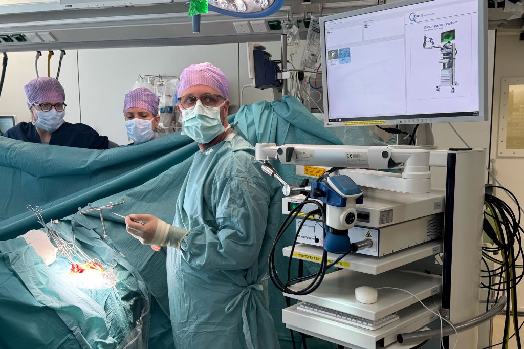
A medical team at Erasmus University Medical Center in the Netherlands uses the new imaging probe with a Quest camera to get a better view of cancerous tumors during non-brain cancer surgery. Photo courtesy of Erasmus University Medical Center
In a significant leap forward for successful cancer surgery, researchers at the University of Missouri and collaborators have developed a new imaging probe to help surgeons more accurately identify and remove aggressive tumours during operations.
The tool is expected to be a critical advancement in the fight against glioblastoma, one of the most difficult-to-treat brain cancers. In the future, it is intended to be expanded for image-guided surgery of various other solid tumours.
Described in a new study in Nature Publishing Group Imaging, the innovation works by pairing a fluorescent dye with a fatty acid molecule that cancer cells readily absorb. When introduced into the body, the compound is taken up by tumour cells, causing them to glow under near-infrared light, revealing cancer that might otherwise remain hidden.
Glioblastoma is considered surgically incurable because the tumour doesn’t stay in one place – it spreads and invades healthy brain tissue in a diffuse, microscopic way. This makes it impossible to remove completely without risking serious damage to brain function.
“Surgery remains one of the primary treatments for many cancers,” Elena Goun, associate professor of chemistry in the College of Arts and Science and one of the lead authors of the study, said. “In breast or prostate cancer, surgeons can often remove the tumour along with surrounding tissue. In brain cancer, that’s simply not possible. You must preserve healthy brain tissue. But if even a few cancer cells are left behind, the disease will return.”
That dilemma is especially acute with glioblastoma, which doesn’t form a neatly contained mass. Instead, it sends out microscopic extensions — finger-like projections that blend into healthy brain tissue and are invisible to the naked eye.
Because of this, surgeons must walk a fine line: removing as much tumour as possible while avoiding harm to vital brain areas. The more thoroughly the tumour is removed, the more effective follow-up treatments like radiation and chemotherapy tend to be.
The new small-molecule probe, known as FA-ICG, is engineered to solve that problem. It links a natural long-chain fatty acid (FA) to indocyanine green (ICG), an FDA-approved near-infrared dye widely used in surgical imaging. This fatty acid-based approach means the probe is highly selective: glioblastoma cells, which thrive on fatty acids, absorb it more than normal brain cells. That makes the cancer stand out more clearly.
The result is a tool that takes advantage of cancer’s altered metabolism to highlight tumour cells from within.
“Surgeons would view a monitor during surgery showing where the probe is lighting up,” Goun explained. “If they still see fluorescent signals, it means cancer is still present and more tissue needs to be removed. When the light disappears, they would know they’ve cleared the area.”
In the operating room, surgeons already use a variety of tools to guide tumour removal – including microscopes, ultrasound and fluorescent dyes. Of those, fluorescent dyes are particularly useful because they make otherwise invisible tumour cells light up under special lighting.
Right now, the only approved imaging dye for glioblastoma surgery is 5-ALA, which fluoresces under blue light. But 5-ALA comes with major limitations: The operating room must be darkened in order to see it, tissue penetration is shallow and the fluorescent signal is often weak and non-specific.
It also comes with side effects, including photosensitivity, meaning patients must avoid bright light exposure after surgery due to the risk of skin and eye damage.
That’s where the FA-ICG probe shines – both literally and functionally.
Compared to 5-ALA, FA-ICG is brighter, works under normal surgical lighting, and offers real-time visualisation under the microscope – no need to turn the lights off mid-surgery. This saves time and makes procedures more efficient. The signal-to-background ratio is also higher, meaning it’s easier to distinguish tumour tissue from healthy brain.
The FA-ICG probe is not only easier to see, it’s also easier to use. Its longer half-life allows more flexibility in scheduling surgeries, and the logistics of administration are simpler than with current probes.
“The upside of fluorescence-guided surgery is that you can make little remnants much more visible using the light emitting properties of these tumour cells when you give them a dye,” said Rutger Balvers, a neurosurgeon at Erasmus University Medical Center in the Netherlands, who is expected to lead human clinical trials of the probe. “And we think that the upside of FA-ICG compared to what we have now is that it’s more select in targeting tumour cells. The visual properties of the probe are better than what we’ve used before.”
Michael Chicoine is a neurosurgeon at MU Health Care and chair of Mizzou’s School of Medicine’s Department of Neurosurgery. While he’s not directly involved in the research, Chicoine understands the potential benefits firsthand.
Currently, he said, MRIs are the gold standard for imaging tumours; however, they’re expensive and time-consuming, especially when required during an operation.
“This fluorescent metabolically linked tool gives you real-time imaging,” he said. “We could merge techniques, using the probe during surgery and saving the MRI for a sort of final exam. It’s definitely an exciting advancement.”
Researchers are also excited about other uses for the probe, including for other types of cancers and for use during follow-up treatments.
“After radiation or chemotherapy, it becomes very difficult to distinguish between scar tissue and active tumor,” Chicoine said. “This probe could give us a definitive answer – helping doctors know whether to continue treatment or adjust it, or consider another surgery. Eliminating the current uncertainty would be really helpful.”
Another promising use of the probe could be in photodynamic therapy either during or after surgery. Since the dye also has light-activated properties that can kill cancer cells, researchers are exploring whether it could double as a treatment tool, not just a diagnostic one.
Clinical trials for use in glioblastoma cases are expected to start in Europe, with strong interest already growing among neurosurgical teams.
The upcoming Phase 1 trial will focus on how patients tolerate the probe, whether there are any side effects at an effective dose and how its performance compares to existing tools. Ultimately, the goal is to make brain tumour surgery safer, helping surgeons remove all cancerous tissues while preserving as much healthy brain tissue as possible.
If results are positive, future studies could expand the use of FA-ICG beyond brain tumours to other cancers with high fatty acid metabolism, such as pancreatic cancer,according to fellow corresponding author Laura Mezzanotte from the Erasmus’ Department of Radiology and Nuclear Medicine.
Mitochondrial pathways help melanoma cells become aggressive, and some currently available drugs target these pathways.
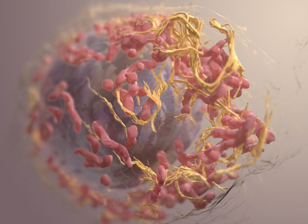
3D structure of a melanoma cell derived by ion abrasion scanning electron microscopy. Credit: Sriram Subramaniam/ National Cancer Institute
Researchers have discovered that the most aggressive melanomas, the deadliest form of skin cancer, overactivate two key processes in mitochondria. Blocking these pathways with currently available drugs effectively killed melanoma cells. The findings are published by Wiley online in CANCER, a peer-reviewed journal of the American Cancer Society.
By mapping the proteins expressed in 151 tumour and normal skin samples, investigators found that the most aggressive melanomas hyper-activate the machinery that builds mitochondrial proteins and the mitochondrial system that turns nutrients into energy.
Remarkably, blocking these pathways effectively halted or killed melanoma cells cultured in lab dishes. Two types of drugs accomplished this: antibiotics, originally designed to block bacterial protein synthesis machinery, which closely resembles the machinery found in mitochondria, and specialised energy-production inhibitors. Importantly, non-cancerous skin cells remained mostly unaffected, highlighting the safety and specificity of these treatment approaches.
“This discovery identifies melanoma’s excessive reliance on mitochondrial energy as its Achilles’ heel, revealing a therapeutic vulnerability that we can exploit with existing drugs,” said senior author Jeovanis Gil, PhD, of Lund University in Sweden. “By pairing mitochondrial blockers with today’s standards of care, we may cut off a major escape route that cancers use to resist therapy and come back.”
Dr Gil added that the mitochondrial-protein signature his team discovered can be measured in routine biopsy material and could serve as a biomarker to identify patients most likely to benefit from mitochondrial-targeted add-on therapies. By enabling clinicians to match treatments to each patient’s tumour biology, these findings mark a step forward for precision medicine in melanoma. Moreover, because mitochondrial rewiring fuels resistance across many cancers, success in melanoma could open the door to similar personalised combination strategies in other hard-to-treat cancers.
A sample of Aspergillus flavus cultured in the Gao Lab. (Credit: Bella Ciervo)
University of Pennsylvania-led researchers have turned a deadly fungus into a potent cancer-fighting compound. After isolating a new class of molecules from Aspergillus flavus, a toxic crop fungus linked to deaths in excavating ancient tombs, the researchers modified the chemicals and tested them against leukaemia cells. The result was a promising cancer-killing compound that rivals FDA-approved drugs and opens up new frontiers in the discovery of more fungal medicines.
“Fungi gave us penicillin,” says Sherry Gao, Presidential Penn Compact Associate Professor in Chemical and Biomolecular Engineering (CBE) and in Bioengineering (BE) and senior author of a new paper in Nature Chemical Biology on the findings. “These results show that many more medicines derived from natural products remain to be found.”
From Curse to Cure
A. flavus, named for its yellow spores, has long been a microbial villain. After archaeologists opened King Tutankhamun’s tomb in the 1920s, a series of untimely deaths among the excavation team fuelled rumours of a pharaoh’s curse. Decades later, doctors theorised that fungal spores, dormant for millennia, could have played a role.
In the 1970s, a dozen scientists entered the tomb of Casimir IV in Poland. Within weeks, 10 of them died. Later investigations revealed the tomb contained A. flavus, whose toxins can lead to lung infections, especially in people with compromised immune systems.
Now, that same fungus is the unlikely source of a promising new cancer therapy.
A Rare Fungal Find
The therapy in question is a class of ribosomally synthesised and post-translationally modified peptides, or RiPPs, pronounced like the “rip” in a piece of fabric. The name refers to how the compound is produced – by the ribosome, a tiny cellular structure that makes proteins – and the fact that it is modified later, in this case, to enhance its cancer-killing properties.
“Purifying these chemicals is difficult,” says Qiuyue Nie, a postdoctoral fellow in CBE and the paper’s first author. While thousands of RiPPs have been identified in bacteria, only a handful have been found in fungi. In part, this is because past researchers misidentified fungal RiPPs as non-ribosomal peptides and had little understanding of how fungi created the molecules. “The synthesis of these compounds is complicated,” adds Nie. “But that’s also what gives them this remarkable bioactivity.”
Hunting for Chemicals
To find more fungal RiPPs, the researchers first scanned a dozen strains of Aspergillus, which previous research suggested might contain more of the chemicals.
By comparing chemicals produced by these strains with known RiPP building blocks, the researchers identified A. flavus as a promising candidate for further study.
Genetic analysis pointed to a particular protein in A. flavus as a source of fungal RiPPs. When the researchers turned the genes that create that protein off, the chemical markers indicating the presence of RiPPs also disappeared.
This novel approach – combining metabolic and genetic information – not only pinpointed the source of fungal RiPPs in A. flavus, but could be used to find more fungal RiPPs in the future.
A Potent New Medicine
After purifying four different RiPPs, the researchers found the molecules shared a unique structure of interlocking rings. The researchers named these molecules, which have never been previously described, after the fungus in which they were found: asperigimycins.
Even with no modification, when mixed with human cancer cells, asperigimycins demonstrated medical potential: two of the four variants had potent effects against leukaemia cells.
Another variant, to which the researchers added a lipid found in bees’ royal jelly, performed as well as cytarabine and daunorubicin, two FDA-approved drugs that have been used for decades to treat leukaemia.
Cracking the Code of Cell Entry
To understand why lipids enhanced asperigimycins’ potency, the researchers selectively turned genes on and off in the leukaemia cells. One gene, SLC46A3, proved critical in allowing asperigimycins to enter leukaemia cells in sufficient numbers.
That gene helps materials exit lysosomes, the tiny sacs that collect foreign materials entering human cells. “This gene acts like a gateway,” says Nie. “It doesn’t just help asperigimycins get into cells, it may also enable other ‘cyclic peptides’ to do the same.”
Like asperigimycins, those chemicals have medicinal properties – nearly two dozen cyclic peptides have received clinical approval since 2000 to treat diseases as varied as cancer and lupus – but many of them need modification to enter cells in sufficient quantities.
“Knowing that lipids can affect how this gene transports chemicals into cells gives us another tool for drug development,” says Nie.
Disrupting Cell Division
Through further experimentation, the researchers found that asperigimycins likely disrupt the process of cell division. “Cancer cells divide uncontrollably,” says Gao. “These compounds block the formation of microtubules, which are essential for cell division.”
Notably, the compounds had little to no effect on breast, liver or lung cancer cells – or a range of bacteria and fungi – suggesting that asperigimycins’ disruptive effects are specific to certain types of cells, a critical feature for any future medication.
Future Directions
In addition to demonstrating the medical potential of asperigimycins, the researchers identified similar clusters of genes in other fungi, suggesting that more fungal RiPPS remain to be discovered. “Even though only a few have been found, almost all of them have strong bioactivity,” says Nie. “This is an unexplored region with tremendous potential.”
The next step is to test asperigimycins in animal models, with the hope of one day moving to human clinical trials. “Nature has given us this incredible pharmacy,” says Gao. “It’s up to us to uncover its secrets. As engineers, we’re excited to keep exploring, learning from nature and using that knowledge to design better solutions.”
Genetic material shed by tumours can be detected in the bloodstream three years prior to cancer diagnosis, according to a study led by investigators at Johns Hopkins.
The study, partly funded by the National Institutes of Health, was published in Cancer Discovery.
Investigators were surprised they could detect cancer-derived mutations in the blood so much earlier, says lead study author Yuxuan Wang, MD, PhD, an assistant professor of oncology at the Johns Hopkins University School of Medicine. “Three years earlier provides time for intervention. The tumours are likely to be much less advanced and more likely to be curable.”
To determine how early cancers could be detected prior to clinical signs or symptoms, Wang and colleagues assessed plasma samples that were collected for the Atherosclerosis Risk in Communities (ARIC) study, a large National Institutes of Health-funded study to investigate risk factors for heart attack, stroke, heart failure and other cardiovascular diseases. They used highly accurate and sensitive sequencing techniques to analyse blood samples from 26 participants in the ARIC study who were diagnosed with cancer within six months after sample collection, and 26 from similar participants who were not diagnosed with cancer.
At the time of blood sample collection, eight of these 52 participants scored positively on a multicancer early detection (MCED) laboratory test. All eight were diagnosed within four months following blood collection. For six of the eight individuals, investigators also were able to assess additional blood samples collected 3.1–3.5 years prior to diagnosis, and in four of these cases, tumour-derived mutations could also be identified in samples taken at the earlier timepoint.
“This study shows the promise of MCED tests in detecting cancers very early, and sets the benchmark sensitivities required for their success,” says Bert Vogelstein, MD, Clayton Professor of Oncology, co-director of the Ludwig Center at Johns Hopkins and a senior author on the study.
“Detecting cancers years before their clinical diagnosis could help provide management with a more favourable outcome,” adds Nickolas Papadopoulos, PhD, professor of oncology, Ludwig Center investigator and senior author of the study. “Of course, we need to determine the appropriate clinical follow-up after a positive test for such cancers.”
The potential role of vitamin D in preventing and treating colorectal cancer (CRC) has attracted growing research interest – especially as CRC rates are rising, particularly among younger adults. This isn’t a new area of study. Low vitamin D levels have long been linked to a higher risk of developing colorectal cancer.
One large study involving over 12 000 participants found that people with low blood levels of vitamin D had a 31% greater risk of developing CRC compared to those with higher levels. Similarly, another study reported a 25% lower CRC risk among individuals with high dietary vitamin D intake.
Data from the Nurses’ Health Study – a long-term investigation of American nurses – showed that women with the highest vitamin D intake had a 58% lower risk of developing colorectal cancer compared to those with the lowest intake.
Now, a review highlights vitamin D’s promise in colorectal cancer prevention and treatment – but also underscores the complexity and contradictions in current research.
While observational data, which follow people’s use of vitamin D, and mechanistic studies, to investigate how vitamin D works in the laboratory, suggest protective effects, this isn’t confirmed by larger trials.
In fact, randomised controlled trials (RCTs), in which some people receive vitamin D and others don’t, the gold standard by which treatments are judged, reveal inconsistent outcomes. This highlights the need for a balanced approach to its integration into public health strategies.
Vitamin D is synthesised in the skin in response to sunlight and exerts its biological effects through vitamin D receptors (VDRs) found throughout the body, including in colon tissue. When activated, these receptors help regulate gene activity related to inflammation, immune response and cell growth – processes central to cancer development and progression.
Preclinical studies have shown that the active form of vitamin D (calcitriol) can suppress inflammation, boost immune surveillance (the immune system’s ability to detect abnormal cells), inhibit tumour blood vessel growth and regulate cell division – a key factor in cancer development, as demonstrated in my recent research.
Epidemiological studies, which track health outcomes across large populations over time, consistently find that people with higher blood levels of vitamin D have a lower risk of developing CRC. This paints a hopeful picture, suggesting that something as simple as getting more vitamin D – via sun exposure, diet, or supplements – could lower cancer risk.
But the story gets more complicated.
Mixed results
When it comes to medical decision-making, randomised controlled trials (RCTs) are the gold standard. These studies randomly assign participants to receive either a treatment (like vitamin D) or a placebo, helping eliminate bias and isolate cause-and-effect relationships.
Unfortunately, RCTs on vitamin D and CRC have produced mixed results.
For example, the VITAL trial – a major RCT involving over 25 000 participants – found no significant reduction in overall colorectal cancer incidence with 2000 IU/day of vitamin D supplementation over several years.
However, a meta-analysis of seven RCTs did show a 30% improvement in CRC survival rates with vitamin D supplements, suggesting potential benefits later in the disease course rather than for prevention.
On the other hand, the Vitamin D/Calcium Polyp Prevention Trial found no reduction in the recurrence of adenomas (pre-cancerous growths) with supplementation, raising questions about who benefits most, and at what dosage.
Adding to the uncertainty is the question of causation. Does low vitamin D contribute to cancer development? Or does the onset of cancer reduce vitamin D levels in the body? It’s also possible that the observed benefits are partly due to increased sunlight exposure, which itself may have independent protective effects.
The big picture
These discrepancies highlight the importance of considering the “totality of evidence” – treating each study as one piece of a larger puzzle.
The biologic plausibility is there. Observational and mechanistic studies suggest a meaningful link between vitamin D and lower CRC risk. But the clinical evidence isn’t yet strong enough to recommend vitamin D as a standalone prevention or treatment strategy.
That said, maintaining sufficient vitamin D levels – at least 30ng/mL – is a low-risk, cost-effective health measure. And when combined with other strategies like regular screening, a healthy diet, physical activity, and personalised care, vitamin D could still play a valuable role in overall cancer prevention.
Vitamin D is not a miracle cure – but it is part of a much broader picture. Its role in colorectal cancer is promising but still being defined. While it’s not time to rely on supplements alone, ensuring adequate vitamin D levels – through sun exposure, diet, or supplements – remains a smart choice for your health.
Colorectal cancer is a complex disease, and tackling it requires an equally nuanced approach. For now, that means focusing on evidence-based lifestyle changes, regular screenings, and staying informed as new research unfolds.
New study highlights the importance of preserving fossilised soft tissues
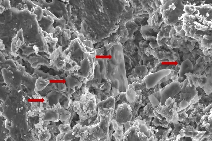
An image of fossilised erythrocyte-like structures. Credit: Anglia Ruskin University
New techniques used to analyse soft tissue in dinosaur fossils may hold the key to new cancer discoveries, according to a new study published in the journal Biology.
Researchers from Anglia Ruskin University (ARU) and Imperial College London analysed dinosaur fossils using advanced paleoproteomic techniques, a method that holds promise for uncovering molecular data from ancient specimens.
The researchers discovered red blood cell-like structures in a fossil while studying a Telmatosaurus transsylvanicus, a duck-billed, plant eating “marsh lizard” that lived between 66-70 million years ago in the Hateg Basin in present-day Romania.
The new study used Scanning Electron Microscopy (SEM) techniques to identify low-density structures resembling erythrocytes, or red blood cells, in the fossilised bone.
The findings raise the possibility that soft tissue and cellular components are more commonly preserved in ancient remains than previously thought.
By identifying preserved proteins and biomarkers, scientists believe they can gain insights into the diseases that affected prehistoric creatures, including cancer, potentially influencing future treatments for humans.
The authors of the new study highlight the necessity of prioritising the collection and preservation of fossilised soft tissue, rather than just dinosaur skeletons, as future advancements in molecular techniques will enable deeper insights into disease evolution.
A separate study had previously identified evidence of cancer in Telmatosaurus transsylvanicus, indicating its deep evolutionary roots.
More precise treatment options with robotic technology
Friday, 20 June 2025:Prostate cancer is a major risk to men’s health, with South African men facing a one in eight chance of developing this most common of male cancers.
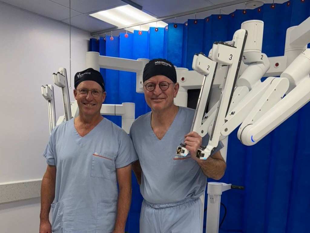
Urologists Dr Hannes Brummer and Dr Johan Coetzee, who practise at Netcare Greenacres Hospital, are encouraging men to prioritise prostate cancer screening this Men’s Health Month.
“Usually, men do not feel any symptoms until prostate cancer has progressed significantly, which is why they need to be proactive about booking their routine prostate cancer screenings,” explains Dr Coetzee.
“With the advanced prostate specific antigen [PSA] screening blood test available from GPs these days, there is so much more opportunity for prostate cancer to be detected earlier when it is still at a highly treatable stage.”
“For men who are diagnosed with prostate cancer following a needle biopsy, the treatment options available have improved to such an extent that there is more hope than ever before. Even where surgery is needed, prostate cancer does not necessarily pose a significant disruption to your life,” Dr Brummer adds.
“A prostate cancer diagnosis can be daunting. We have walked this path with so many men, and robotic assisted surgery offers some important advantages for the removal of cancerous tissue in the prostate gland, in particular the precision of this minimally invasive option.”
Over 1 000 robotic assisted procedures have been performed at Netcare Greenacres Hospital since the introduction of this technology in August 2017.
At Netcare Greenacres Hospital, Dr Brummer and Dr Coetzee use the da Vinci X robotic assisted surgical system to operate through tiny punctures in the skin using slender instruments more dexterous than the human hand.
Dr Brummer and Dr Coetzee emphasise that the surgeon remains in control of the robotic system at all times. With magnified 3D imaging capabilities, including a large fixed-focus area at the highest resolution, the nerves, blood vessels and tumour are visible with great clarity for the intricate procedure.
“This robotic system is especially useful for operating on the prostate, as we can more clearly distinguish the nerves controlling erectile function and urinary continence. In most cases, there is less need for blood transfusion and reduced risk of complications,” Dr Brummer explains.
“Another of the advantages of this robotic technology for prostate tumours is that there is much less tissue damage in this sensitive area. Compared with traditional surgery, this means men usually experience much less downtime with less discomfort after the procedure. This translates into shorter hospital stays and faster recovery with robotic assisted prostatectomies overall.”
General manager of Netcare Greenacres Hospital, Reon van Rensburg, joined the urologists in reinforcing the importance of prostate cancer awareness. “Let’s talk to our brothers, fathers, sons and grandsons about health issues, and get to know your family risk for both prostate cancer and breast cancer.”
Van Rensburg thanked Dr Brummer and Dr Coetzee for their continued dedication to making the world-class minimally-invasive robotic assisted surgical option available for patients local to Gqeberha and from as far afield as Knysna, George and East London, inland regions of the Eastern Cape, and parts of the southern Free State, the Northern Cape and the north-eastern region of the Western Cape.
“This Men’s Health Month, let’s pledge to be decisive about booking those routine health checks. Making the time now and every year could help to save your life in future,” Dr Brummer and Dr Coetzee concluded.