Category: Cancer

Radiotherapy Overcomes Resistance to Immunotherapy in Some Cancers
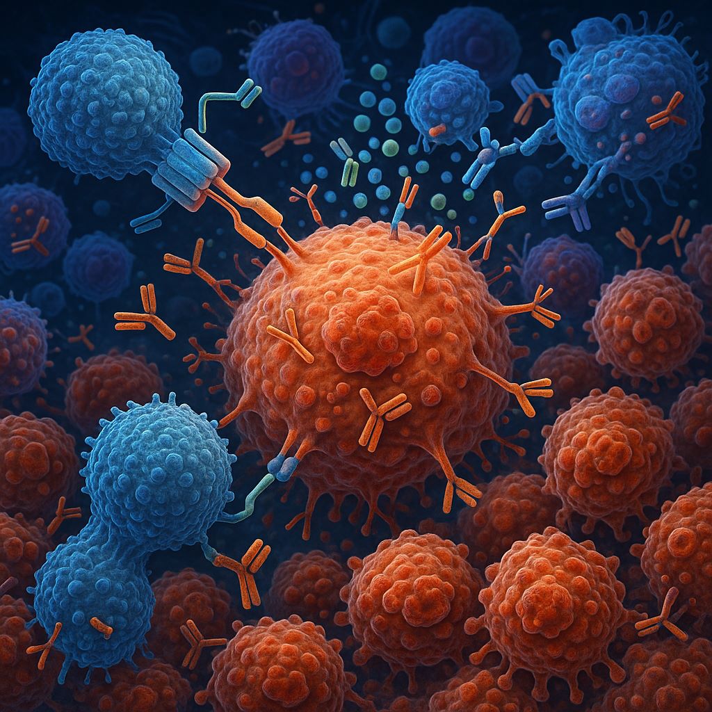
By sparking the immune system into action, radiation therapy makes certain tumours that resist immunotherapy susceptible to the treatment, leading to positive outcomes for patients, according to new research published July 22 in Nature Cancer. Investigators dove deep into the molecular biology of non-small cell lung cancer to pinpoint what happens on a cellular and molecular level over time when the cancer is treated with either radiation therapy followed by immunotherapy or immunotherapy alone.

They found that radiation plus immunotherapy induced a systemic anti-tumour immune response in lung cancers that do not typically respond to immunotherapy. The combination therapy also yielded improved clinical response in patients whose tumours harbour features of immunotherapy resistance. 

Clinically, the results suggest that radiation therapy can help overcome immunotherapy resistance in certain patients. Researchers at the Johns Hopkins Kimmel Cancer Center Bloomberg~Kimmel Institute for Cancer Immunotherapy and the Netherlands Cancer Institute conducted the study, which was supported by the National Institutes of Health. 

“For a fraction of lung cancers where we aren’t expecting therapy responses, radiation may be particularly effective to help circumvent primary resistance to immunotherapy; this could potentially be applicable to acquired resistance, too,” says senior study author Valsamo “Elsa” Anagnostou, MD, PhD, co-director of the Upper Aerodigestive Malignancies Program, director of the Thoracic Oncology Biorepository, leader of Precision Oncology Analytics, co-leader of the Johns Hopkins Molecular Tumor Board and co-director of the Lung Cancer Precision Medicine Center of Excellence at Johns Hopkins. 

Researchers have long sought to better understand why some tumours grow resistant to immunotherapy and how to intercept that resistance. 

Radiation therapy has been proposed as one possible way to induce a systemic immune response because of a unique phenomenon called the abscopal effect. Radiation at the site of a primary tumour typically causes tumour cells to die and release their contents into the local microenvironment. Sometimes, the immune system discovers those contents, learns the tumour’s molecular footprint, then activates immune cells around the body to attack cancer cells at tumour sites that were not the targets of the radiation, including some far away from the primary cancer in the body. 

Because of this effect, radiation therapy could potentially improve how well an immunotherapy works against a cancer, even far from the original radiation site. Yet little has been known about the molecular biology behind the abscopal effect, or how to predict when and in which patients it will occur. 

To study this phenomenon, Anagnostou and colleagues obtained samples from patients with lung cancer at different times throughout their treatment journey and from various locations in the body, not just at the primary tumour site. They collaborated with Willemijn Theelen and Paul Baas at the Netherlands Cancer Institute, who were running a phase II clinical trial on the effect of radiation therapy followed by immunotherapy, specifically the PD-1 inhibitor pembrolizumab. 

With help from Theelen and Baas, Anagnostou’s team analysed 293 blood and tumour samples from 72 patients, obtained at baseline and after three to six weeks of treatment. Patients in the control group received immunotherapy alone, while the experimental group received radiation followed by immunotherapy. 

The team then performed multiomic analyses on the samples (combining different “omics” tools, including genomics, transcriptomics and various cell assays) to deeply characterise what was happening to the immune system systemically and in the local microenvironment at tumour sites that were not directly exposed to radiation. 

In particular, the team focused on immunologically “cold” tumours — those that typically do not respond to immunotherapy. These tumours can be recognised by particular biomarkers: a low mutation burden, no expression of a protein called PD-L1, or the presence of mutations in a signalling pathway called Wnt. 

Following radiation and immunotherapy, the team found that “cold” tumors far from the site of radiation experienced a prominent reshaping of the tumor microenvironment. Anagnostou describes this shift as the tumors “warming up,” transitioning from little or no immune activity to inflamed sites with strong immune activity, including the expansion of new and pre-existing T cells. 

“Our findings highlight how radiation can bolster the systemic anti-tumor immune response in lung cancers unlikely to respond to immunotherapy alone,” says lead study author Justin Huang, who led the multiomic analyses. “Our work underscores the value of international, interdisciplinary collaboration in translating cancer biology insights to clinical relevance.” Huang was awarded the 2025 Paul Ehrlich Research Award in recognition of groundbreaking discoveries by young investigators and their faculty mentors at the Johns Hopkins University School of Medicine.     

With Kellie Smith, PhD, an associate professor of oncology at the Johns Hopkins Kimmel Cancer Center and a Bloomberg~Kimmel Institute for Cancer Immunotherapy researcher, Anagnostou’s team focused on patients who attained long-term survival with combination radiotherapy and immunotherapy, and performed a functional test to find out what the patients’ own T cells were doing in the body. In cell cultures, they confirmed that the T cells expanding in patients who received radiation and immunotherapy were indeed recognizing specific mutation-associated neoantigens from the patients’ tumours. 

Finally, by tracking patient outcomes from the clinical trial, the team observed that patients with immunologically cold tumours that “warmed up” due to radiation therapy had better outcomes than those who did not receive radiation therapy. 

“It was super exciting, and truly made everything come full circle,” says Anagnostou. “We not only captured the abscopal effect, but we linked the immune response with clinical outcomes in tumours where one would not expect to see immunotherapy responses.” 

Using specimens from the same cohorts of patients, the team has recently been working to capture the body’s response to immunotherapy by detecting circulating tumour DNA (ctDNA) in the blood. That work was presented April 28 at the annual meeting of the American Association for Cancer Research in Chicago. 

Source: Johns Hopkins Medicine
A Surprise One-two Punch for a Universal Cancer Vaccine

Photo by Raghavendra V Konkathi on Unsplash

An experimental mRNA vaccine boosted the tumour-fighting effects of immunotherapy in a mouse-model study, bringing researchers one step closer to their goal of developing a universal vaccine to “wake up” the immune system against cancer.

Published in Nature Biomedical Engineering, the University of Florida study showed that like a one-two punch, pairing the test vaccine with common anticancer drugs called immune checkpoint inhibitors triggered a strong antitumor response.

A surprising element, researchers said, was that they achieved the promising results not by attacking a specific target protein expressed in the tumour, but by simply revving up the immune system — spurring it to respond as if fighting a virus. They did this by stimulating the expression of a protein called PD-L1 inside of tumours, making them more receptive to treatment. The research was supported by multiple federal agencies and foundations, including the National Institutes of Health.

Senior author Elias Sayour, MD, PhD, a UF Health paediatric oncologist, said the results reveal a potential new treatment path with broad implications for battling many types of treatment-resistant tumours.

“This paper describes a very unexpected and exciting observation: that even a vaccine not specific to any particular tumour or virus — so long as it is an mRNA vaccine — could lead to tumour-specific effects,” said Sayour, principal investigator at the RNA Engineering Laboratory within UF’s Preston A. Wells Jr. Center for Brain Tumor Therapy.

“This finding is a proof of concept that these vaccines potentially could be commercialised as universal cancer vaccines to sensitise the immune system against a patient’s individual tumour,” said Sayour, a McKnight Brain Institute investigator and co-leader of a program in immuno-oncology and microbiome research.

Until now, there have been two main ideas in cancer-vaccine development: To find a specific target expressed in many people with cancer, or to tailor a vaccine that is specific to targets expressed within a patient’s own cancer.

“This study suggests a third emerging paradigm,” said Duane Mitchell, M.D., Ph.D., a co-author of the paper. “What we found is by using a vaccine designed not to target cancer specifically but rather to stimulate a strong immunologic response, we could elicit a very strong anticancer reaction. And so this has significant potential to be broadly used across cancer patients — even possibly leading us to an off-the-shelf cancer vaccine.”

For more than eight years, Sayour has pioneered high-tech anticancer vaccines by combining lipid nanoparticles and mRNA. Short for messenger RNA, mRNA is found inside every cell — including tumor cells — and serves as a blueprint for protein production.

This new study builds upon a breakthrough last year by Sayour’s lab: In a first-ever human clinical trial, an mRNA vaccine quickly reprogrammed the immune system to attack glioblastoma, an aggressive brain tumor with a dismal prognosis. Among the most impressive findings in the four-patient trial was how quickly the new method — which used a “specific” or personalized vaccine made using a patient’s own tumor cells — spurred a vigorous immune-system response to reject the tumor.

In the latest study, Sayour’s research team adapted their technology to test a “generalized” mRNA vaccine — meaning it was not aimed at a specific virus or mutated cells of cancer but engineered simply to prompt a strong immune system response. The mRNA formulation was made similarly to the COVID-19 vaccines, rooted in similar technology, but wasn’t aimed directly at the well-known spike protein of COVID.

In mouse models of melanoma, the team saw promising results in normally treatment-resistant tumors when combining the mRNA formulation with a common immunotherapy drug called a PD-1 inhibitor, a type of monoclonal antibody that attempts to “educate” the immune system that a tumor is foreign, said Sayour, a professor in UF’s Lillian S. Wells Department of Neurosurgery and the Department of Pediatrics in the UF College of Medicine.

Taking the research a step further, in mouse models of skin, bone and brain cancers, the investigators found beneficial effects when testing a different mRNA formulation as a solo treatment. In some models, the tumors were eliminated entirely.

Sayour and colleagues observed that using an mRNA vaccine to activate immune responses seemingly unrelated to cancer could prompt T cells that weren’t working before to actually multiply and kill the cancer if the response spurred by the vaccine is strong enough.

Taken together, the study’s implications are striking, said Mitchell. “It could potentially be a universal way of waking up a patient’s own immune response to cancer,” Mitchell said. “And that would be profound if generalisable to human studies.”

The results, he said, show potential for a universal cancer vaccine that could activate the immune system and prime it to work in tandem with checkpoint inhibitor drugs to seize upon cancer—or in some cases, even work on its own to kill cancer.

The research team is now working to improve current formulations and move to human clinical trials as rapidly as possible.

Source: University of Florida Health

Breastfeeding’s Protective Effect Against Breast Cancer Explained by Metabolism

Study shows a mother’s mitochondria determine if lactation is protective or not against breast cancer—and points to a possible intervention to increase the benefit to more women

Photo by Wendy Wei

Breastfeeding is often linked with better health for both mothers and babies, but it does not protect all women against breast cancer. The reason remains unknown. Since breast cancer in young women is on the rise, understanding why breastfeeding is protective in some women but not others is critical. 

A new study, led by a team of researchers from The Tisch Cancer Institute at the Icahn School of Medicine at Mount Sinai and published in the journal Nature Communications, addressed this question. 

The study was performed in mice, which exhibit a similar phenomenon: lactation strongly protects some mice against breast cancer, but others are more susceptible. The researchers studied female mice that had the same basic DNA but different types of mitochondria. The team found that the way the body responds to breastfeeding could change depending on the mother’s mitochondria.  

In mice with specific types of mitochondria, the researchers found, lactation allowed a certain group of cells similar to those found in postpartum breast cancer in humans to expand and grow, explains Edmund Jenkins, PhD, Assistant Professor of Medicine (Hematology and Medical Oncology) at the Icahn School of Medicine at Mount Sinai. Dr Jenkins served as the bioinformatics expert on the study. 

“We’ve always thought that breastfeeding is good for all women when it comes to lowering breast cancer risk,” said senior author Doris Germain, PhD, Professor of Medicine (Hematology and Medical Oncology) at the Icahn School of Medicine at Mount Sinai. “But our study shows that it really depends on a woman’s metabolism and how her body responds to lactation at the cellular level.” 

The researchers also discovered that they could change the way the body reacts during breastfeeding by using a natural dietary supplement. In the mice that were at higher risk of developing postpartum breast cancer, this treatment switched their response from harmful to protective. This finding opens the door to a possible way to help more women benefit from breastfeeding by supporting their bodies in the right way.  

“Our research raises the possibility that one day, doctors might be able to identify women whose breastfeeding response puts them at risk and then offer them a simple, natural dietary intervention to change that,” said first author Mrittika Chattopadhyay, PhD, Assistant Professor of Medicine (Hematology and Medical Oncology) at the Icahn School of Medicine at Mount Sinai. 

The team is now studying human breast milk with the goal of identifying milk metabolites that can show whether a woman’s body is reacting to breastfeeding in a way that is helpful or harmful. They also plan to develop a study in humans. One question then will be whether, because this dietary supplement may impact the development of the child, it should be tested only in mothers after they have stopped giving milk to their newborns.  

Source: Mount Sinai

FDA-Approved Drug Halts EBV-Driven Lymphoma by Disrupting a Key Cancer Pathway

Swollen lymph nodes. Credit: Scientific Animations CC0

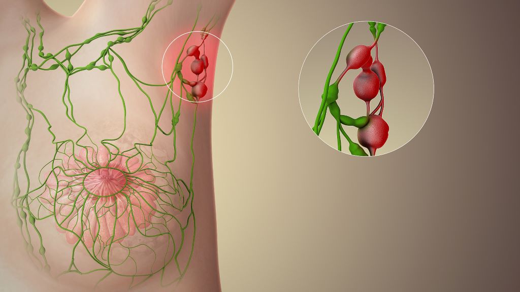
Scientists at The Wistar Institute have discovered that a class of FDA-approved cancer drugs known as PARP1 inhibitors can effectively combat Epstein-Barr virus (EBV)-driven lymphomas. The findings, published in the Journal of Medical Virology, demonstrate that these drugs, which work by blocking the activity of the PARP1 enzyme, can halt tumour growth by interfering with the EBV’s ability to activate key cancer-promoting genes.

“We’ve uncovered a completely different mechanism for how PARP inhibitors work in EBV-positive cancers,” said Italo Tempera, PhD, associate professor at Wistar’s Ellen and Ronald Caplan Cancer Center and senior author of the study. “Instead of preventing DNA damage from repairing itself in the tumours, like these drugs do in other cancers, they essentially cut off the virus’s ability to hijack cellular machinery to drive cancer growth. This opens up exciting possibilities for repurposing existing FDA-approved drugs to treat EBV-associated cancers.”

EBV infects over 90% of the global population. While most people with the virus remain symptom-free, immunocompromised individuals such as people with HIV and transplant recipients have an increased risk of EBV causing several types of cancer, including various lymphomas and carcinomas. Despite the virus’s clear role in driving these malignancies, no specific therapies currently target EBV-driven cancer.

In search of such a therapy, Tempera and his research team focused on PARP1, a cellular protein that is known primarily for its role in DNA repair. In cancer treatment, PARP inhibitors typically work by preventing cancer cells from repairing their DNA, causing them to die. However, Tempera’s team had previously discovered that PARP1 plays a very different role in EBV infection: It helps control which genes are accessible and active, essentially acting as a master regulator of gene expression.

“Think of PARP1 as a key that opens up DNA to make certain genes readable,” explained Tempera. “EBV uses this key to unlock cancer-promoting genes. When we block PARP1, we’re essentially taking away the key so the virus can’t get in and use our DNA for its own purposes.”

Using a mouse model of EBV-driven lymphoma, the researchers treated the animals with BMN 673 (talazoparib/talzenna), a PARP inhibitor that has already been approved for breast cancer treatment. Compared to controls, the treated mice showed an 80% reduction in tumour growth, and the cancer’s ability to spread to other organs was significantly reduced. Further, when the team analysed the tumours, they found no increase in DNA damage in the treated animals – the hallmark of how PARP inhibitors typically work. Instead, they discovered that PARP1 inhibition disrupted a critical partnership between the viral protein EBNA2 and the cellular oncogene MYC.

“EBNA2 is like the conductor of an orchestra, directing cellular genes to play a cancer symphony,” said Tempera. “It specifically turns on MYC, which is one of the most important cancer-promoting genes. When we inhibit PARP1, EBNA2 can’t effectively activate MYC anymore, and the whole cancer program falls apart.”

The findings have significant therapeutic implications. Because PARP inhibitors are already FDA-approved and their safety profiles are well established, the path to clinical application could be accelerated compared to developing entirely new drugs.

The research also suggests this approach might work beyond EBV-associated lymphomas. The team is now investigating whether PARP inhibitors could be effective against other EBV-driven cancers, including nasopharyngeal and gastric carcinomas. Additionally, given EBV’s suspected role in autoimmune diseases, the researchers are exploring whether PARP1’s regulation of viral gene expression might contribute to these conditions.

“This work really showcases the power of understanding fundamental viral biology,” said Tempera. “We’re taking insights from basic virology research and translating them into potential therapies. With further development, this approach could provide new hope for patients with EBV-associated cancers who currently have limited treatment options.”

Source: Wistar Institute

Why Humans Are More Susceptible to Cancers than Other Primates

Photo by Andre Mouton on Unsplash

New research from UC Davis Comprehensive Cancer Center has uncovered an evolutionary change that may explain why certain immune cells in humans are less effective at fighting solid tumours compared to non-human primates. The findings, published in Nature Communications, could lead to more powerful cancer treatments.

The study revealed a tiny genetic difference in an immune protein called Fas Ligand (FasL) between humans and non-human primates. This genetic mutation makes the FasL protein vulnerable to being disabled by plasmin, a tumour-associated enzyme. This vulnerability seems unique to humans and is not found in non-human primates, such as chimpanzees.

“The evolutionary mutation in FasL may have contributed to the larger brain size in humans,” said Jogender Tushir-Singh, senior author for the study and an associate professor in the Department of Medical Microbiology and Immunology. “But in the context of cancer, it was an unfavourable tradeoff because the mutation gives certain tumours a way to disarm parts of our immune system.”

Tumour environment neutralises key immune protein

FasL is an immune cell membrane protein that triggers apoptosis, which activated immune cells, including CAR-T cells, make use of to kill cancer cells.

The UC Davis team discovered that in human genes, a single evolutionary amino acid change — serine instead of proline at position 153 — makes FasL more susceptible to being cut and inactivated by plasmin.

Plasmin is a protease enzyme that is often elevated in aggressive solid tumours like triple negative breast cancer, colon cancer and ovarian cancer.

This means that even when human immune cells are activated and ready to attack the tumour cells, one of their key apoptosis tools, FasL, can be neutralised by the tumour environment, reducing the effectiveness of immunotherapies.

The findings may help explain why CAR-T and T-cell-based therapies can be effective in blood cancers but often fall short in solid tumours. Blood cancers often do not rely on plasmin to metastasise, whereas tumours like ovarian cancer rely heavily on plasmin to spread the cancer.

Plasmin inhibitors may enhance immunotherapy

Significantly, the study also showed that blocking plasmin or shielding FasL from cleavage can restore its cancer-killing power. That finding may open new doors for improving cancer immunotherapy.

By combining current treatments with plasmin inhibitors or specially designed antibodies that protect FasL, scientists may be able to boost immune responses in patients with solid tumours.

“Humans have a significantly higher rate of cancer than chimpanzees and other primates. There is a lot that we do not know and can still learn from primates and apply to improve human cancer immunotherapies,” said Tushir-Singh. “Regardless, this is a major step toward personalising and enhancing immunotherapy for the plasmin-positive cancers that have been difficult to treat.”

Source: UC Davis Cancer Center

SAHPRA‘s Position on Anti-cancer Medications in South Africa

Photo by Marcelo Leal on Unsplash

Pretoria, 15 July 2025 – The South African Health Products Regulatory Authority (SAHPRA) was notified of the Lancet Global Health 2025; 13: e1250, an investigational study and its findings on substandard anti-cancer medications in Sub-Saharan African countries, including Ethiopia, Kenya, Malawi, and Cameroon. This study did not include South Africa. The seven (7) medicines/dosage forms mentioned in the study are cisplatin, oxaliplatin, methotrexate, doxorubicin, cyclophosphamide, ifosfamide, and leucovorin. The specific brands mentioned/shown in the article are neither registered nor marketed in South Africa. 

SAHPRA, in terms of the Medicines and Related Substances Act 101 of 1965, as amended, and its General Regulations, requires medicines marketed in the country to meet prescribed requirements and adhere to set standards. Every batch of medicine produced must undergo testing to ensure that the integrity of the product is consistent with approved specifications before the release for sale, and imported medicines must additionally comply with the Guideline for Post-Importation Testing. 

SAHPRA commenced internal processes to verify whether any of the South African-registered cancer medicines with the mentioned Active Pharmaceutical Ingredients (API) might have been affected or implicated. The cancer products registered and marketed in South Africa were not implicated/affected by the investigational study and its findings on substandard anti-cancer medicines. SAHPRA conducts risk-based post-market surveillance (PMS), sampling, and testing on high-risk medical products. 

SAHPRA is satisfied that the marketed and registered cancer medicines meet the appropriate specifications; therefore, no substandard cancer medicines were detected. 

“SAHPRA is committed to the three pillars of quality, safety, and efficacy. I am satisfied that our rigorous regulatory processes have borne fruit and that all patients, especially cancer patients, can rest assured that their health and well-being are not compromised,” indicated SAHPRA CEO, Dr Boitumelo Semete-Makokotlela. 

Student Designs a Prostate Checking Device to Replace the Digital Exam

Pro check, designed by Loughborough University student Devon Tyso.

A Loughborough University student has developed a new medical device that could transform how prostate health is assessed and monitored.

Devon Tyso, a Product Design and Technology student, has designed ‘PRO check’, an innovative tool designed to replace the traditional digital rectal examination (DRE), which involves a doctor manually assessing the prostate with a finger.

According to Devon, the current approach is heavily reliant on a clinician’s subjective judgement and experience, and many see the method as ‘intrusive’.

“As one in seven men will get prostate cancer, it’s vital to detect abnormalities early and track changes over time,” said Devon, “The current examination method involves a lot of guesswork.

“PRO check provides objective, measurable data and allows prostate health to be visualised – enabling more accurate diagnosis, and improved long-term monitoring.

“Having a device conduct the exam may also feel less invasive, which may encourage more men to get checked, potentially catching issues earlier.”

How the device works

Designed for use by GPs during routine prostate assessments, PRO check allows doctors to evaluate the size and texture of the prostate — two key indicators of potential health issues — in a more objective and consistent way than the traditional digital rectal examination.

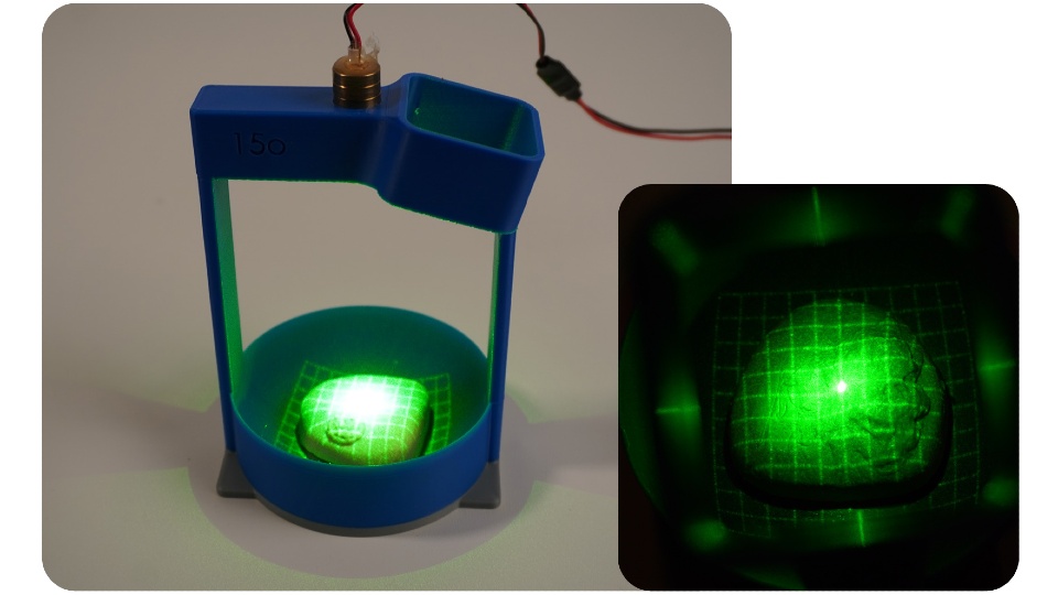
The device is a handheld probe, and it is covered with a condom before being inserted into the body. Once in position, the condom inflates to different pressures, pressing against the surface of the prostate, causing it to compress. A laser grid is projected onto the inner surface of the condom so the shape of the underlying prostate can be captured.

Stereoscopic cameras capture images of the laser grid, tracking where the gridlines intersect and how these intersections shift as pressure changes. This information is then fed into mathematical equations to create 3D images — or ‘topographical representations’ — that reveal the prostate’s shape and surface structure under different pressures.

Studying the prostate’s surface details could help clinicians identify areas requiring further investigation. Healthy prostate tissue is typically soft and compressible, so regions that appear stiff or resist pressure could indicate potential abnormalities and warrant further investigation.

The device can also produce data on prostate volume – one of the measurements used to calculate prostate-specific antigen (PSA) density, which helps assess prostate cancer risk. Devon says currently volume estimates are often based on a clinician’s best judgement.

In addition, data from PRO check can be used to generate a compressibility-versus-pressure graph – a novel data type not currently available in clinical practice. This graph shows how the prostate compresses at different pressure levels, which Devon hopes could offer new insights into prostate health and complement existing diagnostic tools.

PRO check is designed to integrate with artificial intelligence, enabling automatic extraction of video data, real-time calculations, and the generation of 3D images for live display on a laptop or tablet during the examination.

The idea is that all examination data from PRO check would be stored on the patient’s records, helping to build a personalised prostate health profile that can be tracked and monitored over time.

Inspiration

Devon’s inspiration for PRO check came from a mix of personal experience – after his grandfather’s prostate cancer diagnosis – and unexpected technical research.

“It really hit home how common prostate issues are after my family member was found to have an enlarged prostate,” said the 22-year-old from Cardiff, “I realised nearly everyone I spoke to about it knew someone affected by it.

“When I started looking into prostate examinations, I kept thinking ‘how can a doctor remember what your prostate felt like four months ago?’ and how horrible it must be just be told whether you’re fine or not without seeing any data or anything visual.”

While researching non-invasive ways to assess tissue structure inside the body, Devon came across a technique used by NASA to map the surface of asteroids — projecting laser grids onto them, capturing images with satellite-mounted cameras, and analysing the gridline intersections to reveal the contours of the surface.

“I saw that NASA were mapping surface heights on a massive scale, and I thought – if they can do that in space, why can’t we use similar principles to examine something here on Earth?” said Devon, “I’ve basically used the exact same technique and scaled it down for PRO check.”

Prototypes

Devon designed PRO check as part of his final year project – which was exhibited at the School of Design and Creative Arts’ 2025 Degree Show – and has prototyped several of its key components.

He has built and tested two working prototypes. The first demonstrates how a laser grid and camera can be setup to map the surface of the prostate.

Devon designed a custom rig that enabled him to capture images of a laser grid projected onto different silicone prostate models — representing a healthy gland, a small tumour, a large tumour, and an enlarged prostate — from an optimal angle using a smartphone camera.

First prototype of pro checkPRO check prototype one demonstrated how laser gridlines and a camera can be used to image the surface of the prostate.
The second prototype features electronics that inflate a small balloon at controlled pressures, regulated by a pressure-sensing chip. Devon consulted three healthcare professionals to measure the pressure typically applied during prostate exams and replicated those levels in his design.

Devon tested the prototype using the silicone prostate models but encased them in a sponge disc to simulate surrounding tissue.

Devon manually extracted data on the gridline intersections from the camera footage and applied mathematical equations to generate 3D images of the prostate surfaces and surrounding tissue under different pressures.

Next steps

Devon hopes to collaborate with medical professionals and product developers to turn PRO check into a fully realised medical device.

When speaking about his ultimate goal, Devon said: “I’d love to see this used in GP surgeries across the UK one day.

“With early detection being so critical, anything that helps men get checked sooner and more comfortably – and provides reliable data and visualisations – has huge potential. I really believe this could make a difference.”

Further information on PRO check can be found on the Degree Show website.

Source: Loughborough University

Study Finds 1 in 6 Cancer Medications in Sub-Saharan Africa Are Defective

Source: Unsplash CC0

Serious quality defects were found in a significant number of cancer medications from sub-Saharan Africa, according to new research from the University of Notre Dame.

For the study published in The Lancet Global Health, researchers collected different cancer medications from Cameroon, Ethiopia, Kenya and Malawi and evaluated whether each drug met regulatory standards. Researchers considered a variety of factors, including appearance, packaging, labelling and, most importantly, the assay value.

The assay value is the quantity of active pharmaceutical ingredient (API) found in each drug. To meet safety standards, most products should be within a range of 90 to 110% of the right amount of API. Researchers measured the API content of each product and compared that number to what was designated on the medication packaging.

“It is important that cancer medications contain the right amount of the active ingredients so the patient gets the correct dose,” said Marya Lieberman, professor of chemistry and biochemistry at Notre Dame and lead author of the study. “If the patient’s dose is too small, the cancer can survive and spread to other locations. If the patient’s dose is too high, they can be harmed by toxic side effects from the medicine.”

One in six cancer medications tested was found to contain the incorrect quantity of API, with tested medications having APIs ranging from 28 to 120%. The study evaluated 251 samples of cancer medications collected from major hospitals and private markets in all four countries.

The study, funded by the National Cancer Institute of the National Institutes of Health, is among the first to evaluate cancer drug quality in sub-Saharan Africa. Currently, sub-Saharan Africa has no pharmaceutical regulatory laboratories carrying out chemical analyses for cancer drugs according to the standards required for regulatory purposes.

Yet, the need for cancer drugs is growing.

“We found bad-quality cancer medications in all of the countries, in all of the hospital pharmacies and in the private markets,” said Lieberman, an affiliate of Notre Dame’s Eck Institute for Global Health and Harper Cancer Research Institute. “We learned that visual inspection, which is the main method for detecting bad-quality cancer drugs in sub-Saharan Africa today, only found one in 10 of the bad products.”

In their study, the researchers explained how a combination of high demand for cancer medications, lack of regulatory capacity, and poor manufacturing, distribution and storage practices likely created a problematic environment throughout sub-Saharan Africa. They also argue that given these factors and the global supply chain for pharmaceuticals, substandard cancer medications are likely present in other low and middle-income countries as well.

Lieberman and her team identified several strategies that could help the global community address poor-quality cancer medications:

  • Provide inexpensive technologies at the point of care to screen for bad-quality cancer medicines and create policies for how to respond to products that fail screening tests.
  • Help regulatory agencies in low and middle-income countries get safety equipment and training so they can analyze the quality of cancer medicines in their markets, conduct root-cause investigations when products fail testing, take quick regulatory actions enabled by lab data and share data about bad-quality products.
  • Perform cost-benefit analyses of interventions that tackle common problems (such as medications being out of stock, unsafe shipping, storage or dispensing practices, and lack of availability or affordability of medications) to help policymakers and funders get the most impact on patient outcomes from their available resources.
  • Work with care providers to develop site-specific response policies and messaging for patients and engage regulators, donors and other resources.

Lieberman and her lab are developing a user-friendly technology called the chemoPAD for screening cancer medications. This low-cost paper device could potentially help hospitals, pharmacies and health care professionals in low and middle-income countries monitor drug quality without restricting a patient’s access to the medication.

“This is all part of a bigger project aimed at developing the ChemoPAD as a point-of-care testing device that we can use, something that’s more accurate in detecting poor-quality products than just visual inspection,” Lieberman said.

“There are lots of medicines where the regulators don’t have enough resources to verify the quality, and some manufacturers take advantage of that to cut corners. There are also problems with distribution systems, so even if a product is good quality when it leaves the manufacturer, it may be degraded during shipping or storage. These products flow into low and middle-income countries, and they get used on patients. I want to change that.”

Source: University of Notre Dame

Attempts Underway to Fix Gap in SA’s Plan to Fight Cancer

A cancer patient receiving care at a public health facility in Gauteng. (Photo: Rosetta Msimango/Spotlight)

By Chris Bateman

Experts say cancer patients in the public sector in South Africa are dying for avoidable reasons like dysfunctional referral systems and a lack of medical imaging and treatment. We look at efforts to get the country’s battle with cancer back on track.

Many people with cancer in Gauteng have not been able to access the treatment and care they require in recent years. Though activists and the provincial government are at odds about what should, or should have been, done about it, nobody is denying that there is a problem.

At the same time, there have also been issues at a national level, with South Africa’s key cancer strategy having lapsed. The National Cancer Strategic Framework for South Africa 2017 – 2022 was previously extended to also cover 2023. Medical Brief recently reported that a new strategy is on the verge of being signed by the Director-General of Health.

The committee meant to advise the minister on cancer has also lapsed. Dr Busisiwe Ndlovu, the top government official in charge of non-communicable diseases (NCDs), said that the term of the Ministerial Advisory Committee on Cancer expired in early 2024, and new members were pending the approval of Health Minister Dr Aaron Motsoaledi. She was speaking at the KwaZulu-Natal leg of a cancer research and innovation strategy workshop in May. These consultative meetings are taking place across the country’s provinces. It aims to shape a national research and innovation strategy based on the World Health Organization’s cancer control pillars: prevention, early detection and diagnosis, treatment, and palliative care and survivorship.

The scale of the problem

While researchers anticipate that rates of infectious diseases like HIV and tuberculosis in South Africa will decline in the coming decade or two, rates of NCDs, including diabetes and cancers, are expected to increase. According to the WHO, an estimated one in five people will develop some form of cancer in their lifetimes. Increases in developing countries are expected to be particularly steep.

According to a StatsSA report published in 2023, and based on National Cancer registry (NCR) numbers and StatsSA’s mortality data, cancer-related deaths in the country increased by 29% from 2008 to 2018. They reported that 85 000 people were diagnosed with cancer in 2019 and that 44 000 died of cancers in 2018. Experts previously told Spotlight that the estimate of cancer cases may be an undercount of as much as 40%.

The most common cancers in men were prostate, colorectal, and lung – around one in four cancer diagnoses in men were for prostate cancer. Bronchus and lung cancer accounted for just under 19% of cancer-related deaths in men, while prostate cancer accounted for around 17%.

Among women, the most diagnosed cancers were breast cancer at around 23% of diagnoses and cervical cancer at around 16% of diagnoses. Cervical cancer accounted for just under 18% of all cancer deaths in women and breast cancer for 17%.

The NCR recorded 87 853 new laboratory-confirmed cancer cases in 2023, although this figure likely underestimated the true burden as it excluded clinically or radiologically diagnosed cancers, Dr Judith Mwansa-Kambafwile, senior epidemiologist with the NCR told attendees at the Durban workshop.

In a paper published in the South African Journal of Oncology in 2022, researchers calculate that cancer incidence (new cases per year) in South Africa could double from around 62 000 in 2019 to 121 000 in 2030. This is due to two factors: firstly, South Africa’s population is aging and cancers generally become more common as people age. And secondly, the risk of cancers is generally increasing for people of all ages. The researchers focused on only the five most common types of cancer, but an NCR report shows a very wide variety of cancers are being diagnosed in the country.

Since not all cancers are diagnosed, the real numbers are likely substantially higher than reported. There is also no single repository of all cancer diagnoses in the country – for the above quoted article researchers used both data from Discovery Health Medical Scheme and from the NCR.

The data gap

Cancer statistics in South Africa has been largely based on pathology results, which is to say blood or biopsies that were tested in the lab. Other types of diagnoses, such as those based on symptoms and scans have not always been counted systematically. One recent initiative aimed at addressing this data gap is a patient-led registry that feeds information into the NCR.

Mwansa-Kambafwile, explained that the NGO, Living with Cancer, was driving the patient-led registry, aimed at cross referencing and supplementing patient records with her NCR’s own patient database. Leaflets in oncologists’ reception rooms encouraged patients to upload their pathology/histology test results onto the Living with Cancer website via a standard online National Department of Health form. A national shopping mall campaign in May was aimed at boosting awareness.

“Living with Cancer had a Memorandum of Understanding with us and in addition, links cancer survivors with the same type of cancer to one another in support groups online where they can share experiences and knowledge,” she added.

Dr Mazvita Muchengeti, who heads up work on the NCR at the National Health Laboratory Services which is part of the National Institute for Communicable Diseases (NICD), previously told Spotlight that cancer was made a reportable disease under the National Health Act in 2011. While compulsory reporting has improved data on cancer cases, she added: “There is an increase in the number of reported cancers; this does not necessarily translate to an increase in cancer, we are just counting cancer cases better because reporting is now compulsory.”

Another new strategy

In light of the country’s cancer burden, a group of organisations is leading the development of a new National Cancer Research and Innovation Strategy. This collective includes the Nuclear Medicine Research Infrastructure at the University of Pretoria, the South African Medical Research Council, and the Department of Science, Technology and Innovation, in partnership with the National Department of Health.

They are hosting provincial workshops to help understand the current state of cancer research in South Africa, identify key challenges, set national priorities, and develop a strong, future-focused strategy. These workshops are part of a broader plan to make sure the strategy is inclusive, based on evidence, and meets the country’s needs.

This research and innovation strategy differs from the health department’s National Cancer Strategic Framework, which guides provinces as to what the cancer priorities are.

‘Integrated cancer care approach’

At the Durban workshop, Ndlovu, emphasised the need for an integrated cancer care approach across all levels of the healthcare system. She noted the importance of streamlined referral pathways and urgent attention to waiting times, care packages, registry improvements, and financing. The expired national cancer strategy required urgent evaluation and revision, Ndlovu added.

A clear pattern emerging from these workshops is one whereby cancers are often diagnosed too late, and patients frequently struggle to access timely, appropriate care.

Also at the Durban workshop, Professor Jeannette Parkes, Head of Radiation Oncology at Groote Schuur Hospital and the University of Cape Town, outlined the many systemic barriers to early detection. These included socio-cultural factors, urban-rural divides, and broken referral systems.

“We have a massive issue with accessing imaging services, biopsy support, pathology services, and their costs,” she said.

Parkes, who is also President of the College of Radiation Oncology of South Africa and clinical director of the Access to Care Cape Town programme, said early cancer detection was better in the private sector because patients could access and afford the necessary systems and diagnostic technology. The remaining 85% of the population depended on the public sector, in particular overburdened primary healthcare clinics but also on all levels of care.

“There’s a bias towards urban versus rural areas and too often a failure to refer. The referral pathway is problematic and differs from province to province and in various settings. We have a massive issue with regards to accessing imaging services, while biopsy support and pathology services and their costs are also a big issue,” she told the workshop.

Late diagnosis

At the Johannesburg meeting, late diagnosis was singled out as a particular problem when it comes to cervical cancer. Dr Mary Kawonga, public health specialist with the Gauteng Department of Health and Wits School of Public Health, said that 16% of women screened at Charlotte Maxeke Academic Hospital’s drainage district had pre-cancerous lesions, underlining the lack of preventative care. “Patients often only begin treatment on their sixth visit,” she said, citing the failure of diagnostic tools, referral inefficiencies, and poor implementation of available technologies.

Dr Mariza Vorster, Head of Nuclear Medicine at the University of KwaZulu-Natal and Inkosi Albert Luthuli Academic Hospital, said that insufficient specialists and excessive patient loads result in unacceptable turnaround times for diagnosis.

Clinicians often get blamed for delays, but as Dr Sheynaz Bassa, Head of Radiation Oncology at Steve Biko Academic Hospital, pointed out, many patients wait weeks or months to afford transport to care facilities. “By the time they get to us, they’re already in crisis mode,” she said. “Peripheral clinics and hospitals must improve referral systems before we can make real progress.”

Salomé Meyer, Director of Cancer Alliance, alleged that survivorship care is almost entirely absent in both the public and private sectors. “Supportive and palliative care often ends when treatment stops. Survivors are left without co-ordinated care,” she said.

Apart from improving screening and referral systems, other recommendations emerging from the workshops included better coordination between clinicians and the NCR, leveraging mobile technology like the health department’s Mom Connect app to reduce clinic visits and fast-track referrals. Greater community involvement in setting research priorities, using mobile clinics to conduct cancer screening in rural areas, and increasing awareness for breast self-examination. More research into the genetic factors relating to cancers in South Africa was also argued for.

Call for new cancer institute

Meyer has been leading a call for South Africa to establish a National Cancer Institute (NCI).

“An NCI would develop clear guidelines on treatment protocols, workforce allocation, and facility requirements,” she said. With South Africa transitioning toward a National Health Insurance system, Meyer said an NCI would help plan resource allocation based on cancer projections, enabling smarter investments in infrastructure, technology, and staffing.

The lapsed National Cancer Strategic Framework lacked province-specific detail, leaving provinces to adapt guidelines as they saw fit, often leading to fragmented service delivery, she added. Meyer said decentralisation was essential. “We can no longer restrict cancer treatment to tertiary hospitals. Many district and regional facilities could provide diagnostics and some treatments if properly resourced,” she said.

A reset of South Africa’s disease monitoring and research infrastructure has been on the cards for some time. The NICD was set to be replaced by the new National Public Health Institute of South Africa (NAPHISA) after the NAPHISA Act became law in 2020. Five years later, NAPHISA has not yet been established. On the face of it, NAPHISA would be a natural home for an entity like the proposed NCI were it to be created.

–  Additional reporting by Marcus Low

Republished from Spotlight under a Creative Commons licence.

Read the original article.

Celebrate Christmas in July with PinkDrive

Cold Nights, Warm Hearts, Festive Vibes – and Support for Early Detection

Think log fires, festive cheer, a three-course dinner and dancing to a live band – all wrapped up in the joy of giving. On Saturday, 5th July 2025, PinkDrive will host a Christmas in July dinner at the Indaba Hotel in Johannesburg, a night of holiday cheer and hope in action: raising funds for a life-changing cause. And you’re invited.

PinkDrive is a non-profit (NPC) committed to prolonging lives through the early detection of gender-related cancers. It delivers essential health services to thousands of South Africans every year by bringing mobile mammography units directly to communities that need them most, from rural villages to peri-urban areas across the nine provinces. These trucks provide clinical breast exams, mammograms, pap smears, and PSA testing, helping to detect cancer early in areas where healthcare access is often limited or unavailable.

Like many non-profits, PinkDrive depends on the support of corporate partners, sponsors, government, and the public to sustain its vital work, among them, Lee-Chem Laboratories. “This is a cause that is close to our hearts,” says Bhavna Sanker, Marketing Manager at Lee-Chem Laboratories. “Each year, we proudly support PinkDrive through our Mandy’s brand sponsorship, focusing on spreading awareness, sharing survivor stories, and making sure people understand their healthcare options.”

As part of their continued support, every guest at the Christmas in July function will receive a goodie bag from Lee-Chem, filled with products from their Mandy’s brand. “We are truly honoured to have Lee-Chem and the Mandy’s brand as valued sponsors of our Christmas in July Dinner fundraiser,” comments Nelius du Preez, Operations Manager at PinkDrive NPC. “Their continued support, alongside our other generous sponsors, makes events like this possible and helps us not only raise vital funds but also amplify the message of early detection and health education across South Africa.”

The fundraiser will spotlight the resilience of breast cancer survivors, with Deputy Minister of Electricity and Energy, Samantha Graham-Maré, sharing her personal journey with cancer. PinkDrive CEO and Founder, Noelene Kotschan, a passionate advocate for early detection prolonging  lives, will also address guests, and your charming host for the evening? A dashing Mister Global SA finalist will take the mic as MC, steering the evening from heartfelt reflections to lively fundraising with raffles and auctions featuring holiday packages, original artworks, and other exclusive prizes.

“We call the event ‘a night of giving back’ because it is occasions like these that allow us to keep our mobile health units on the road, reaching men and women who might otherwise not have access to screening services,” says du Preez. “Together with compassionate partners like Lee-Chem, we are driving change and prolonging lives, one screening at a time. And we are grateful to every corporate and individual who supports the event by purchasing tickets,” he adds.

All proceeds from the evening will be ringfenced to build and operate a new mobile mammography unit, expanding PinkDrive’s reach to screen more South Africans and detect cancer early, when treatment is most effective. “We encourage the public to join us in this mission by purchasing tickets and supporting the evening’s fundraising efforts,” concludes Sanker.

Tickets for the Christmas in July dinner are R600 per person and available now at pinkdrive.org. Don’t miss this chance to dress up, give back, and help bring hope where it’s needed most.