

Tag: cancer research

South African Scientists Make Breakthrough in Decoding Cancer’s Most Effective Survival Strategy
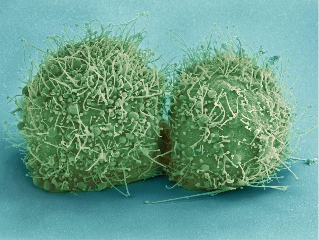
Scanning electron micrograph of just-divided HeLa cells. Zeiss Merlin HR-SEM. Credit: National Center for Microscopy and Imaging Research

Kevin Naidoo, University of Cape Town

In the intricate biology of the human body, organs such as the breast, the colon and the lungs are lined with a defensive barrier known as the epithelium. At the heart of this barrier sits a remarkable protein called Mucin-1 (MUC1). In a healthy body, MUC1 is like a sentinel.

It stands on the cell wall, draped in a complex “armour” of long chains of sugar molecules (carbohydrates), where it serves as a physical shield against bacteria, viruses and toxins. Crucially, it communicates with the immune system, telling our natural defences when the body is under threat.

But in the case of cancer, this guardian exchanges its sugar coat armour for shorter sugar chains and so turns into a traitor. It stops sending danger signals to the immune system and instead binds to the immune cells, creating an anti-inflammatory microenvironment that promotes tumours.

The team I lead at the Scientific Computing Research Unit at the University of Cape Town is home to computer modelling experts and experimental chemical biology research scientists. The molecular details of this MUC1 alteration, which contributes to the transformation of normal cells into tumour cells, were recently published in Nature Communications, and provide a new look at exactly how this process happens.

By developing a novel “test-tube” synthetic biology approach, we modelled and decoded the molecular assembly line reorganisation that allows cancer to “redecorate” MUC1, turning it from a protective shield into a cloak of invisibility. We used our own computational chemistry algorithms to map the exact sugar coating positions that create a tumour-promoting environment.

Understanding the location and nature of the MUC1 sugars that prevent the immune system from detecting tumours provides the foundation for our laboratory and others in the field to develop cancer vaccines, biomarkers and therapeutics.

This South African-led discovery represents a major leap forward in our ability to decode one of cancer’s most effective survival strategies.

The problem: a malignant makeover

In a normal cell, the sugar molecules attached to MUC1 are long and complex. The process of attaching sugars is called glycosylation. In cancer cells, however, this process goes haywire. The sugar molecules are often cut short or altered, creating “aberrant” structures like the Tn and sialyl-Tn (sTn) antigens. These are specific types of sugar-protein combinations that are tags for tumour cells.

These altered sugars do two dangerous things: they allow the tumour to evade detection by the immune system, and they actively trigger the process of turning a normal cell into a cancerous one.

Because MUC1 is found in so many different types of cancer, the US National Cancer Institute has ranked it as the most accesible target.

To stop the cascading effect of the MUC1 changes from normal to tumour cells, scientists first had to understand exactly how the “assembly line” breaks down.

The discovery: relocating the factory

Our research team set out to do something ambitious: recreate the transition from a healthy sugar coating to a cancerous one in a laboratory setting.

In normal cells, the enzymes that build these sugar chains (long molecules) live in a part of the cell called the Golgi apparatus, the cell’s “packaging and delivery centre”. We built an in vitro (test-tube) model to simulate what happens when these conditions change. We discovered that in tumour cells, the enzymes responsible for starting the sugar chains are relocated to another part of the cell, the endoplasmic reticulum, essentially the cell’s “factory floor”.

This relocation changes everything. Here, the enzymes are no longer inhibited by the usual cellular checks and balances. They take over the sugar sites on the MUC1 protein, creating the foundation for the cancerous Tn antigen.

To take the study even further, we used quantum chemistry. We simulated the behaviour of atoms and molecules at the most fundamental level to find out where these changes are most likely to happen. We identified a specific location on the MUC1 protein, known as the T13 site, which cancer enzymes prefer. This specific interaction is what drives the massive increase in the sTn antigen seen in malignant tumours.

Why this matters: from lab to patient

Understanding the “how” and the “where” of these sugar changes is the first step towards stopping them. The research didn’t stop at the test tube; the team is already looking at what this means for patients.

The next phase of the research, as detailed in a recent paper in Glycobiology, involves building a sophisticated “systems biology” computational model. A model can connect the changes in the MUC1 sugar coating to the behaviour of immune cells. For example, scientists found that when these cancerous sugars interact with macrophages (a type of white blood cell), they trigger the release of specific signals that tell the tumour to grow and spread.

We are refining these details for various types of cancer. We are comparing common forms of breast cancer with more aggressive, currently untreatable types to see if the “sugar code” differs between them.

By using this accurate, atomic-level data to build computer models of the entire biological system, we hope to identify new drugs that can block these signals. The goal is to move towards precision medicine: treatments that can strip away cancer’s sugar shield, allowing the patient’s own immune system to finally see and destroy the tumour.

Kevin Naidoo, Professor of Scientific Computing and Physical Chemistry, University of Cape Town

This article is republished from The Conversation under a Creative Commons license. Read the original article.
Dinosaur Fossils Could Hold Key to Cancer Discoveries

New study highlights the importance of preserving fossilised soft tissues

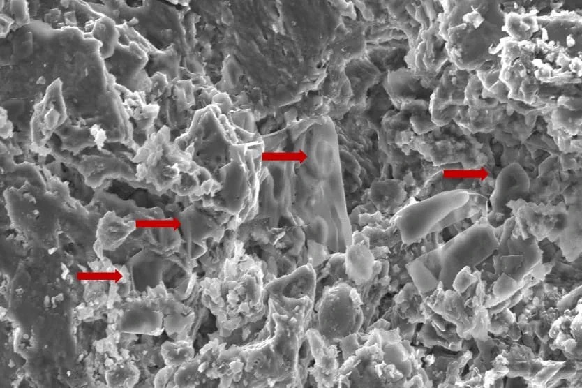
An image of fossilised erythrocyte-like structures. Credit: Anglia Ruskin University

New techniques used to analyse soft tissue in dinosaur fossils may hold the key to new cancer discoveries, according to a new study published in the journal Biology.

Researchers from Anglia Ruskin University (ARU) and Imperial College London analysed dinosaur fossils using advanced paleoproteomic techniques, a method that holds promise for uncovering molecular data from ancient specimens.

The researchers discovered red blood cell-like structures in a fossil while studying a Telmatosaurus transsylvanicus, a duck-billed, plant eating “marsh lizard” that lived between 66-70 million years ago in the Hateg Basin in present-day Romania.

The new study used Scanning Electron Microscopy (SEM) techniques to identify low-density structures resembling erythrocytes, or red blood cells, in the fossilised bone.

The findings raise the possibility that soft tissue and cellular components are more commonly preserved in ancient remains than previously thought.

By identifying preserved proteins and biomarkers, scientists believe they can gain insights into the diseases that affected prehistoric creatures, including cancer, potentially influencing future treatments for humans.

The authors of the new study highlight the necessity of prioritising the collection and preservation of fossilised soft tissue, rather than just dinosaur skeletons, as future advancements in molecular techniques will enable deeper insights into disease evolution.

A separate study had previously identified evidence of cancer in Telmatosaurus transsylvanicus, indicating its deep evolutionary roots.

Source: Anglia Ruskin University

Non-genetic Theories of Cancer Address Inconsistencies in Current Paradigm

A recent essay argues for re-considering cancer as a genetic disease

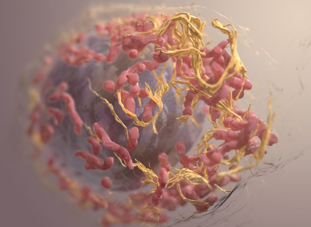
3D structure of a melanoma cell derived by ion abrasion scanning electron microscopy. Credit: Sriram Subramaniam/ National Cancer Institute

It’s time for researchers to reconsider the current paradigm of cancer as a genetic disease, argued Sui Huang from the Institute for Systems Biology, USA, and colleagues in a new essay published March 18th in the open-access journal PLOS Biology.

The prevailing theory on the origin of cancer is that an otherwise normal cell accumulates genetic mutations that allow it to grow and reproduce unchecked. This paradigm has driven large-scale cancer genome sequencing projects, such as The Cancer Genome Atlas, to identify cancer-driving mutations and develop drugs designed to target aberrant proteins and pathways.

In their new essay, Huang and colleagues argue that this somatic mutation theory of cancer is unproductive. They point to inconsistencies in the sequencing data that contradict the current theory, including the fact that many cancers have no known driver mutations while some normal tissues can harbour cancer-causing mutations.

They propose a broader, more “holistic” view that embraces organismal biology and theory. Specifically, they encourage considering alternative paradigms that encompass non-genetic processes involved in tumorigenesis. For example, they explain the concepts of cancer as a result of disruptions in gene regulatory networks (Huang) — or of tissue organisation, a theory that considers the disturbance of the field generated by neighbouring cells and surrounding tissue (Soto-Sonnenschein). The authors argue that these alternative explanations will guide experiments to advance our understanding of the origins of cancer.

The authors add: “A full embrace of the idea that the origin of cancer lies beyond the realm of genetic mutations will open new vistas on cancer treatment and prevention.  Accepting that not all carcinogens are mutagens will strengthen public health policies aimed to prevent exposure to environmental non-mutagenic factors that may promote cancer, such as food additives and plastics and many other toxicants that alter tissue homeostasis.”

Provided by PLOS

How Cancer Reprograms Immune Cells to Join the Enemy

Squamous cancer cell being attacked by cytotoxic T cells. Image by National Cancer Institute on Unsplash

Cancer has been described as “a wound that does not heal,” implying that the immune system is unable to wipe out invading tumour cells. A new discovery reported in PNAS confirms that a key molecule can reprogram immune cells into turncoats that promote cancer growth.

Studying the behaviour of these “pro-tumour” immune cells is important because they could be targets for therapies that block their harmful activity, said Minsoo Kim, PhD, corresponding author of the study and a research leader at the Wilmot Cancer Institute.

Kim led a team of scientists investigating the dynamic interactions that occur between cells in the tumor environment, and the underlying factors that cause the harmful transformation of immune cells from good to bad.

They found that PAF (platelet-activating factor) is the key molecule that controls the destiny of the immune cells. PAF not only recruits cancer-promoting cells, but it also suppresses the immune system’s ability to fight back. In addition, they found that multiple cancers rely on the same PAF signals.

“This is what could be most significant,” said Kim. “Because if we find a treatment that could interfere with PAF, it could potentially apply to many types of cancer.”

Much of the team’s work focused on pancreatic cancer cells. It is one of the most deadly cancers, with a five-year survival rate of about 12%, and is notoriously hard to treat because pancreatic tumours are surrounded by a toxic stew of proteins and other tissues that protect the cancer from the immune system’s natural role to attack invaders. They also studied breast, ovarian, colorectal, and lung cancer cells, using advanced 3D imaging technology to watch the behaviour of immune cells as they swarmed to the cancerous region.

Source: University of Rochester Medical Center