Author: ModernMedia

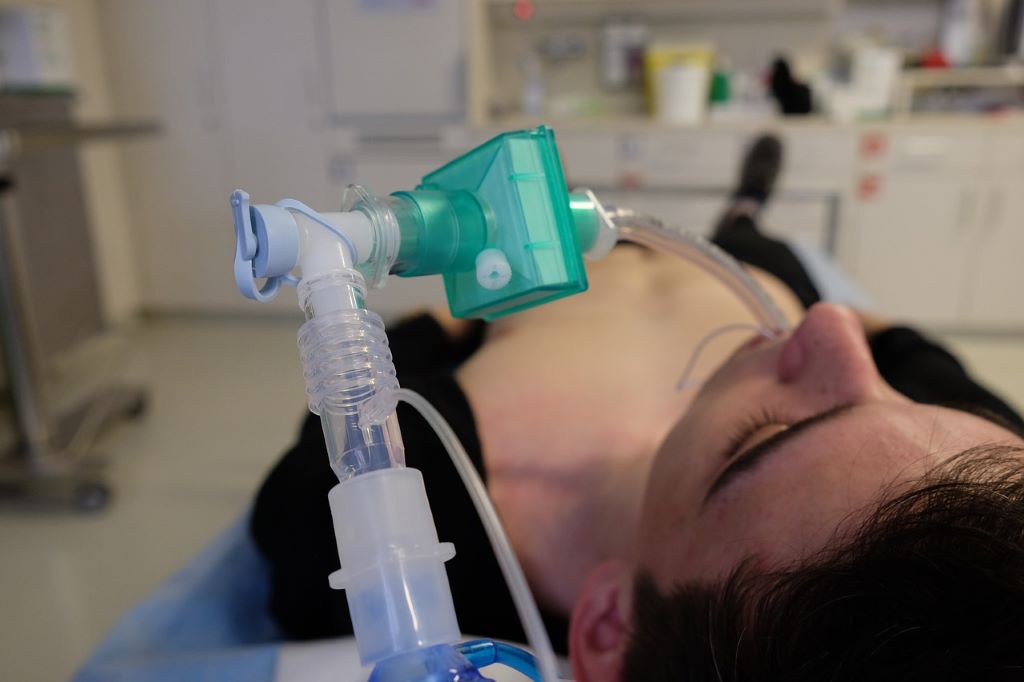
New Ultrasound Helmet Enables Deep Brain Stimulation in People Without Surgery

Illustration of new ultrasound device. Credit: Morgan Roberts.

Scientists have long been looking for a way to modulate brain function, which could improve our understanding of how the brain works and help to treat neurological diseases, using non-invasive methods that don’t involve surgery.

One technology that could help is transcranial ultrasound stimulation (TUS), which was recently discovered to be able to modulate the activity of neurons (the brain’s key communication cells) by delivering gentle mechanical pulses that influence how these cells send signals.

But to date current systems have struggled to reach deeper areas of the brain with sufficient precision to target specific brain structures. Conventional TUS systems often affect broader regions than intended, limiting their utility for targeted neuromodulation.

The study, published in Nature Communications, introduces a new ultrasound device capable of influencing deep brain regions without surgery for the first time, targeting areas around 1000 times smaller than conventional ultrasound devices can pinpoint and 30 times smaller than previous deep brain ultrasound devices.

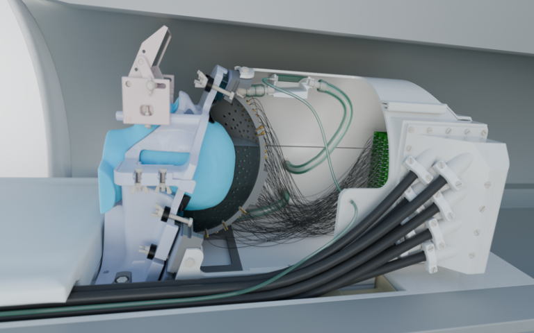
The new technology features 256 elements configured within a special helmet to send focused beams of ultrasound to specific parts of the brain in order to turn neuronal activity up or down. It also includes a soft plastic face mask which helps to target the ultrasound waves more precisely by keeping the head still.

The research team demonstrated the system’s capabilities on seven human volunteers by targeting a part of the thalamus, a small structure in the centre of the brain that helps to relay sensory and motor information, called the lateral geniculate nucleus (LGN). The LGN is involved in processing visual information.

In the first experiment, participants looked at a flashing checkerboard, which sent signals to the brain through the eyes. During stimulation with the ultrasound device, a functional magnetic resonance imaging (fMRI) scan showed significantly increased activity in the participants’ visual cortex, confirming precise targeting of the LGN.

A second experiment revealed sustained decreases in visual cortex activity for at least 40 minutes after ultrasound stimulation, highlighting the system’s potential for inducing lasting changes in brain function.

Though participants did not consciously perceive any changes in what they were seeing during the experiments, the brain scans revealed significant changes in neural activity. The ultimate goal is to harness these effects to produce clinically beneficial outcomes, such as stopping hand tremors.

Professor Bradley Treeby, senior author of the study from UCL Medical Physics and Biomedical Engineering, said: “This advance opens up opportunities for both neuroscience research and clinical treatment. For the first time, scientists can non-invasively study causal relationships in deep brain circuits that were previously only accessible through surgery.

“Clinically, this new technology could transform treatment of neurological and psychiatric disorders like Parkinson’s disease, depression, and essential tremor, offering unprecedented precision in targeting specific brain circuits that play key roles in these conditions.

“The ability to precisely modulate deep brain structures without surgery represents a paradigm shift in neuroscience, offering a safe, reversible, and repeatable method for both understanding brain function and developing targeted therapies.”

In addition to its research applications, the system could pave the way for new clinical interventions. Deep brain stimulation (DBS), currently used to treat conditions like Parkinson’s disease, requires invasive surgery and carries associated risks. The new ultrasound system offers a non-invasive alternative with comparable precision, potentially allowing clinicians to test areas of the brain that could be used to treat disease before surgery or even replace surgical approaches altogether.

Recognising this clinical potential, several members of the research team have recently founded NeuroHarmonics, a UCL spinout company developing a portable, wearable version of the system. The company aims to make precise, non-invasive deep brain therapy accessible for both clinical treatment and broader therapeutic applications.

Dr Eleanor Martin, first author of the study from UCL Medical Physics and Biomedical Engineering, said: “We designed the system to be compatible with simultaneous fMRI, enabling us to monitor the effects of stimulation in real time. This opens up exciting possibilities for closed-loop neuromodulation and personalised therapies.”

The researchers emphasise that further studies are needed to fully understand the mechanisms underlying TUS-induced neuromodulation. However, the results mark a significant milestone in the development of safe, effective, and targeted brain stimulation technologies.

Source: University College London

Long-term Alcohol Use Suspends Liver Cells in Limbo, Preventing Regeneration

Photo by Pavel Danilyuk on Pexels

Excessive alcohol consumption can disrupt the liver’s unique regenerative abilities by trapping cells in limbo between their functional and regenerative states, even after a patient stops drinking, as described in a new study from researchers at University of Illinois Urbana-Champaign and collaborators.

This in-between state is a result of inflammation disrupting how RNA is spliced during the protein-making process, the researchers found, providing scientists with new treatment pathways to explore for the deadly disease. The researchers published their findings in the journal Nature Communications.

The liver has a remarkable ability to regenerate itself after damage or partial removal. However, it loses that ability in patients with alcohol-associated liver disease – the leading cause of liver-related mortality worldwide, resulting in roughly 3 million deaths annually.

“We knew that the liver stops functioning and stops regenerating in patients with alcohol-related hepatitis and cirrhosis, even when a patient has discontinued consuming alcohol, but we didn’t know why,” said U. of I. biochemistry professor Auinash Kalsotra, who co-led the study with Duke University School of Medicine professor Anna Mae Diehl. “The only real life-saving treatment option once a patient reaches the liver failure stage in those diseases is transplantation. But if we understood why these livers were failing, maybe we could intervene.”

Both the Kalsotra and Diehl labs havestudied the molecular and cellular underpinnings of liver regeneration. Over the last five years, they found that in order to regenerate, liver cells reprogram their gene expression to revert to fetal-like progenitor cells, multiply and then reverse the process back to become mature functioning cells again. Armed with this knowledge, the group turned to the question of how those mechanisms were disrupted in alcohol-associated liver disease.

The researchers compared samples of healthy livers and samples of livers with alcohol-associated hepatitis or cirrhosis obtained from Johns Hopkins University Hospital through an initiative supported by the National Institute for Alcohol Abuse and Alcoholism, part of the National Institutes of Health.

The first thing the researchers noticed in diseased livers was that, although damaged cells had begun the process of reverting to the regenerative state, they did not complete the process and instead remained in transitional limbo.

“They are neither functional adult cells nor proliferative progenitor cells. Since they are not functioning, more pressure builds on the remaining cells. So they try to regenerate, and they’re all ending up in this unproductive quasi-progenitor state, and that’s what is causing liver failure,” said U. of I. graduate students Ullas Chembazhi and Sushant Bangru, the co-first authors of the study.

To figure out why the cells were getting stuck in this state, the team investigated which proteins were being made by the liver cells and, in turn, the RNA molecules carrying the instructions for those proteins from the DNA to the cell’s protein-building machinery.

While most studies focus only on the total amounts of RNA or protein in a cell, Kalsotra’s team used deep RNA sequencing technology and computational analyses to zoom in on the splicing of RNA fragments, a key step in stitching together different parts of genetic instructions to make proteins.

“In comparing the samples, we saw RNA was getting mis-spliced broadly in alcohol-related liver disease, across thousands of genes, and it was affecting major functions of proteins,” said Kalsotra.

The researchers found a possible driver of the RNA mis-splicing: Alcohol-damaged liver cells had a deficiency of the protein ESRP2, which binds to RNA to splice it properly.  

“Proteins function at a very specific place in the cell, and that is directed by sequences within the protein that take the protein to that particular spot. We found that, in many cases, the sequence that dictates where the protein localizes within a cell was mis-spliced. That’s why it was important that we did the multiple analyses we did,” said Kalsotra. “There was the same amount of RNA and protein, but the protein was not at the right place to function. Due to mis-splicing, key proteins that are required for productive liver regeneration were getting stuck in the cytoplasm, when they needed to be in the nucleus.”

To verify that ESRP2 deficiency was a likely culprit, the researchers studied mice without the gene that produces ESRP2. They displayed similar liver damage and regeneration failure to that seen in patients with advanced alcohol-related hepatitis.

But why was ESRP2 missing from liver cells from patients with alcohol-related hepatitis? Upon investigation, the researchers found that liver support cells and immune cells, drawn to the liver tissue damaged by alcohol processing, released high amounts of inflammatory and growth factors. Those factors suppress ESRP2 production and activity.

To verify this finding, the researchers treated liver cell cultures with a molecule that inhibits the receptor for one of the inflammation-promoting factors. ESRP2 levels recovered and splicing activity was corrected, pointing to the pathway as a possible treatment target.

“I’m hopeful these findings will become a launching pad for future clinical studies. We can use these mis-spliced RNAs as diagnostic markers or develop treatments that can curb the inflammation. And if we can correct the splicing defects, then maybe we can improve recovery and restore damaged livers,” Kalsotra said.

Source: University of Illinois Urbana-Champaign

How ‘Brain Cleaning’ While We Sleep May Lower Our Risk of Dementia

Photo by Cottonbro on Pexels

Julia Chapman, Macquarie University; Camilla Hoyos, Macquarie University, and Craig Phillips, Macquarie University

The brain has its own waste disposal system – known as the glymphatic system – that’s thought to be more active when we sleep.

But disrupted sleep might hinder this waste disposal system and slow the clearance of waste products or toxins from the brain. And researchers are proposing a build-up of these toxins due to lost sleep could increase someone’s risk of dementia.

There is still some debate about how this glymphatic system works in humans, with most research so far in mice.

But it raises the possibility that better sleep might boost clearance of these toxins from the human brain and so reduce the risk of dementia.

Here’s what we know so far about this emerging area of research.

Why waste matters

All cells in the body create waste. Outside the brain, the lymphatic system carries this waste from the spaces between cells to the blood via a network of lymphatic vessels.

But the brain has no lymphatic vessels. And until about 12 years ago, how the brain clears its waste was a mystery. That’s when scientists discovered the “glymphatic system” and described how it “flushes out” brain toxins.

Let’s start with cerebrospinal fluid, the fluid that surrounds the brain and spinal cord. This fluid flows in the areas surrounding the brain’s blood vessels. It then enters the spaces between the brain cells, collecting waste, then carries it out of the brain via large draining veins.

Scientists then showed in mice that this glymphatic system was most active – with increased flushing of waste products – during sleep.

One such waste product is amyloid beta (Aβ) protein. Aβ that accumulates in the brain can form clumps called plaques. These, along with tangles of tau protein found in neurons (brain cells), are a hallmark of Alzheimer’s disease, the most common type of dementia.

In humans and mice, studies have shown that levels of Aβ detected in the cerebrospinal fluid increase when awake and then rapidly fall during sleep.

But more recently, another study (in mice) showed pretty much the opposite – suggesting the glymphatic system is more active in the daytime. Researchers are debating what might explain the findings.

So we still have some way to go before we can say exactly how the glymphatic system works – in mice or humans – to clear the brain of toxins that might otherwise increase the risk of dementia.

Does this happen in humans too?

We know sleeping well is good for us, particularly our brain health. We are all aware of the short-term effects of sleep deprivation on our brain’s ability to function, and we know sleep helps improve memory.

In one experiment, a single night of complete sleep deprivation in healthy adults increased the amount of Aβ in the hippocampus, an area of the brain implicated in Alzheimer’s disease. This suggests sleep can influence the clearance of Aβ from the human brain, supporting the idea that the human glymphatic system is more active while we sleep.

This also raises the question of whether good sleep might lead to better clearance of toxins such as Aβ from the brain, and so be a potential target to prevent dementia.

How about sleep apnoea or insomnia?

What is less clear is what long-term disrupted sleep, for instance if someone has a sleep disorder, means for the body’s ability to clear Aβ from the brain.

Sleep apnoea is a common sleep disorder when someone’s breathing stops multiple times as they sleep. This can lead to chronic (long-term) sleep deprivation, and reduced oxygen in the blood. Both may be implicated in the accumulation of toxins in the brain.

Sleep apnoea has also been linked with an increased risk of dementia. And we now know that after people are treated for sleep apnoea more Aβ is cleared from the brain.

Insomnia is when someone has difficulty falling asleep and/or staying asleep. When this happens in the long term, there’s also an increased risk of dementia. However, we don’t know the effect of treating insomnia on toxins associated with dementia.

So again, it’s still too early to say for sure that treating a sleep disorder reduces your risk of dementia because of reduced levels of toxins in the brain.

So where does this leave us?

Collectively, these studies suggest enough good quality sleep is important for a healthy brain, and in particular for clearing toxins associated with dementia from the brain.

But we still don’t know if treating a sleep disorder or improving sleep more broadly affects the brain’s ability to remove toxins, and whether this reduces the risk of dementia. It’s an area researchers, including us, are actively working on.

For instance, we’re investigating the concentration of Aβ and tau measured in blood across the 24-hour sleep-wake cycle in people with sleep apnoea, on and off treatment, to better understand how sleep apnoea affects brain cleaning.

Researchers are also looking into the potential for treating insomnia with a class of drugs known as orexin receptor antagonists to see if this affects the clearance of Aβ from the brain.

If you’re concerned

This is an emerging field and we don’t yet have all the answers about the link between disrupted sleep and dementia, or whether better sleep can boost the glymphatic system and so prevent cognitive decline.

So if you are concerned about your sleep or cognition, please see your doctor.

Julia Chapman, Clinical Trials Lead and Postdoctoral Research Fellow, Woolcock Institute of Medical Research and Conjoint Lecturer, Macquarie University; Camilla Hoyos, Senior Lecturer in the Centre for Sleep and Chronobiology, Macquarie University, and Craig Phillips, Associate Professor, Macquarie Medical School, Macquarie University

This article is republished from The Conversation under a Creative Commons license. Read the original article.

Innovative Soft Robotic Intubation Device Could Save Lives

Credit: Pixabay CC0

Endotracheal intubation is a difficult task for highly trained individuals and under the best of circumstances. In the field and in the ER, where seconds matter, emergency medical personnel face many unknowns and wildly challenging conditions which lower their chances of success.

But what if successful endotracheal intubation could be less reliant on ideal conditions and years of specialised training? In a paper published in Science Translational Medicine, UC Santa Barbara researchers  David Haggerty, Elliot Hawkes and collaborators demonstrate a non-electronic soft robotic device that quickly and autonomously guides a soft tube into the trachea. Initial device testing with highly trained users yielded a 100% success rate, and a 96% overall success rate with prehospital medical providers (EMTs and paramedics).

“Current intubation tools require extensive anatomical knowledge, training, skill and ideal conditions to be highly successful,” said recent UC Santa Barbara Ph.D. graduate David Haggerty, a former researcher in the lab of mechanical engineering professor Elliot Hawkes. Current technology calls for the rescuer to first visualize the tracheal opening then manually direct a tube through the serpentine anatomy of the airway into the trachea. The challenge of this procedure increases in prehospital settings due to various factors including inadequate light and nonideal body position in addition to potential injuries and fluid in the airway.

This project is supported in part with funds from the National Science Foundation

From rigid tools to soft robotics

One of the main challenges to successful intubation is the body itself, and the mechanisms it has evolved to prevent food and foreign bodies from entering the lungs. The epiglottis is a small fleshy flap that closes over the trachea and guides food and liquid into the adjacent esophagus with each swallow. Conventional practice typically requires the rescuer to push a metal laryngoscope into the back of the mouth behind the tongue to lift the epiglottis out of the way in order to make room for the breathing tube.

But even with the epiglottis out of the way, the path the endotracheal tube must take is a twisted one, as it has to bend toward the front of the neck where the trachea is located, otherwise air could be delivered to the stomach via the esophagus, instead of to the lungs.

“Traditional tools, which you push from the base, are fundamentally limited in navigating delicate, tortuous anatomy,” Hawkes explained. “They must be relatively stiff so you can push them, and can only get around bends by deflecting off the sensitive tissue.”

The researchers’ device upends that paradigm with a soft, inflatable tube that everts from its tip. Called the soft robotic intubation system (SRIS), it consists of a curved “introducer” that slides into place at the back of the throat and stops at the esophagus. With that in place, a soft, pre-inflated tube is threaded through the introducer, emerging near its tip at the opening of the trachea. As the user advances the tube, it everts from its tip, carrying inside it a soft breathing tube as it enters the trachea. “So instead of trying to push this tube and bend it to get into this complex configuration, we can just mechanically create that complex configuration as we go,” Haggerty said. Once the endotracheal tube is at its destination, the user can inflate a cuff at its distal end to seal the opening and begin ventilation. The introducer can be removed, leaving the breathing tube in place.

Introducing a soft, growing tube eliminates friction with the surrounding tissue and minimizes injury due to excessive or misplaced force. It also automatically conforms to its environment, one of the major benefits of soft robotics. “This growing paradigm naturally accounts for minor variations in anatomical placement, size, shape or configuration,” Haggerty said, and because of this, users need not have extensive skill or a perfect understanding of the environment in order to navigate it.

In tests with mannequins and cadavers, the SRIS proved itself both effective, and, importantly, rapid, with a 100% success rate at a procedure duration of just seconds for expert users. For nonexpert users – the primary target of this technology – a five-minute training session was all that was needed to deliver an 87% success rate for first-pass attempts, translating into an 96% overall success rate, with a significantly lower procedure duration – 21 seconds versus 44 seconds – than state-of-the-art video laryngoscope intubation.

The next step for the researchers is to conduct clinical trials in order to get approvals from the Food and Drug Administration for clinical use. “We have good reason to believe it’s efficacious based on the data, but cadavers don’t say ‘ow’,” Haggerty said. They need to complete more testing to ascertain the device’s safety and effectiveness in a variety of airways and external conditions, he added. If successful, this device could benefit the millions of emergency intubations that occur each year in the US, and find application in military medical care. That’s in addition to its potential to save lives in global populations that have poor or no access to essential health services.

Source: University of California – Santa Barbara

Giant DNA Elements Discovered Inside the Oral Microbiome

Functional characterisation of Inocle. A schematic describing what Inocles do and where they’re found. It shows the kinds of roles its genes might have, and how those jobs could be connected to things happening in the human body. ©2025 Kiguchi et al. CC-BY-ND

Researchers at the University of Tokyo and other institutions have made a surprising discovery hiding in people’s mouths: Inocles, giant DNA elements that had previously escaped detection. These appear to play a central role in helping bacteria adapt to the constantly changing environment of the mouth. The findings provide fresh insight into how oral bacteria colonise and persist in humans, with potential implications for health, disease and microbiome research.

You might think that modern medical science knows everything there is to know about the human body. But even within the last decade, small, previously unknown organs have been discovered, and there’s one area of human biology that is currently going through a research renaissance, the microbiome. This includes familiar areas such as the gut microbiome, but also the oral microbiome. Inspired in part by recent discoveries of extraneous DNA in the microbiome of soil, Project Research Associate Yuya Kiguchi and his team turned their sights to a large set of saliva samples collected by the Yutaka Suzuki Lab of the Graduate School of Frontier Sciences at the University of Tokyo. They wondered if they might find something similar in human saliva.

“We know there are a lot of different kinds of bacteria in the oral microbiome, but many of their functions and means of carrying out those functions are still unknown,” said Kiguchi. “By exploring this, we discovered Inocles, an example of extrachromosomal DNA – chunks of DNA that exist in cells, in this case bacteria, but outside their main DNA. It’s like finding a book with extra footnotes stapled to it, and we’re just starting to read them to find out what they do.”

Detecting Inocles was not easy, as conventional sequencing methods fragment genetic data, making it impossible to reconstruct large elements. To overcome this, the team applied advanced long-read sequencing techniques, which can capture much longer stretches of DNA. A key breakthrough came from co-first author Nagisa Hamamoto, who developed a method called preNuc to selectively remove human DNA from saliva samples, greatly improving the quality of sequencing long sections of other DNA. This allowed the researchers to assemble for the first time complete Inocle genomes, which turned out were hosted by the bacteria Streptococcus salivarius, though identifying the host itself was a difficult matter.

“The average genome size of Inocle is 350 kilobase pairs, a measure of length for genetic sequences, so it is one of the largest extrachromosomal genetic elements in the human microbiome. Plasmids, other forms of extrachromosomal DNA, are at most a few tens of kilobase pairs,” said Kiguchi. “This long length endows Inocles with genes for various functions, including resistance to oxidative stress, DNA damage repair and cell wall-related genes, possibly involved in adapting to extracellular stress response.”

The team aims to develop stable methods for culturing Inocle-containing bacteria. This will allow them to investigate how Inocles function, whether they can spread between individuals, and how they might influence oral health conditions such as cavities and gum disease. Since many Inocle genes remain uncharacterised, researchers will use a mixture of laboratory experiments and also computational simulations such as AlphaFold to predict and model the roles Inocles may play.

“What’s remarkable is that, given the range of the human population the saliva samples represent, we think 74% of all human beings may possess Inocles. And even though the oral microbiome has long been studied, Inocles remained hidden all this time because of technological limitations,” said Kiguchi. “Now that we know they exist, we can begin to explore how they shape the relationship between humans, their resident microbes and our oral health. And there’s even some hints that Inocles might serve as markers for serious diseases like cancer.”

Source: University of Tokyo

Missing Gene on Y Chromosome Linked to Male Infertility

Chromosomes. Credit: NIH

University of Hawaiʻi at Mānoa scientists have uncovered a direct link between a missing Y chromosome gene and male infertility. Their new research reveals that deleting this single gene in mice not only caused infertility but also disrupted hundreds of other genes vital for healthy sperm. The findings, published August 27 in Cell Death and Differentiation, offer significant implications for understanding reproductive health.

The role of Zfy

The study, led by Monika Ward of UH Mānoa’s John A. Burns School of Medicine and the Yanagimachi Institute for Biogenesis Research (YIBR), focused on the Zfy gene, found on the Y chromosome in both humans and mice. In mice, there are two versions of this gene: Zfy1 and Zfy2.

Using CRISPR gene-editing, the team created mice missing one or both versions. Males without both, known as Zfy double knockouts, were completely infertile, with severely abnormal or absent sperm.

“This work really pushes forward our understanding of how this important Zfy gene works,” said Ward. “We identified pathways and other genes that are affected and we can now study how exactly Zfy regulates them.”

To continue investigations, the researchers turned to assisted reproduction techniques pioneered at UH, including intracytoplasmic sperm injection (ICSI) and round spermatid injection (ROSI). This allowed them to examine the molecular consequences of Zfy loss.

When one gene disrupts hundreds

The results revealed that without Zfy, hundreds of genes became misregulated – some too active, others too weak. Many of these genes are responsible for sperm production, DNA packaging, and cell survival.

As a result, sperm precursor cells in the testes died off early, and the sperm that did form carried fragile DNA that wasn’t properly condensed.

The study details can be found in an article published in Cell Death and Differentiation, a leading peer-reviewed journal.

Source: University of Hawai’i

Immune Ageing Found to Drive – Not Be Driven by – Rheumatoid Arthritis

Rheumatoid arthritis. Credit: Scientific Animations CC4.0

Features of immune system ageing can be detected in the earliest stages of rheumatoid arthritis (RA), even before clinical diagnosis, a new study has found which provides at-risk individuals with hope for early intervention.

The research led by academics at the University of Birmingham, delivered through the National Institute for Health and Care Research (NIHR) Birmingham Biomedical Research Centre, and published in the journal eBioMedicine shows that individuals with joint pain or undifferentiated arthritis already exhibit signs of a prematurely aged immune system, suggesting that immune ageing may play a direct role in the development of RA.

The study involved 224 participants across various stages of RA development and was funded by FOREUM and the European League Against Rheumatism (EULAR). It represents one of the most comprehensive analyses of immune ageing in RA to date.

Researchers found that patients with early immune ageing features were more likely to develop RA. These findings could lead to the development of predictive tools that identify at-risk individuals and enable timely treatment.

“We’ve discovered that immune ageing isn’t just a consequence of rheumatoid arthritis—it may be a driver of the disease itself,” said Dr Niharika Duggal, senior author of the study and Associate Professor in Immune Ageing at the University of Birmingham. “We found that people in the early stages of rheumatoid arthritis ie, before a clinical diagnosis show signs of faster immune system ageing.

“These findings suggest we might be able to intercept the disease development in at-risk individuals and prevent it from developing by using treatments that slow ageing, such as boosting the body’s natural process for clearing out damaged cells (autophagy).”

Key Findings

  • Hallmarks of immune ageing, including reduced naïve T cells and thymic output, were observed in patients with early joint symptoms.
  • An elevated IMM-AGE score revealed accelerated immune ageing in patients before RA diagnosis.
  • Elevated levels of inflammatory markers (such as IL-6, TNFα, and CRP) were found in preclinical stages.
  • Advanced ageing features, including senescent T cells and inflammatory Th17 cells, appeared only after RA was fully established.

The study suggests that targeting ageing pathways could offer new strategies to prevent RA. Future research should determine whether geroprotective drugs such as spermidine (autophagy booster), senolytics (clearance of senescent cells) and metformin (attenuates inflammation and boosts autophagy) may help slow or halt disease progression in high-risk individuals.

Source: University of Birmingham

Considering Sex Hormones Led to Better Identification of Genes Linked to Type 2 Diabetes

Genomic and hormone study of white Europeans finds 22 additional disease-related variants

Ball and stick 3D model of testosterone. Source: Wikimedia CC0

Researchers identified almost two dozen previously unknown genetic variants linked to type 2 diabetes by including participants’ hormone levels in their analysis. Yan V. Sun of Emory University, USA, and colleagues reports these findings in the open-access journal PLOS Genetics.

Type 2 diabetes affects an increasing number of people worldwide, and more often affects men than women. The disease is caused by a mix of genetic and lifestyle factors, but little is known about how someone’s environment – both inside and outside the body – interacts with their genes to impact a person’s risk of developing the disease.

In the new study, researchers performed genome-wide interaction studies to investigate whether a person’s hormone levels interact with their genetic variants to affect their risk of developing type 2 diabetes. They grouped males and females independently and considered measurements of three types of sex hormones – total testosterone, bioavailable testosterone and sex-hormone binding globulin. The information came from white European participants in the UK Biobank, which contains biological samples and health data from half a million people.

The researchers used statistical analyses to identify relevant variants in the genomes of individuals with and without type 2 diabetes. By taking into account hormone levels, the analysis was able to identify 22 spots on the genome that increased a person’s risk for type 2 diabetes. These variants had not been reported previously in the most recent genomic study for type 2 diabetes.

The new study suggests that a person’s hormone levels may be interacting with their genes to increase their odds of having type 2 diabetes. For future studies, the researchers recommended that additional hormone measurements for each participant and more diverse cohorts should be included. They conclude that this approach, which includes environmental factors in genomic studies, may help us to identify additional disease-related genes and gain a better understanding of the mechanisms behind complex diseases.

The authors add, “We found that sex hormone levels contribute to differences in genetic risk factors for type 2 diabetes in men and women. By analyzing data for men and women separately, we identified new genetic associations with type 2 diabetes.”

The lead analyst, Amonae Dabbs-Brown notes, “I actually used to work at the CDC developing methods to measure some of these sex hormones. It’s really exciting to see what happens downstream. Maybe one day I’ll even get to see how these analyses are used in the clinic!”

Provided by PLOS

In your coverage, please use this URL to provide access to the freely available paper in PLOS Geneticshttps://plos.io/3ViXDKH

Contact: Rob Spahr [rob.spahr@emory.edu]

Citation: Dabbs-Brown A, Liu C, Hui Q, Wilson PW, Zhou JJ, Gwinn M, et al. (2025) Identification of gene-sex hormone interactions associated with type 2 diabetes among men and women. PLoS Genet 21(9): e1011470. https://doi.org/10.1371/journal.pgen.1011470

Author countries: United States

Funding: This work is supported in part by funding from the National Institutes of Health (HL154996 to YVS, DK139632 to YVS, and HL156991 to YVS). The funders had no role in study design, data collection and analysis, decision to publish, or preparation of the manuscript. YVS received salary support from the National Institutes of Health.

Competing interests: The authors have declared that no competing interests exist.

Red Meat Aggravates IBD by Altering the Gut Microbiome

Photo by Jose Ignacio Pompe on Unsplash

Epidemiological studies have revealed a strong correlation between red meat consumption and the development of inflammatory bowel disease. In a new study published in Molecular Nutrition and Food Research that was conducted in mice, red meat consumption caused an imbalance of bacteria in the intestinal microbiota. 

Investigators fed mice various kinds of red meat, including pork, beef, and mutton, for two weeks, and then they induced colitis with 2.5% dextran sulfate sodium. Intake of these three red meat diets exacerbated colonic inflammation. Analyses revealed an overproduction of pro-inflammatory cytokines and infiltration of immune cells in the colon of mice fed red meat diets. 

These diets led to a marked decrease in the relative abundance of StreptococcusAkkermansiaFaecalibacterium, and Lactococcus bacterial strains, coupled with an increase in Clostridium and Mucispirillum.  

“This study contributes to improving food innervation approaches for inflammatory bowel disease treatment and indicates a close crosstalk among diet, gut microbiota, and intestinal immunity,” said co–corresponding author Dan Tian, MD, PhD, of Capital Medical University, in China. 

Source: Wiley

Study Explains Why Influenza is More Deadly for Older People

Photo by JD Mason on Unsplash

Scientists have discovered why older people are more likely to suffer severely from the flu, and can now use their findings to address this risk. In a new study published in PNAS, experts discovered that older people produce a glycosylated protein called apoplipoprotein D (ApoD), which is involved in lipid metabolism and inflammation, at much higher levels than in younger people. This has the effect of reducing the patient’s ability to resist virus infection, resulting in a more serious disease outcome.

The team established that highly elevated ApoD production with age in the lung drives extensive tissue damage during infection to reduce the protective antiviral type I interferon response.

The research was an international collaboration led by scientists from the China Agricultural University, University of Nottingham, Institute of Microbiology (Chinese Academy of Sciences), National Institute for Viral Disease Control and Prevention (Chinese Centre for Disease Control and Prevention) and the University of Edinburgh.

Aging is a leading risk factor in influenza-related deaths. Furthermore, the global population is aging at an unprecedented rate in human history, posing major issues for healthcare and the economy. So we need to find out why older patients often suffer more severely from influenza virus infection.”

Professor Kin-Chow Chang from the School of Veterinary Medicine and Science at the University of Nottingham, and co-author on the paper

In this new study, the team investigated the mechanisms behind increased severity of influenza virus infection with age using an aging-mouse model and appropriate donor human tissue sections.

They identified ApoD as an age-related cell factor that impairs the activation of the immune system’s antiviral response to influenza virus infection by causing extensive breakdown of mitochondria (mitophagy) resulting in greater production of virus and lung damage during infection. Mitochondria are essential for cellular production of energy and for induction of protective interferons.

ApoD is therefore a target for therapeutic intervention to protect against severe influenza virus infection in the elderly which would have a major impact on reducing morbidity and mortality in the aging population.

Professor Chang, added: “There is now an exciting opportunity to therapeutically ameliorate disease severity of the elderly from influenza virus infection by the inhibitory targeting of ApoD.”

Source: University of Nottingham