A test that rapidly detects signs of inflammatory bowel disease (IBD) in stool samples could improve future diagnosis and monitoring of the condition, a study suggests.
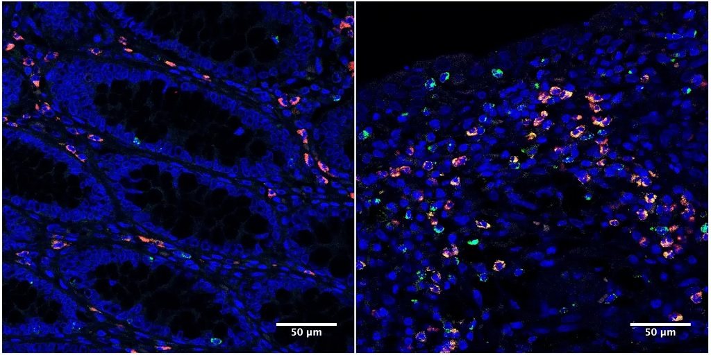
Microscopy images of colon tissue samples from IBD patients under remission (left) and with active disease (right). Granzyme A, indicated by green fluorescence, is elevated in the intestinal tract of IBD patients with active disease. Credit: Emily Thompson.
Scientists have developed a tool to measure the activity of a molecule linked to gut inflammation within faecal samples. The optical tool, known as a luminescent reporter, lights up when it detects the molecule, with higher readouts indicating increased activity and inflammation.
The new technique could boost the accuracy of stool sample tests for IBD, reducing the need for invasive, expensive procedures, experts say.
Gut inflammation
IBD is a chronic illness where the body’s immune system mistakenly attacks the digestive tract, leading to long-lasting inflammation. Diagnosis and monitoring of the condition often rely on colonoscopies, where a small camera is used to examine the gut.
Current IBD stool tests measure general markers of inflammation, such as the protein calprotectin, so a positive result requires further investigation to confirm the source.
University of Edinburgh researchers studied gut tissue from IBD patients and identified high levels of an enzyme – a molecule that speeds up chemical reactions in cells – called granzyme A (GzmA) in inflamed gut tissue compared with non-inflamed tissues.
Enzyme activity
GzmA is released by T cells; in IBD, T cells mistakenly see the gut as a threat and become overactive, which can lead to tissue damage and inflammation.
The research team developed a luminescent reporter to measure the activity of GzmA in stool samples. The reporter tool was tested on 150 samples from both IBD and healthy patients.
Combining the new reporting tool with the current common testing of faecal calprotectin levels was more successful in identifying IBD in patients than using faecal calprotectin scores alone.
Researchers say the ability to identify gut-specific inflammation is a step forward for IBD diagnosis, but caution further research is needed before it can be used in a clinical setting.
Spin-out company
The tool will form part of the assets of a new company in the process of spinning out of the University of Edinburgh, called IDXSense, supported by Edinburgh Innovations, the University’s commercialisation service.
The technique could also support the development of personalised IBD treatments in the future, with the ability to rapidly and accurately monitor gut inflammation levels in response to different therapies, experts say.
In a groundbreaking study published in the Proceedings of the National Academy of Sciences (PNAS), scientists at Pacific Northwest Research Institute (PNRI) have overturned a long-held belief in genetics: that inheriting two harmful variants in the same gene always worsens disease. Instead, the team found that, in many cases, two harmful variants can actually restore normal protein function.
The research focused on a human enzyme called argininosuccinate lyase (ASL), which plays a critical role in removing toxic ammonia from the body. Variants in ASL that decrease its activity cause one of the urea cycle disorders, a set of rare and potentially life-threatening metabolic diseases.
By experimentally measuring the functional impact of several thousand individual variants and variant combinations, PNRI researchers discovered that over 60% of pairs that were individually damaging could, together, bring enzyme activity back to healthy levels.
“This work shows that genetic variants don’t act independently in many important cases,” said Michelle Tang, PhD, PNRI Staff Scientist and lead author of the study. “For a defined group of genes, the default assumptions we use to predict disease risk simply don’t hold.”
This phenomenon is known as intragenic complementation. It occurs when damage caused by one variant is offset by another variant in a different part of the same protein. The mechanism was first proposed in 1964 by Francis Crick and Leslie Orgel, but until now had not been tested systematically or shown to be common or predictable at scale.
To make these interactions predictable, the research team developed an AI-based model that accurately forecasted whether two variants would restore protein function. The model achieved nearly 100% accuracy in predicting intragenic complementation in ASL as well as in a second human enzyme, fumarase, suggesting these rules apply broadly across the human genome.
“We’ve shown that, in many cases, two damaging variants can work together to restore protein function,“ said Aimée Dudley, PhD, PNRI Senior Investigator who led the study. “This kind of genetic interaction is not an isolated exception, but a widespread and underappreciated way that variants can interact, especially in rare disease contexts.”
The researchers estimate that approximately 4% of human genes have the structural features that allow this type of interaction. For these genes, standard genetic predictions can overestimate disease risk, particularly for people who carry two different variants in the same gene.
Vegetarian and vegan diets can support healthy growth when carefully planned with appropriate supplementation, finds a major new meta-analysis – the most comprehensive study to-date of plant-based diets in children.
A team of researchers, from Italy, USA and Australia, analysed data from over 48 000 children and adolescents worldwide who followed different dietary patterns, examining health outcomes, growth and nutritional adequacy. They found that vegan and vegetarian diets can be nutrient-rich and support healthy growth, but also carry a risk of deficiencies if key nutrients are not obtained through fortified foods or supplements.
The peer-reviewed study, published in Critical Reviews in Food Science and Nutrition, also suggests that plant-based diets may offer additional health benefits for children – including improved cardiovascular risk profiles – compared with omnivorous diets that include meat, fish and other animal-derived foods.
This large meta-analysis is the most comprehensive study to date of plant-based diets in children under 18 years of age, examining data from 59 studies across 18 countries. It compared lacto-ovo-vegetarian (which include dairy products and eggs, but exclude meat, fish and poultry) and vegan diets (which exclude all animal-derived foods) with omnivorous diets across a wide range of nutritional and health outcomes in 7280 lacto-ovo-vegetarians, 1289 vegans and 40 059 omnivores.
The study found that vegetarian children consumed more fibre, iron, folate, vitamin C and magnesium than omnivores, but they had lower intakes of energy, protein, fat, vitamin B12 and zinc. While evidence on vegan diets was more limited, similar patterns emerged.
“Notably, vitamin B12 didn’t reach adequate levels without supplementation or fortified foods, and calcium, iodine and zinc intakes were often at the lower end of recommended ranges, making them important nutrients to consider for children on plant-based diets,” explains the study co-author Dr Jeannette Beasley, an Associate Professor in the Departments of Nutrition and Food Studies and Medicine at New York University.
“Vegan children, in particular, had especially low calcium intake.”
Health benefits
Despite these risks, both vegan and vegetarian children displayed more favourable cardiovascular health profiles than omnivores, with lower total and low-density lipoprotein (LDL) cholesterol – the “unhealthy” form of cholesterol.
Growth and body composition measures indicated that children on plant-based diets tended to be leaner than omnivores: vegetarian children were slightly shorter and lighter, with lower body mass index (BMI), fat mass and bone mineral content. Vegan children also had shorter stature and lower BMI scores.
“Our analysis of current evidence suggests that well-planned and appropriately supplemented vegetarian and vegan diets can meet nutritional requirements and support healthy growth in children,” states lead-author Dr Monica Dinu, who focuses on exploring how nutrition shapes health and well-being at the Department of Experimental and Clinical Medicine, at the University of Florence, in Italy.
Parents: take an informed approach
Plant based diets remain entirely achievable for children and can offer environmental advantages as well as health benefits. The authors stress that families should not be discouraged from choosing vegetarian or vegan diets for ethical, environmental or health reasons. Instead, they recommend that parents approach these diets with informed planning and, where possible, seek support from clinicians such as dietitians and paediatric health professionals. With attention to a few key nutrients, these diets can fully meet children’s needs during periods of rapid growth while reducing nutritional risks.
“We hope these findings offer clearer guidance on both the benefits and potential risks of plant-based diets, helping the growing number of parents choosing these diets for health, ethical or environmental reasons,” Dr Dinu adds.
More research needed, but balance is key
The authors also emphasise the need for clear, evidence-based guidance to support families with planning healthy plant-based diets for children, who may have higher nutritional needs during periods of rapid growth and development.
However, the researchers caution that these results are limited by the cross-sectional design of most included studies, variability in methods and populations, and challenges in accurately assessing children’s dietary intake.
“In conclusion,” says fellow co-author Dr Wolfgang Marx, from the Food & Mood Centre, at Deakin University, Australia, “while well-planned vegetarian and vegan diets are nutritionally adequate and beneficial for adults, there is far less clarity about their suitability for children – leading to inconsistent or even conflicting advice for parents.
“Our findings suggest that a balanced approach is essential, with families paying close attention to certain nutrients – particularly vitamin B12, calcium, iodine, iron and zinc – to ensure their children get everything they need to thrive.”
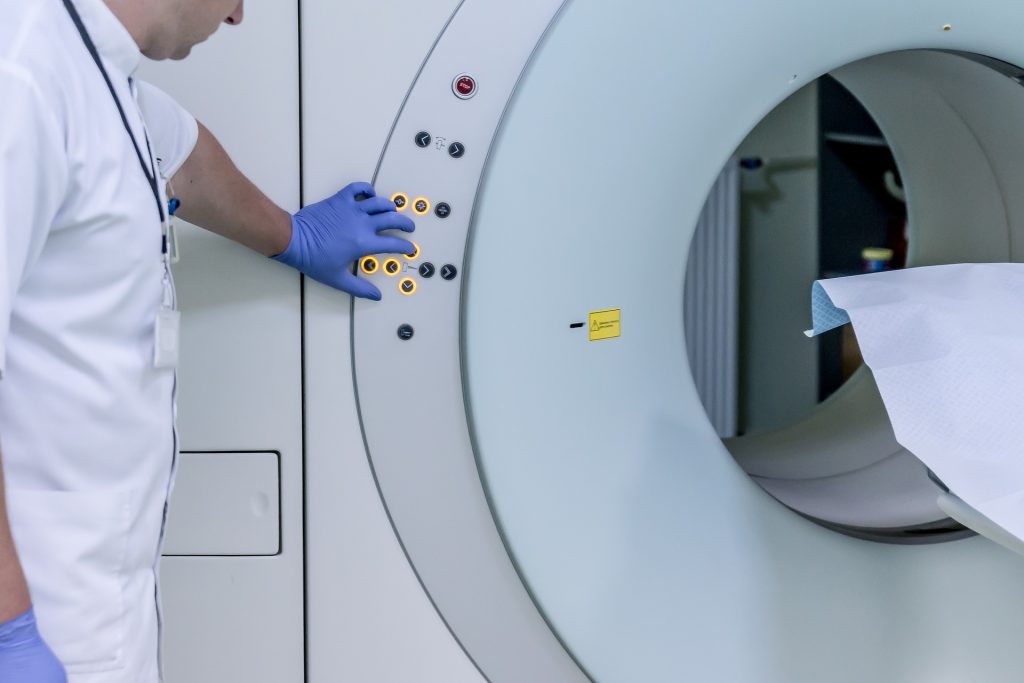
Adding breast magnetic resonance imaging (MRI) to a diagnostic mammogram did not reduce five-year cancer recurrence rates for patients with stage I/II hormone receptor (HR)-negative breast cancer, according to researchers at The University of Texas MD Anderson Cancer Center.
The Phase III Alliance A011104/ACRIN6694 trial found that five-year locoregional recurrence rates were 6.8% in patients who received an MRI as part of a diagnostic work-up and 4.3% in those who did not. These data were presented today at the San Antonio Breast Cancer Symposium (SABCS) by principal investigator Isabelle Bedrosian, MD, professor of Breast Surgical Oncology (Abstract GS2-07).
“We have long assumed that finding more breast cancer on an MRI and removing it with surgery would help lower the chance of a patient’s cancer coming back,” Bedrosian said. “When you look at our findings alongside earlier trials, the message is clear: adding MRI before surgery doesn’t improve results for patients – and may not have to be used as a standard part of the diagnostic process.”
No additional MRI benefit in this group
The trial enrolled 319 patients between 2014 and 2018 with newly diagnosed stage I or II HR-negative breast cancer. These patients were eligible for lumpectomy and did not have germline BRCA1/2 mutations, bilateral breast cancer or a history of prior breast cancer. All patients had undergone diagnostic mammography with or without ultrasound prior to trial enrolment. Patients were randomly assigned to undergo additional imaging by breast MRI (161 patients) or to receive no further imaging (158 patients).
Not only did breast MRI not impact five-year recurrence rates, but there were also not significant differences between groups for five-year distant recurrence-free survival nor overall survival.
A small subset of patients with tumour subtypes (HR- HER2+ and HR-HER2-) and those over the age of 50 at diagnosis also showed no benefit to MRI.
Pre-op MRI not finding anything important
Breast MRI is a common part of the diagnostic evaluation because it can reveal cancer that mammography might not detect. However, the evidence that it improves surgical outcomes for patients has been limited.
“We believe the reason MRI did not reduce recurrence rates may be twofold,” Bedrosian said. “It is possible that MRI didn’t uncover many lesions that mammography hadn’t already found, or perhaps identifying and surgically removing those additional lesions was not important to reducing risk of the cancer coming back. It’s possible that in the group that did not receive MRI, radiation and chemotherapy effectively treated the occult areas of disease”.
Experts are now analysing how often breast MRI identified additional lesions in the trial population to better understand why breast MRI did not impact oncologic outcomes.
Study limitations
Limitations included that most patients involved in the trial had breast cancer that hadn’t spread to their lymph nodes, which may partly explain why recurrence rates were low overall. Despite being open to women of all ages, the study enrolled mostly older women who may have been less likely to benefit from breast MRI.
First-of-its-kind study offers evidence that microbes from different primate species influence physiology in ways linked to brain size and function
Source: Pixabay
Humans have the largest relative brain size of any primate, but little is known about how mammals with larger brains evolved to meet the intense energy demands required to support brain growth and maintenance.
A new study from Northwestern University provides the first empirical data showing the direct role the gut microbiome plays in shaping differences in the way the brain functions across different primate species.
“Our study shows that microbes are acting on traits that are relevant to our understanding of evolution, and particularly the evolution of human brains,” said Katie Amato, associate professor of biological anthropology and principal investigator of the study, which was published in PNAS.
The study builds upon previous findings from Amato’s lab that showed the microbes of larger-brained primates, when introduced in host mice, produced more metabolic energy in the microbiome of the host – a prerequisite for larger brains, which are energetically costly to develop and function. This time, the researchers wanted to look at the brain itself to see if the microbes from different primates with different relative brain sizes would change how the brains of host mice functioned.
What they found
In a controlled lab experiment, the researchers implanted gut microbes from two large-brain primate species (human and squirrel monkey) and one small-brain primate species (macaque) into microbe-free mice.
Within eight weeks of making changes to the hosts’ microbiomes, they observed that the brains of mice with microbes from small-brain primates were indeed working differently than the brains of mice with microbes from large-brain primates.
In the mice with large-brain primate microbes, the researchers found increased expression of genes associated with energy production and synaptic plasticity, the physical process of learning in the brain. In the mice with smaller-brain primate microbes, there was less expression of these processes.
“What was super interesting is we were able to compare data we had from the brains of the host mice with data from actual macaque and human brains, and to our surprise, many of the patterns we saw in brain gene expression of the mice were the same patterns seen in the actual primates themselves,” Amato said. “In other words, we were able to make the brains of mice look like the brains of the actual primates the microbes came from.”
Another surprising discovery the researchers made was a pattern of gene expression associated with ADHD, schizophrenia, bipolar and autism in the genes of the mice with the microbes from smaller-brained primates.
While there is existing evidence showing correlations between conditions like autism and the composition of the gut microbiome, there is a lack of data showing the gut microbes contribute to these conditions.
“This study provides more evidence that microbes may causally contribute to these disorders —specifically, the gut microbiome is shaping brain function during development,” Amato said. “Based on our findings, we can speculate that if the human brain is exposed to the actions of the ‘wrong’ microbes, its development will change, and we will see symptoms of these disorders, i.e., if you don’t get exposed to the ‘right’ human microbes in early life, your brain will work differently, and this may lead to symptoms of these conditions.”
Implications and next steps
Amato sees clinical implications for further exploration of the origins of some psychological disorders and for taking an evolutionary perspective on the way microbes affect brain physiology.
“It’s interesting to think about brain development in species and individuals and investigating whether we can look at cross-sectional, cross-species differences in patterns and discover rules for the way microbes are interacting with the brain, and whether the rules can be translated into development as well.
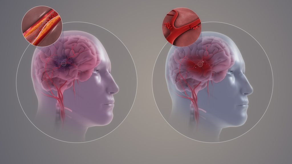
Ischaemic and haemorrhagic stroke. Credit: Scientific Animations CC4.0
When a person suffers a stroke, physicians must restore blood flow to the brain as quickly as possible to save their life. But, ironically, that life-saving rush of blood can also trigger a second wave of damage — killing brain cells, fuelling inflammation and increasing the odds of long-term disability.
Now, Northwestern University scientists have developed an injectable regenerative nanomaterial that helps protect the brain during this vulnerable window.
In a new preclinical study, the team delivered a single intravenous dose, immediately after restoring blood flow, in a mouse model of ischemic stroke, the most common type of stroke. The therapy successfully crossed the blood-brain barrier — a major challenge for most drugs — to reach and repair brain tissue. The material significantly reduced brain damage and showed no signs of side effects or organ toxicity.
Published in the journal Neurotherapeutics, the findings suggest the new therapy could eventually complement existing stroke treatments by limiting secondary brain injury and supporting recovery.
“Current clinical approaches are entirely focused on blood flow restoration,” said co-corresponding author Dr Ayush Batra, associate professor at Northwestern and a neurocritical care physician with Northwestern Medicine. “Any treatment that facilitates neuronal recovery and minimises injury would be very powerful, but that holy grail doesn’t yet exist. This study is promising because it’s leading us down a pathway to develop these technologies and therapeutics for this unmet need.”
The injectable therapy is based on supramolecular therapeutic peptides (STPs), a platform developed by Northwestern’s Samuel I. Stupp. A study published in 2021 in the journal Science demonstrated the use of an STP technology — nicknamed “dancing molecules” — because of the highly dynamic nature of its therapeutic agents that could reverse paralysis and repair tissue in mice after a single injection at the site of severe spinal cord injury. The new study found scientists can administer similar dynamic assemblies of molecules intravenously, without requiring surgery or an invasive injection directly into the brain.
“One of the most promising aspects of this study is that we were able to show this therapeutic technology, which has shown incredible promise in spinal cord injury, can now begin to be applied in a stroke model and that it can be delivered systemically,” said Stupp, co-corresponding author. “This systemic delivery mechanism and the ability to cross the blood-brain barrier is a significant advance that could also be useful in treating traumatic brain injuries and neurodegenerative diseases such as ALS.”
Study mimicked real-world stroke treatment
Acute ischaemic stroke is a devastating condition and is one of the leading causes of morbidity and mortality worldwide, Batra said, severely impacting a patient’s quality of life and engagement in society.
“It has not only a significant personal and emotional burden on patients, but also a financial burden on families and communities,” he said. “Reducing this level of disability with a therapy that could potentially help in restoring function and minimising injury would really have a powerful long-term impact.”
The findings are highly relevant for future clinical applications because the scientists tested the approach in a mouse model that closely mimics real-world ischemic stroke treatment, Batra said. They first blocked blood flow to simulate a major ischaemic stroke and then restored it (a process called reperfusion), just as whem doctors restore blood flow acutely for ischaemic stroke patients.
The scientists monitored the mice for seven days and didn’t observe any significant side effects or biocompatibility issues such as toxicity or immune system rejection. They used advanced imaging techniques, such as real-time intravital intracranial microscopy seen in this video, to confirm the therapy localised to the stroke injury site. Compared to untreated mice, those treated with the “dancing molecules” had significantly less brain tissue damage, reduced signs of inflammation and reduced signs of excessive, damaging immune response.
Stupp said the therapy has pro-regenerative and anti-inflammatory properties, both of which contributed to the positive results.
“You get an accumulation of harmful molecules once the blockage occurs and then suddenly you remove the clot and all those ‘bad actors’ get released into the bloodstream, where they cause additional damage,” Stupp said. “But the dancing molecules carry with them some anti-inflammatory activity to counteract these effects and at the same time help repair neural networks.”
Dynamic ‘dancing molecules’ can be dialed down in concentration
The secret behind Stupp’s “dancing molecules” breakthrough therapeutic is tuning the collective motion of molecules, so they can find and properly engage constantly moving cellular receptors. The treatment sends signals that encourage nerve cells to repair themselves. For example, it can help nerve fibres (called axons) grow again and reconnect with other nerve cells, restoring lost communication through neural plasticity.
In previous studies, scientists injected the dancing molecules as a liquid, and when used to treat spinal cord injury, the therapy immediately gels into a complex network of nanofibres that mimic the dense, extracellular matrix of the spinal cord. By matching the matrix’s structure, mimicking the motion of biological molecules and incorporating signals for receptors, the synthetic materials are able to communicate with cells.
In the new study, the scientists dialled down the concentration of supramolecular peptide assemblies to prevent possible clotting as the therapy enters the bloodstream. Smaller aggregates of peptides easily crossed the blood-brain barrier. Once enough molecules cross, larger nanofibre assemblies can form in brain tissue to produce a more potent therapeutic effect, Stupp said.
“We chose for this stroke study one of the most dynamic therapies we had in terms of its molecular structure so that supramolecular assemblies would have a better probability of crossing the blood-brain barrier,” Stupp said.
Optimiaing therapeutic targeting
The fact that seemingly effective therapies cannot cross the blood-brain barrier has plagued the neuroscience field for decades, Batra said. This new therapy could change that.
When a physician acutely restores blood flow to a region of the brain in a stroke patient, the blood-brain barrier permeability is locally increased, naturally creating a transient opening and opportunity for therapeutic intervention, Batra said.
“Add to that a dynamic peptide that is able to cross more readily, and you’re really optimising the chances that your therapy is going where you want it to go,” Batra said.
Next steps
Further studies will need to assess whether this treatment can support longer-term, functional recovery, Batra said. For instance, many stroke patients suffer from significant cognitive decline throughout the subsequent year after a stroke. The new therapy is primed to address that secondary injury, Batra said, but the studies will require a longer follow-up period and more sophisticated behavioral testing.
In addition, the team is interested in testing whether additional regenerative signals could be incorporated into the therapeutic peptides to produce even better results.
Mycobacterium tuberculosis drug susceptibility test. Photo by CDC on Unsplash
A University of Virginia-led team of researchers has made a discovery that may change sepsis treatment for patients in Africa.
Over the course of five years, the researchers studied patients with HIV-related sepsis in eastern Africa, discovering that the most common cause of sepsis was tuberculosis and that treating it immediately, even before a tuberculosis diagnosis was made, significantly improved survival rates.
Sepsis, or critical illness due to infection, is the leading global cause of death, responsible for an estimated one-fifth of deaths worldwide.
“We designed a trial with colleagues in Tanzania and Uganda to look specifically at people living with HIV, who suffer higher rates of sepsis and are more likely to die when they contract it,” said Dr Scott Heysell, director of the UVA Center for Global Health Equity and the co-lead investigator of the study. “Over half of the people enrolled in this trial were ultimately found to have tuberculosis and, if they immediately received tuberculosis treatment, they were significantly more likely to survive.”
Funded by a grant from the National Institutes of Health, the research, dubbed the “ATLAS study,” was done by a team of nearly 30 doctors, nurses, pharmacists, study coordinators and statisticians, including leading HIV and tuberculosis physician-scientists, Dr Stellah Mpagama from Kibong’oto Infectious Diseases Hospital in Tanzania, and Dr Conrad Muzoora, from the Mbarara University of Science and Technology in Uganda.
“The trial is the culmination of almost 20 years of collaborative work with colleagues in Uganda and Tanzania to better understand, diagnose and manage sepsis,” said co-lead investigator Dr Christopher Moore, professor of medicine and global health equity at the UVA School of Medicine. “The results of ATLAS have broad and significant implications for the treatment of sepsis in Africa, an all too common and deadly illness, which sadly is likely to become even more common with the advent of global public health funding cuts.”
It is often difficult to diagnose tuberculosis, so the team had to use newer and more exhaustive testing, according to Heysell.
“It is a tragedy to be on the front lines and witness the excessive mortality and morbidity from sepsis and tuberculosis, particularly among people with HIV,” said Dr Tania Thomas, a contributing researcher and associate professor of infectious diseases and international health at UVA. “These are treatable conditions, but time is rarely on our side. Until we have more accurate rapid diagnostic tests for tuberculosis, we are pleased to demonstrate that the strategy of immediate tuberculosis treatment can improve survival.”
The team has received additional NIH funding this year to continue its work through a new trial at four hospitals in Tanzania and Uganda to test whether the use of hydrocortisone to reduce inflammation and improve blood pressure, and/or an immediate treatment for tuberculosis and other bacterial pathogens, will improve 28-day mortality from HIV-related sepsis.
“In programmatic settings, tuberculosis treatment was mostly the same as for people without HIV, even though their health needs are more complex,” said Dr Mpagama. “Many of these patients have multiple infections at the same time, which makes their care more challenging.”
The research is part of UVA’s Center for Global Health Equity’s effort to establish meaningful, two-sided research partnerships in Eastern Africa, according to Heysell, who is working to increase educational and research opportunities outside of the US for UVA students. This includes coordinating clinical electives for medical students and other health science students in hospitals and clinics abroad.
To that end, emergency medicine professor Dr Amita Sudhir has been promoted to inaugural director for global health training within the center. Her goal will be to increase abroad opportunities for medical students within existing partnering organisations.
South Africa’s water quality monitoring gap explained
By Robert Erasmus, Managing Director at Sanitech
Source: Unsplash CC0
Access to safe and clean water remains a critical concern in South Africa. Recent incidents, including a highly contaminated water sample from Secunda that showed extreme levels of E. coli, have highlighted the urgent need for faster and more reliable water quality monitoring across the country. Public health depends on the safety of the water flowing through our rivers, dams and municipal systems, yet current monitoring processes often struggle to detect contamination before it reaches communities. Improving these systems will require a combination of practical testing methods, independent oversight and community involvement.
Why traditional testing cannot keep up South Africa’s public water testing framework is accurate but slow. When contamination is suspected, samples must be collected, couriered to an accredited laboratory and cultured to detect biological threats such as E. coli. While potential of Hydrogen (pH) and chlorine levels can be measured quickly on site, biological tests take one to two weeks because the organisms must be grown before results can be confirmed. This delay leaves a dangerous gap in which water quality could deteriorate without immediate detection.
The process is also costly. A single accredited test, including logistics, can cost around R5 000, which makes frequent testing inaccessible for households and many community organisations. As a result, many people rely on the assumption that water from the tap is safe. When contamination does occur, individuals may fall ill without realising the cause because there is no real time feedback on water quality.
How in-house testing can speed up detection Although accredited labs are still required for official reporting, new approaches are emerging that can help organisations identify risks earlier. Some companies are now investing in equipment that allows them to carry out basic testing in house. These tests are not accredited but they give fast, useful readings that act as early warning indicators. If an organisation detects abnormal results, it can immediately escalate the matter to an accredited lab instead of waiting for contamination to spread.
Routine pH and chlorine monitoring also plays a valuable role. These tests are inexpensive, easy to perform and can be carried out continuously within businesses or local facilities. While they cannot detect biological contamination, they help ensure that the chemical balance of the water stays within safe limits. When combined with monthly or cyclical biological testing, this creates a more proactive monitoring system.
This approach recently proved critical in Secunda, where a business conducting its own branch-level testing discovered that municipal water entering the site was contaminated with sewage. The in-house test flagged the issue quickly, prompting further investigation. Without this internal programme, the problem might have gone unnoticed for far longer.
Why collaboration improves water safety A stronger water monitoring system cannot rely on public authorities alone. Partnerships between municipalities, private companies and communities can help improve both the speed and reliability of responses. Independent testing at business level introduces greater transparency and can highlight water quality issues that may otherwise go unreported. When patterns of poor quality emerge, communities gain evidence to push for corrective action.
Transparency also drives accountability. If businesses in a region consistently report poor water quality, it becomes more difficult for the problem to remain hidden. Public pressure increases and municipalities have a clearer picture of where urgent interventions are needed. This type of shared visibility is essential for strengthening trust and promoting faster action.
Communities have an important role as well. Residents are often the first to notice discolouration, odour or unusual cloudiness in their tap water. Reporting these signs to employers or organisations with the means to test can lead to early detection. Raising issues solely through political channels may not always lead to immediate investigation, but involving local businesses can create quicker pathways to testing and response.
A path toward safer and more reliable water A safer water future for South Africa will depend on strengthening both formal and informal monitoring systems. Accredited labs remain vital for official results, yet in house testing, routine checks and community reporting can highlight risks long before formal samples are processed. When contamination is confirmed, solutions like filtration, Ultraviolet (UV) treatment or proper chlorination can be deployed quickly to restore safety.
What this shows is simple: the safety of tap water cannot be taken at face value. Consistent monitoring and transparent reporting are key to safeguarding public health. With better coordination between public bodies, private organisations and communities, South Africa can build a water monitoring system that identifies problems early and protects every household.
Pretoria, 8 January 2026 – The South African Health Products Regulatory Authority (SAHPRA) has been made aware of products in the market containing Zinc picolinate (as a source material for zinc) and/or Selenium intended for use in children.
The safety concerns related to children are as follows:
Zinc picolinate, at any supplemental dose, can cause side effects which include indigestion, diarrhoea, headache, nausea, and vomiting. As the bio-availability of Zn from Zn-picolinate is variable due to multiple factors, the risk of side effects may be higher and unpredictable, and it is unsuitable as a source of elemental zinc supplementation in children; and
Selenium, when supplemented to children, represents a safety concern considering the potential differences in selenium daily intake between different population groups. While selenium intake is a viable requirement for children in areas of famine or dietary restriction, the potential adverse effects of selenium overdose are of concern when provided in general supplements/medicines intended for children.
The products currently on the market are marketed and sold, among others, as “Immune boosters” for children, with the main active ingredients being Zinc (when derived from Zinc picolinate) and/or Selenium intended for use in children. These products are indicated for supporting the treatment of colds, flu, diarrhoea, and skin-related conditions, rendering the products in question medicines that require registration by SAHPRA.
Any medicine sold that contains Zinc picolinate or Selenium intended for use in children does not qualify as a Category D (complementary) medicine. As such, their sale as a Category D medicine is illegal. Therefore, with effect from the date of publication of this notice, all selenium and zinc picolinate-containing products intended for use in children shall be subject to registration as a medicine falling into Category A, as defined in Section 14(2) of the Medicines and Related Substances Act, 101 of 1965, and need to be submitted to SAHPRA for registration. The sale of Category D (complementary) medicines containing Zinc picolinate or Selenium and intended for use in children must be withdrawn from the market within six (6) months of the date of this publication.
Advice for health professionals and distributors:
SAHPRA requests that Health professionals cease all distribution, selling, and/or dispensing and remove all selenium and zinc picolinate-containing products intended for use in children from stores, storage facilities, and shelves.
Members of the public are urged to return products containing Zinc Picolinate and Selenium when intended for use in children, to their pharmacist, supplying warehouse, or distributor.
Reporting side effects
Public and healthcare professionals are encouraged to report any side effects after using a health product by using the Med Safety App. Your report will contribute to our monitoring of these health products.
Your guide to safe, effective, and natural hair restoration in 2026
Hair restoration is one of the fastest-growing aesthetic procedures worldwide. The International Society of Hair Restoration Surgery (ISHRS) reports hundreds of thousands of procedures performed globally each year, with demand climbing steadily. As more people seek confidence-boosting solutions to start the new year, South African specialists warn that choosing the wrong clinic can turn a life-changing decision into lasting damage.
Dr Kashmal Kalan, Medical Director at Alvi Armani South Africa, explains: “January brings a sense of renewal. Many people reassess their goals, and hair restoration has become one of the most transformative ways to invest in yourself. It’s no longer just about fitness or weight loss – hair and skin now play a central role in personal confidence.”
A successful hair restoration journey begins long before the procedure and continues well beyond it. At Alvi Armani, every patient undergoes a thorough, personalised consultation. The team evaluates hair loss patterns, donor density, scalp condition, hair type, and personal goals. Advanced AI-assisted microscopic analysis helps ensure patients are suitable and that the procedure is planned for optimal, natural results.
On procedure day, patients enter a calm, controlled environment. Hairline design is finalised, Follicular Unit Extraction (FUE) is performed with precision, and grafts are implanted to follow the natural flow of hair. Recovery is gradual, with initial shedding giving way to new growth. Density and texture refine over 12-18 months, and ongoing check-ins ensure progress stays on track.
Hairline design is the most artistic aspect of the process. Age, facial symmetry, ethnicity, and donor capacity all influence the final outcome. “We aim for perfection within imperfection. The goal is a hairline that complements the face naturally. No one should be able to tell a transplant took place.”
Strategic density planning is equally critical. Every follicle in the donor area is finite, and poor planning can create gaps or thin patches. This can leave permanent aesthetic imbalance. Reputable clinics plan for decades, not just the first few months. Patients should also understand that growth is gradual, and progressive hair loss may require more than one procedure to achieve the desired result.
Alvi Armani ensures every procedure is doctor-led and supported with ongoing care, including stabilisation medications, regenerative therapies, and annual check-ups. Dr Kalan cautions against so-called “dark clinics” offering prices too good to be true, often operating in unhygienic or mobile facilities. “These clinics treat hair restoration as a commodity rather than medicine. They overharvest donor areas, produce unnatural results, and leave patients needing urgent repairs. Repair procedures now make up roughly a quarter of our cases.”
Beyond procedural excellence, Alvi Armani educates patients on lifestyle choices that support lasting results, from nutrition and scalp care to ongoing therapies. While the process requires patience, the rewards – confidence, natural appearance, and the security of a clinic that plans carefully for the future – make it worthwhile.
For anyone considering hair restoration in 2026, the advice is clear: invest in quality from the start. With the right clinic, personalised planning, and medical oversight, patients can achieve safe, natural results that endure for years to come.